Observational Study
. 2025 Apr 9;12(1):e003196.
doi: 10.1136/openhrt-2025-003196.

Prevalence of ventricular parasystole in patients with cardiac sarcoidosis: correlation between parasystole and inflammation in ventricular fibrillation

Affiliations
Observational Study

Prevalence of ventricular parasystole in patients with cardiac sarcoidosis: correlation between parasystole and inflammation in ventricular fibrillation

Takashi Ikee et al. Open Heart. .

Abstract

Background: Ventricular parasystole is strongly associated with ventricular fibrillation (VF) in patients with non-ischaemic cardiomyopathy. However, the relationship between ventricular parasystole and cardiac sarcoidosis (CS) remains unclear. The purpose of this study was to examine the prevalence of parasystole in patients with CS.

Methods: This was a retrospective observational study of 214 consecutive patients diagnosed with CS (mean age: 69±12 years, 104 males, median follow-up period: 6.8 years (IQR: 3.2-10.7) in our centre. We investigated parasystole in the patients who developed ventricular arrhythmia (VA) using 9886 ECGs, 280 Holter ECGs and 6391 implantable cardioverter defibrillator interrogation records. Classic parasystole was defined as three ventricular ectopic beats with the same morphology, occurring at integer-multiple intervals but with different coupling intervals (CI) on ECG. New parasystole was defined as two ventricular ectopic beats with a CI difference of more than 120 ms. We also analysed the correlation between inflammation sites and parasystole morphology observed on a 12-lead ECG.

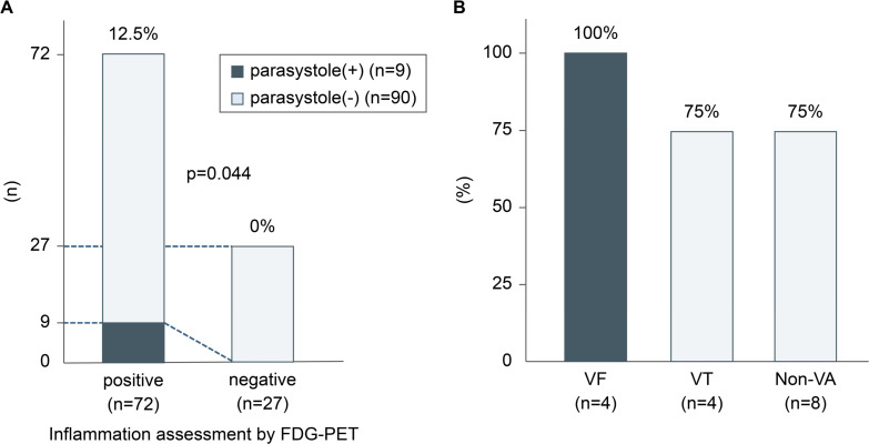
Results: VA was identified in 95 patients (33.7%), and 22 developed VF (23.2%). Parasystole was observed in 12 of the 22 patients with VF (classic: 5, new: 7), 20 of 73 with ventricular tachycardia (classic: 5, new: 15) and 44 of 118 without VA (classic: 16, new: 28). Parasystole was significantly more common in the VF group than in the non-VF group (p=0.049). The site of inflammation observed on 18F-fluorodeoxyglucose positron emission tomography performed within 3 months after the development of VA and the origin of parasystole matched in all four patients with VF who had 12-lead ECG records of parasystole. Inflammation was correlated with the origin of parasystole.

Conclusion: Ventricular parasystole was detected in one-third of patients with CS in this study, especially those with VF. The presence of parasystole and inflammation may predict the occurrence of VF in patients with CS.

Keywords: Inflammation; Myocarditis; Tachycardia, Ventricular; Ventricular Fibrillation; Ventricular Premature Complexes.


Conflict of interest statement

Competing interests: KK received speaker honoraria from Daiichi Sankyo Company, and Medtronic Japan, and research grants from Medtronic Japan and JSR. KM received funding/grants from Medtronic, Biosense Webster, Abbott and Boston and honoraria/speakers’ bureaus from Medtronic, Biosense Webster, Abbott and Boston outside the submitted work, and is affiliated with a department endowed by Medtronic outside the submitted work.

Figures

Figure 1
Figure 1. Definition of ventricular parasystole. (A) Classic parasystole was defined as PVCs with the same morphology and coupling intervals different from the previous QRS complex within the same continuous ECG recording. At least three PVCs appear at intervals measured using callipers, occurring multiple times in a fixed interval. (B) New parasystole was defined as PVCs with the same morphology and higher coupling interval variability than that expected of non-parasystolic PVCs (defined as at least a 120 ms difference between the shortest and longest coupling intervals within the same continuous ECG strip), even if the presence of three beats at a constant interval could not be demonstrated. PVC, premature ventricular contraction.
Figure 2
Figure 2. Prevalence of parasystole in each group. (A) Total prevalence (classic and new) of parasystole in the VF and non-VF groups. (B) Total prevalence of parasystole in the VF, VT and non-VA groups. (C) Prevalence of classic parasystole in the VF, VT and non-VA groups. VA, ventricular arrhythmia; VF, ventricular fibrillation; VT, ventricular tachycardia.
Figure 3
Figure 3. Flowchart of the identification of the patients who had data on the identification of inflammatory sites and origin of parasystole on FDG-PET. Parasystole was observed in 12 patients in the VF group (classic: 5, new: 7), 20 patients in the VT group (classic: 5, new: 15) and 45 patients in the non-VA group (classic: 16, new: 29). Among these, patients without 12-lead ECG data were excluded (6 in the VF group, 11 in the VT group and 27 in the non-VA group). Additionally, two patients in the VF group who showed diffuse PET uptake were excluded. Four and nine patients in the VT and non-VA groups, respectively, did not undergo PET scans within 3 months. In addition, one patient from each group showed no inflammation on FDG-PET. Finally, the relationship between the site of inflammation detected on FDG-PET and the origin of parasystole was evaluated in four patients in the VF group, four in the VT group and eight in the non-VA group. FDG-PET, 18F-fluorodeoxyglucose-positron emission tomography; VA, ventricular arrhythmia; VF, ventricular fibrillation; VT, ventricular tachycardia.
Figure 4
Figure 4. (A) Correlation between the timing of parasystole and the period of inflammation detected on FDG-PET in patients with VF. (B) Origin of premature ventricular contractions of a parasystole matching the inflammatory site detected on FDG-PET. FDG-PET, 18F-fluorodeoxyglucose-positron emission tomography; VA, ventricular arrhythmia; VF, ventricular fibrillation; VT, ventricular tachycardia.

References

    1. Lehtonen J, Uusitalo V, Pöyhönen P, et al. Cardiac sarcoidosis: phenotypes, diagnosis, treatment, and prognosis. Eur Heart J. 2023;44:1495–510. doi: 10.1093/eurheartj/ehad067. - DOI - PMC - PubMed
    1. Kandolin R, Lehtonen J, Airaksinen J, et al. Cardiac sarcoidosis: Epidemiology, characteristics, and outcome over 25 years in a nationwide study. Circulation Wolters Kluwer Health. 2015;131:624–32. doi: 10.1161/CIRCULATIONAHA.114.011522. - DOI - PubMed
    1. Kusano KF, Satomi K. Diagnosis and treatment of cardiac sarcoidosis. Heart. 2016;102:184–90. doi: 10.1136/heartjnl-2015-307877. - DOI - PubMed
    1. Segawa M, Fukuda K, Nakano M, et al. Time Course and Factors Correlating With Ventricular Tachyarrhythmias After Introduction of Steroid Therapy in Cardiac Sarcoidosis. Circ: Arrhythmia and Electrophysiology. 2016;9 doi: 10.1161/CIRCEP.115.003353. - DOI - PubMed
    1. Peretto G, Sala S, Rizzo S, et al. Ventricular arrhythmias in myocarditis: Characterization and relationships with myocardial inflammation. J Am Coll Cardiol. 2020;75:1046–57. doi: 10.1016/j.jacc.2020.01.036. - DOI - PubMed

Publication types

MeSH terms

LinkOut - more resources