. 2025 May 2;39(9-10):634-651.
doi: 10.1101/gad.352311.124.

CDK4 loss-of-function mutations cause microcephaly and short stature

Affiliations

CDK4 loss-of-function mutations cause microcephaly and short stature

Aitana Verdu Schlie et al. Genes Dev. .

Abstract

Cell number is a major determinant of organism size in mammals. In humans, gene mutations in cell cycle components result in restricted growth through reduced cell numbers. Here we identified biallelic mutations in CDK4 as a cause of microcephaly and short stature. CDK4 encodes a key cell cycle kinase that associates with D-type cyclins during G1 of the cell cycle to promote S-phase entry and cell proliferation through retinoblastoma (RB) phosphorylation. CDK4 and CDK6 are believed to be functionally redundant and are targeted jointly by chemotherapeutic CDK4/6 inhibitors. Using molecular and cell biology approaches, we show that functional CDK4 protein is not detectable in cells with CDK4 mutations. Cells display impaired RB phosphorylation in G1, leading to G1/S-phase transition defects and reduced cell proliferation, consistent with complete loss of cellular CDK4 enzymatic activity. Together, these findings demonstrate that CDK4 is itself required for cell proliferation, human growth, and brain size determination during development.

Keywords: cell cycle; centrosome; cyclin-dependent kinase; microcephalic dwarfism; microcephaly.


Figures

Figure 1.
Figure 1.
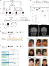
Individuals with biallelic CDK4 variants display microcephaly and short stature. (A) Family pedigrees with segregation of CDK4 variants. (Square) Male, (circle) female, (filled symbols) individuals with microcephaly, (strikethrough) deceased. WT Reference (+), variants v1 and v2, and zygosity are indicated for each studied individual. (B) Diagram of CDK4 transcript (top) and protein (bottom); coding exons are depicted as black rectangles. Red lines indicate variant location. (SS) Splice site disrupted. (C) Altered splicing predictions for the c.218G > A substitution generated using Alamut. (Blue rectangles) Strength of splice donor predictions for individual splice algorithms, (blue triangle) predicted donor splice site. (D) Growth parameters at birth and at last assessment (postnatal). (W) Weight, (OFC) orbito–frontal circumference. Z-scores show standard deviations from population mean for age and sex. Dashed lines indicate a 95% confidence interval for the general population. Individual subject data points from families A (circles) and B (squares) are graphed, and mean values are plotted. (E) MRI scan of age-matched control (4 years 8 months) and affected individuals with a CDK4 variant. Coronal FLAIR projection shows simplified parietal and temporal gyri, reduced white matter volume, and the absence of brain malformations. Scale bars, 10 cm. (See also Supplemental Figure S1C for additional MRI projections.) (F) Photographs of all affected individuals.
Figure 2.
Figure 2.
Transcriptional consequences of CDK4 mutations. (A) Transcript analysis by RT-PCR of RNA extracted from primary fibroblasts. Agarose gel electrophoresis of RT-PCR products using CDK4 5′ and 3′ UTR primers. A full-length (t1) transcript of 912 bp and a shortened (t2) one were seen in P1 (v2), whereas P2 (v1) exhibited a predominant smaller transcript (t3). (B) Schematics of detected transcripts and their relative quantification (percent transcript) based on qPCR results presented in C; the corresponding predicted proteins are shown at the right. Supplemental Figure S2 presents Sanger sequences of cloned CDK4 transcripts after RT-PCR. (C) qPCR analysis of WT control (C1 and C2) and patient-specific CDK4 transcripts relative to control. Primer locations for each qPCR reaction are indicated above each bar graph. n = 3 experiments; mean ± SEM; two-tailed t-tests.
Figure 3.
Figure 3.
Full-length CDK4 protein is undetectable in patient fibroblasts. (A,B) Immunoblots of total cell extracts obtained from exponentially growing control (C1 and C2) and patient (P1 and P2) fibroblasts without (A) and with (B) CDK4 complementation. α-Tubulin was used as the loading control. A rabbit monoclonal antibody to C-terminal CDK4 was used; a different mouse CDK4 antibody raised against full-length CDK4 was used in Figure 5A. A smaller ∼12 kDa molecular weight band was variably detected in P1 with this antibody (Supplemental Fig. S2D) that might correspond to the 46 amino acid truncated nonfunctional protein predicted from RNA studies. (C) CDK6 and Cyclin D1 levels were unchanged in patient fibroblasts compared with wild-type controls.
Figure 4.
Figure 4.
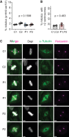
CDK4 mutations do not alter mitosis. (A) Percentage of mitotic cells (p-Histone H3 ser10-positive) in control (C1 and C2) and patient (P1 and P2) fibroblasts as measured by flow cytometry. Data points are from three independent experiments (two for C1); one-way ANOVA with Tukey post test; mean ± SEM. (B) Quantification of metaphase cells with more than two centrosomes, expressed as percentage. Numbers of cells analyzed were as follows: C1, 79; C2, 94; P1, 150; and P2, 101. Two-tailed t-test; mean ± SEM; measurements were pooled from two independent experiments. (C) Representative confocal images of control (C1 and C2) and patient (P1 and P2) fibroblasts fixed and stained for DAPI (gray), α-tubulin (green), and pericentrin (magenta). Scale bars, 5 µm.
Figure 5.
Figure 5.
CDK4 mutations impair G1-to-S progression and lead to reduced cell proliferation. (A) Western blot of control and patient-derived fibroblasts with and without WT CDK4 complementation. (B, left) Growth curves of control and patient-derived fibroblasts with and without WT CDK4 complementation. (Right) Bar graph showing quantification of doubling times; one-way ANOVA with Tukey post test. P-values are indicated; mean ± SEM. (C) Cell cycle distribution (G0/G1, S, and G2/M) derived from BrdU and DNA (DAPI) flow cytometry scatter plots show fewer cells in S phase (BrdU+) in patient-derived fibroblasts compared with controls. n = 3 independent experiments; mean ± SEM. Gates are shown on representative plots at the right. (D) Cell cycle distribution after complementation of patient-derived fibroblasts with CDK4. Reduced G0/G1 and increased S-phase populations consistent with rescue of a G1/S progression defect. n = 3 independent experiments; mean ± SEM. (See also Supplemental Fig. S4A.) (E) Quantification of DNA synthesis rate (BrdU mean fluorescence intensity [MFI] of gated population in the red rectangle) from experiments depicted in C.
Figure 6.
Figure 6.
CDK4 mutations impair retinoblastoma phosphorylation in G1. (AE) Quantitative image-based cytometry (QIBC). (A) Gating strategy for cell cycle stages by DNA content (DAPI) and EdU incorporation. (B) Representative DAPI versus pRB-ser807/811 scatter plots demonstrate impaired RB phosphorylation in G1 in CDK4-deficient cells (P1) relative to control 1. (C) Representative scatter plot of total RB levels demonstrating equivalent levels of RB between C1 and P1. Data points for C and D individual cells: n > 1500 cells/sample in each independent experiment. (D,E) Quantification of pRB-ser807/811 (D) and total RB (E) fluorescence intensity per nucleus show significantly reduced pRB-ser807/811 and normal total RB levels in G0/G1 in CDK4-deficient fibroblasts relative to controls. Mean ± SEM; n ≥ 4 independent experiments with 72 images/condition, totaling ≥1500 cells/sample or condition in each experiment analyzed.

References

    1. Asif M, Abdullah U, Nurnberg P, Tinschert S, Hussain MS. 2023. Congenital microcephaly: a debate on diagnostic challenges and etiological paradigm of the shift from isolated/non-syndromic to syndromic microcephaly. Cells 12: 642. 10.3390/cells12040642 - DOI - PMC - PubMed
    1. Baker SJ, Poulikakos PI, Irie HY, Parekh S, Reddy EP. 2022. CDK4: a master regulator of the cell cycle and its role in cancer. Genes Cancer 13: 21–45. 10.18632/genesandcancer.221 - DOI - PMC - PubMed
    1. Bracken AP, Ciro M, Cocito A, Helin K. 2004. E2f target genes: unraveling the biology. Trends Biochem Sci 29: 409–417. 10.1016/j.tibs.2004.06.006 - DOI - PubMed
    1. Chen S, Francioli LC, Goodrich JK, Collins RL, Kanai M, Wang Q, Alföldi J, Watts NA, Vittal C, Gauthier LD, et al. 2024. A genomic mutational constraint map using variation in 76,156 human genomes. Nature 625: 92–100. 10.1038/s41586-023-06045-0 - DOI - PMC - PubMed
    1. Chung M, Liu C, Yang HW, Köberlin MS, Cappell SD, Meyer T. 2019. Transient hysteresis in CDK4/6 activity underlies passage of the restriction point in G1. Mol Cell 76: 562–573.e4. 10.1016/j.molcel.2019.08.020 - DOI - PMC - PubMed

MeSH terms

LinkOut - more resources