. 2025 Apr 11;16(1):3437.
doi: 10.1038/s41467-025-58693-7.

Molecular basis for the phosphorylation of bacterial tyrosine kinase Wzc

Affiliations

Molecular basis for the phosphorylation of bacterial tyrosine kinase Wzc

Yun Yang et al. Nat Commun. .

Abstract

The regulation of polymerisation and translocation of biomolecules is fundamental. Wzc, an integral cytoplasmic membrane tyrosine autokinase protein serves as the master regulator of the biosynthesis and export of many bacterial capsular polysaccharides and exopolysaccharides. Such polysaccharides play essential roles in infection, defence, and some are important industrial products. Wzc comprises a large periplasmic domain, two transmembrane helices and a C-terminal cytoplasmic kinase domain with a tyrosine-rich tail. Wzc regulates polymerisation functions through cycling the formation and dissociation of an octameric complex, driven by changes in the phosphorylation status of the tyrosine-rich tail. E. coli Wzc serves a model for a wider family of polysaccharide co-polymerases. Here, we determine structures of intermediate states with different extents of phosphorylation. Structural and computational data reveal the pre-ordering of the tyrosine-rich tail, the molecular basis underlying the unidirectionality of phosphorylation events, and the underlying structural dynamics on how phosphorylation status is transmitted.


Conflict of interest statement

Competing interests: The authors declare no competing interests.

Figures

Fig. 1
Fig. 1. Wzc and the Wzx-Wzy pathway.
a Schematic model for the assembly and export of capsular polysaccharide (CPS) in E. coli K30, the prototype for Wzy-dependent CPS biogenesis. The repeat unit of E.coli K30 CPS is synthesized on an undecaprenol diphosphate carrier lipid at the cytoplasmic side of the inner membrane (IM) by a phosphoglycosyltransferase (PGT; WbaP) and glycosyltransferases (GT(s); WbaZ, WcaO and WcaN), and flipped to the periplasmic face of the membrane by Wzx. The repeat units are polymerized by Wzy, and the polymers are exported by Wza. Polymerization and export are regulated by Wzc (a PCP-2a family autokinase), which cycles between a phosphorylated state and dephosphorylated state due the activity of the Wzb phosphatase. Wzi is involved in the surface attachment of CPS. b The gene cluster for the synthesis and export of the CPS of E. coli K30 (GenBank Acc. AF104912.3). c A cartoon of a Wzc monomer, comprising the periplasmic domain containing motif 1–3, two transmembrane helices, the BY-kinase domain, and the C-terminal tyrosine-rich tail (Y-tail) (adapted from).
Fig. 2
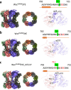
Fig. 2. Cryo-EM maps of Wzc mutants.
a WzcK540M2YE. b WzcK540M3YE. c WzcK540M3YE_N711Y. In each panel, on the left is shown the coulombic map of the entire molecule viewed from the side. The middle image shows the coulombic map of kinase domains only, viewed from the periplasm. The coulombic map of helix α23 and the tyrosine tail as they insert into the active site of the neighbouring kinase is shown on the right.
Fig. 3
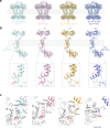
Fig. 3. Conformational snapshots of Wzc.
a The structures of the octamers reveal shifts in periplasmic and transmembrane domains relative to WzcK540M. b Superposition using the kinase domain of the monomer from each snapshot shows the movement in the transmembrane helices and periplasmic domain. Zoomed insets highlight conformational changes involving helices α1 and α23. c Structure of the C-terminal region beginning at residue 700 and helix α1 is highlighted, with the kinase domain of neighbouring monomer coloured grey.
Fig. 4
Fig. 4. Comparison of the structures of Wzc octamers.
a Side view, b top view comparing the structures of WzcK540M (PDB ID: 7NHR) and WzcK540M4YE (PDB ID: 7NIB). WzcK540M (PDB ID: 7NHR) is coloured gray, and WzcK540M4YE (PDB ID: 7NIB) is coloured by chain. c Structure comparison of the four states. The octameric cytoplasmic kinase domain is used for the structural alignment.
Fig. 5
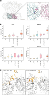
Fig. 5. Molecular dynamics analysis of phosphorylation.
a The portion of the structure used in the MD simulation and the metric used in comparison. The metric was defined as the distance between the carbon beta of the tyrosine (or glutamic acid) and the carbon alpha of D564, located in the active site of the adjacent subunit. b Boxplots of this metric for each structure. The boxplots represent the variation of the distance over time and the replicates (n = 3000 MD simulations frames. The box bounds the interquartile range divided by the median, with the whiskers extending to a maximum of 1.5 times the interquartile range beyond the box). See Supplementary Table 2 for the summary of the MD simulations, Supplementary Fig. 7 for the time trace. c Comparison between the initial and final frames of the 3YE (2pY) showing the displacement of E713 from the active site and the binding of Y708. Helix α1 is shown for reference but was not included in the simulations. Source data are provided as a Source Data file.
Fig. 6
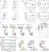
Fig. 6. Mechanism of the transmembrane signal transduction.
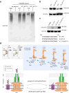
a The θTMH1,θTMH2,θα1 angles are defined as the angles relative to the axis normal to the membrane, and θTMH1TMH2 is the angle between TMH1 and TMH2. b Boxplot of the metrics for each system. The boxplots represent the variation of the angles over time and the replicates (n = 1500 MD simulations frames. The box bounds the interquartile range divided by the median, with the whiskers extending to a maximum of 1.5 times the interquartile range beyond the box). c Superposition (using the kinase domain) of the initial (white) and final frames (coloured) reveals structural shifts for each state of Wzc. d Time trace of the minimum distance between the residues D28 and K701. e Close up view showing the changes in the interactions between K701 and D28. See Supplementary Table 2 for the summary of the MD simulations. Source data are provided as a Source Data file. f Immunoblot (anti-K30 CPS) of whole cell lysates separated by SDS-PAGE. The wildtype E. coli strain E69 (serotype O9a:H12:K30) was used as a positive control, and E. coli strain CWG285 (Δwzc) was used as a negative control. WT, D28A, K701A, D28A/K701A represent plasmids encoding wild-type Wzc, Wzc with the D28A mutation, Wzc with the K701A mutations, and Wzc with the D28A/K701A mutations respectively. As CPS does not migrate strictly according to size, protein standard is not useful to indicate the molecular weight. These data are representative of three biological replicates.
Fig. 7
Fig. 7. The YxY motif regulates the activity of Wzy.
a Immunoblot (anti-K30 CPS) of whole cell lysates separated by SDS-PAGE. The wildtype E. coli serotype K30 strain (E69) was used as a positive control, and E. coli CWG285 (Δwzc) was used as a negative control. WT, WT_N711Y, 3YE, 3YE_N711Y represent plasmids encoding wild-type Wzc and derivatives with the N711Y, Y715E/Y717E/Y718E and Y715E/Y717E/Y718E/N711Y mutations, respectively. As CPS does not migrate strictly according to size, protein standard is not useful to indicate the molecular weight. These data are representative of three biological replicates. b Corresponding western immunoblot of purified Wzc proteins probed with anti-pTyr antibody (top) and anti-hexahistidine antibody (bottom). The experiment represents one of three technical replicates. c Western immunoblot of purified Wzc proteins using anti-pTyr antibody (top) and anti-hexahistidine antibody (bottom). The mutations are identified above each lane. The C-terminal sequences of the tyrosine-rich portion of YF_713Y, YF_N711Y and YF_713Y/N711Y are shown below. The data shows one of three technical replicates. d The ratchet mechanism. The YxY motif acts as a clamp stabilizing the octamer but also ordering the C-terminal tail (top). The tyrosine-rich tails in the octamer are phosphorylated and move unidirectionally through the active sites of adjacent monomers (top right). Phosphorylation progressively destabilizes the octamer which, after the fourth phosphorylation, disassembles (top right and bottom). Wzc regulates the polymerization activity of Wzy (purple) and is thought to form a complex with the translocon Wza (green). The cycling of the structural arrangement of the octamer is driven by (auto)phosphorylation and dephosphorylation (catalysed by Wzb) and the cycling is required for the production and export of CPS (bottom).

References

    1. Whitfield, C., Wear, S. S. & Sande, C. Assembly of bacterial capsular polysaccharides and exopolysaccharides. Annu. Rev. Microbiol.74, 521–543 (2020). - PubMed
    1. Ernst, C. M. et al. Adaptive evolution of virulence and persistence in carbapenem-resistant Klebsiella pneumoniae. Nat. Med.26, 705–711 (2020). - PMC - PubMed
    1. Le Bas, A. et al. Structure of WzxE the lipid III flippase for enterobacterial common antigen polysaccharide. Open Biol.15, 240310 (2025). - PMC - PubMed
    1. Meitil, I. K. S., Gippert, G. P., Barrett, K., Hunt, C. J. & Henrissat, B. Diversity of sugar-diphospholipid-utilizing glycosyltransferase families. Commun. Biol.7, 285 (2024). - PMC - PubMed
    1. Woodward, R. et al. In vitro bacterial polysaccharide biosynthesis: defining the functions of Wzy and Wzz. Nat. Chem. Biol.6, 418–423 (2010). - PMC - PubMed

MeSH terms

LinkOut - more resources