. 2025 Jun;57(6):1367-1373.
doi: 10.1038/s41588-025-02159-5. Epub 2025 Apr 10.

Mutations in the small nuclear RNA gene RNU2-2 cause a severe neurodevelopmental disorder with prominent epilepsy

Affiliations

Mutations in the small nuclear RNA gene RNU2-2 cause a severe neurodevelopmental disorder with prominent epilepsy

Daniel Greene et al. Nat Genet. 2025 Jun.

Abstract

The major spliceosome includes five small nuclear RNA (snRNAs), U1, U2, U4, U5 and U6, each of which is encoded by multiple genes. We recently showed that mutations in RNU4-2, the gene that encodes the U4-2 snRNA, cause one of the most prevalent monogenic neurodevelopmental disorders. Here, we report that recurrent germline mutations in RNU2-2 (previously known as pseudogene RNU2-2P), a 191-bp gene that encodes the U2-2 snRNA, are responsible for a related disorder. By genetic association, we identified recurrent de novo single-nucleotide mutations at nucleotide positions 4 and 35 of RNU2-2 in nine cases. We replicated this finding in 16 additional cases, bringing the total to 25. We estimate that RNU2-2 syndrome has a prevalence of ~20% that of RNU4-2 syndrome. The disorder is characterized by intellectual disability, autistic behavior, microcephaly, hypotonia, epilepsy and hyperventilation. All cases display a severe and complex seizure phenotype. We found that U2-2 and canonical U2-1 were similarly expressed in blood. Despite mutant U2-2 being expressed in patient blood samples, we found no evidence of missplicing. Our findings cement the role of major spliceosomal snRNAs in the etiologies of neurodevelopmental disorders.


Conflict of interest statement

Competing interests: The authors affiliated with deCODE genetics/Amgen Inc. (B.O.J., K. Stefansson and P.S.) are employed by the company. The other authors declare no competing interests.

Figures

Fig. 1
Fig. 1. Discovery and replication of RNU2-2 as an etiological gene for a new NDD.
a, BeviMed PPAs between each of RNU4-2 and RNU2-2 (previously known as RNU2-2P) and NDA. All other noncoding genes and pseudogenes had PPA < 0.5. Only two RNU2-2 variants had conditional PPP > 0.5: n.4G>A and n.35A>G. Prob., probability. b, Distribution of phenotypic homogeneity scores for 100,000 randomly selected sets of nine participants chosen from 9,112 unrelated NDA-coded participants. The score corresponding to the nine identified cases with one of the two RNU2-2 variants with PPP > 0.5 is indicated with a red line. c, Scatter plot of log10 expression of RNU2-1 against that of RNU2-2 in whole-blood samples from a random subset of 500 participants in the NGRL and in four blood cell types from 204 NBR participants. TPM, transcripts per million. d, Top, numbers of participants with a rare allele at each of the 191 bases of RNU2-2, stratified by affection status and inheritance information of the carried allele. The two variants with PPP > 0.5 are indicated with green arrows. The color-coded track shows the aggregated (over distinct alleles at a position) minor allele count (aMAC) in gnomAD v.4.1.0 (gn.) at each position, and the black bars show the numbers of distinct alternate alleles in gnomAD at each position (multiple insertions and multiple deletions at a given position each count as one). Variants failing quality control (QC) in gnomAD are not shown in this subpanel. Bottom, data corresponding to nucleotide positions 1 to 41 in greater detail, including gnomAD-QC-failing variant n.35A>T. Above and below the RNU2-2 cDNA sequence (Seq.), the alternate alleles in 100KGP participants and the distinct alleles in gnomAD are shown, respectively; ‘+’ indicates insertions, and the variant that failed QC in gnomAD is indicated. e, Pedigrees for participants with a rare alternate allele n.4 or n.35 in RNU2-2. Pedigrees used for discovery have a ‘G’ prefix and are labeled in black. Pedigrees used for replication in the IMPaCT-GENóMICA, URDCat and ENoD-CIBERER aggregate collection; the 100KGP; the NBR; Erasmus MC UMC; the GMS; Radboud UMC; deCODE or the ZOEMBA study have an ‘I’, ‘M’, ‘N’, ‘R’, ‘S’, ‘W’, ‘Y’ or ‘Z’ prefix, respectively, and are labeled in blue. Hom., homozygous; ref., reference.
Fig. 2
Fig. 2. Prevalence in the 100KGP.
Of the 9,112 unrelated NDA-coded cases in the 100KGP, the numbers solved through pathogenic or likely pathogenic variants in a gene are shown, provided at least nine cases were diagnosed. For RNU2-2, the number of NDA-coded cases in the 100KGP with one of the recurring de novo variants is shown.
Fig. 3
Fig. 3. Phenotypic enrichment in the 100KGP.
Graph showing the ‘is-a’ relationships among HPO terms present in at least three of the nine NDA-coded RNU2-2 cases in the discovery collection or significantly enriched among them relative to 9,112 unrelated NDA-coded participants of the 100KGP. The significantly overrepresented terms are highlighted. For each term, the number of cases with the term and the proportion that number represents out of nine is shown. For each overrepresented term, the proportion of NDA-coded participants with the term and the proportion of NDA-coded RNU2-2 cases with the term are represented as the horizontal coordinate of the base and the head of an arrow, respectively. *, Only eight of the nine (89%) of the cases had the ‘Seizure’ HPO term in the NGRL, but epilepsy was confirmed in the case without the HPO term by inspecting the individual’s electronic health record and the numbers attached to ‘Seizure’ were updated accordingly.
Fig. 4
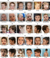
Fig. 4. Clinical photographs.
Clinical photographs of individuals from pedigrees G1, G4, S3, R1 and I1–6. The individuals in these cases show common features of long palpebral fissures with slight eversion of the lateral lower lids, long eyelashes, broad nasal root, large low set ears, wide mouth and wide spaced teeth. The approximate ages of the individuals when the photographs were taken are shown. Photographs of individual M2, who has Radio–Tartaglia syndrome in addition to RNU2-2 syndrome, are included in the Supplementary Note. We have obtained specific consent from the families to publish these clinical photographs. m, months; yr, years.
Extended Data Fig. 1
Extended Data Fig. 1. Effect on PPAs of relaxing the CADD score threshold.
Histograms of the posterior probability of association (PPA) between the 41,132 canonical Ensembl transcripts not annotated as being protein-coding and neurodevelopmental abnormality (NDA), with and without filtering out variants with a CADD v1.6 score <10. The CADD v1.6 scores for n.4 G > A, n.35 A > G and n.35 A > C were 7.7, 9.4 and 9.1, respectively. The more recent CADD v1.7 gives scores >10 for these variants.
Extended Data Fig. 2
Extended Data Fig. 2. RNA-seq coverage in the RNU2-2 locus.
Coverage of uniquely aligned RNA-seq reads from the whole blood of five RNU2-2 cases in the NGRL and in four blood cell types of an exemplar participant in the NBR demonstrating that RNU2-2 (previously annotated as the pseudogene RNU2-2P) is expressed abundantly in blood cells.
Extended Data Fig. 3
Extended Data Fig. 3. Location of the pathogenic variants in U2-2 snRNA within the major spliceosome.
Assembly of the spliceosome A complex is initiated by binding of the intronic 5′ splice site (5′SS) to the U1 snRNA and the intronic branch site sequence to the U2-2 snRNA through Watson-Crick pairing of cognate ribonucleotides. The branch site sequence is depicted as the human YNYUNAY consensus motif (Y means C or T; N means any ribonucleotide), which interacts with the GUAGUA sequence at positions 33 to 38 in the U2-2 snRNA (depicted in red). The spliceosome pre-B complex is formed by incorporation of the U4/U6.U5 tri-small nuclear ribonucleoprotein (snRNP) complex that contains the U4, U5 and U6 snRNAs. This requires interactions between U5 snRNA and the 5′ and 3′ exons and further interactions between nucleotides near the 3′ end of the U6 snRNA and a cognate CGCUUCUCG sequence (nucleotides 3–11) close to the 5′ end of the U2-2 snRNA (depicted in blue). Tethering of U4/U6.U5 tri-snRNP to U2-2 within the spliceosome pre-B complex enables displacement of U1 to enable a new interaction between U6 snRNA with the 5′SS and reconfiguration of U4/U6.U5 tri-snRNP to form the catalytically active spliceosome B complex, which is a prerequisite for the splicing reaction. The critical U6 snRNA region that interacts with the intronic 5′SS is maintained in correct orientation by conserved regions in the adjacent U4 snRNA (depicted in orange), which are the sites of destabilizing variants responsible for the recently described RNU4-2 syndrome. The variants responsible for RNU2-2 syndrome occur at critical interaction sites between U2-2 snRNA near r.4 and U6 snRNA and between U2-2 snRNA near r.35 and intronic branch sites. These interactions are necessary for intron recognition and the correct assembly of the catalytically active spliceosome B complex.
Extended Data Fig. 4
Extended Data Fig. 4. Mosaicism analysis.
a, For each of the three rare variants at positions n.4 and n.35 of RNU2-2 called in the discovery collection, truncated bar charts showing the distribution of the proportions of reads supporting the alternate allele over participants, partitioned into 0% and all left-open intervals of size 4% up to 100%. In contrast to n.4 G > A and n.35 A > G, the reads in the eight participants with the n.35 A > T heterozygous call exhibit a strong skew in favor of the reference allele. Furthermore, seven participants with a homozygous reference call at n.35 have at least 8% of aligned reads at that position supporting the ‘T’ allele, suggesting that n.35 A > T is not a germline variant, but rather a low-frequency somatic mosaic variant. b, Histogram of age at enrollment of participants in the discovery collection. The purple points show the age at enrollment of study participants with at least 8% of aligned reads supporting the ‘T’ allele at n.35. These participants are significantly older than expected by chance (P = 1.3 × 10⁻³, Kolmogorov-Smirnoff test). To comply with Genomics England’s rules on identifiability, all ages of at least 95 years are included in the same x = 95 bin. c, Sanger sequencing traces from an NDA case (in pedigree N1) with the n.4 G > A call, an unaffected participant with the n.35 A > T call, and a control with neither call, showing that n.4 G > A is a germline variant while n.35 A > T is a likely somatic mosaic variant.
Extended Data Fig. 5
Extended Data Fig. 5. Predicted effects of the mutants on duplex binding stability.
a, Differential binding stability (ΔΔG) values between U2-2 and U6-1 for the A4 mutant allele compared to the reference G4 allele and between U2-2 and each of 16 branch site sequences consistent with the human YUNAY motif. Each of the substitutions reduces the predicted free energies of association relative to the corresponding reference allele. b, For each of the alleles observed at r.4 of RNU2-2 (the reference G4 and the mutant A4), a graphical representation of Watson-Crick interactions between the U6-interacting region in U2-2 (encompassing UCGCU at r.2–6) and the corresponding U6-1 snRNA region. Hydrogen bonding between cognate nucleotides is depicted with dotted lines. c, For each of the germline alleles observed at r.35 (the reference A35 and the mutant G35 and C35 alleles), a graphical representation of Watson-Crick interactions between the branch site recognition region in U2-2 (GUAG at n.33–36) and an example branch site sequence (CUUAU). Hydrogen bonding between cognate nucleotides is depicted with dotted lines.
Extended Data Fig. 6
Extended Data Fig. 6. Read pileups in the replication collections.
Sequencing read pileups for cases identified in the replication collections. The reads supporting the reference allele are in blue and those supporting the variant allele are in red.
Extended Data Fig. 7
Extended Data Fig. 7. Allele-specific expression of RNU2-2 in cases.
Coverage of RNA-seq reads from whole blood aligned to the genome near RNU2-2 in five cases. The coverage levels of reads containing alternate alleles at heterozygous sites are shown in red. The locations of the mutant alleles at n.4 and n.35 are indicated with green arrows. The aligned reads overlapping heterozygous sites show that both alleles are expressed robustly in the cases in pedigrees G6, M1 and S3. The cases in pedigrees G1 and G5 were heterozygous only at n.4, where coverage was too low to assess allele-specific expression.
Extended Data Fig. 8
Extended Data Fig. 8. Aberrant splicing analysis.
a, Histogram of the number of differentially expressed genes controlling FDR at 0.05 with the Benjamini-Hochberg procedure for randomly selected sets of five from 500 RNA-seq samples (five cases with implicated variants in RNU2-2 and 495 unexplained unrelated NDD cases). The number of such genes for the five cases is shown with a red line. b, Histogram of the proportion of unique RNA-seq alignments that contain a splice junction in the 500 RNA-seq samples. The proportions corresponding to the samples from the five cases with implicated variants in RNU2-2 are shown with red bars. c, Histogram of the mean (over randomly selected sets of five samples) rank of normalized splice junction (SJ) usage of the splice junction with the lowest (left) and highest (right) mean rank. The red lines correspond to the lowest and highest mean ranks for the five RNU2-2 cases. d, One-sided P values obtained by permutation of case labels within the 500 NGRL samples for the lowness of the sum of ranks of normalized numbers of reads supporting groups of splice junctions ranked from high to low (the upward facing blue triangles) and low to high (the downward facing red triangles), assigning the maximum rank in the event of ties. The splice junctions were grouped by: dinucleotide pairs at the splice sites (for N ≥ 5), quantile of GC content in the region encompassed by the splice junction, and quantile of splice junction length. The dashed line at y = 0.05/102 indicates the P value significance threshold to control the family-wise error rate at 0.05.

Update of

References

    1. Martin, A. R. et al. PanelApp crowdsources expert knowledge to establish consensus diagnostic gene panels. Nat. Genet.51, 1560–1565 (2019). - PubMed
    1. He, H. et al. Mutations in U4atac snRNA, a component of the minor spliceosome, in the developmental disorder MOPD I. Science332, 238–240 (2011). - PMC - PubMed
    1. Edery, P. et al. Association of TALS developmental disorder with defect in minor splicing component U4atac snRNA. Science332, 240–243 (2011). - PubMed
    1. Merico, D. et al. Compound heterozygous mutations in the noncoding RNU4ATAC cause Roifman syndrome by disrupting minor intron splicing. Nat. Commun.6, 8718 (2015). - PMC - PubMed
    1. Farach, L. S. et al. The expanding phenotype of RNU4ATAC pathogenic variants to Lowry Wood syndrome. Am. J. Med. Genet. A176, 465–469 (2018). - PMC - PubMed

Substances