. 2025 May;103(5):571-582.
doi: 10.1007/s00109-025-02541-0. Epub 2025 Apr 10.

ONC201 exerts oncogenic effects beyond its mitochondria-disturbing role in neuroblastoma subsets

Affiliations

ONC201 exerts oncogenic effects beyond its mitochondria-disturbing role in neuroblastoma subsets

Jyun-Hong Jiang et al. J Mol Med (Berl). 2025 May.

Abstract

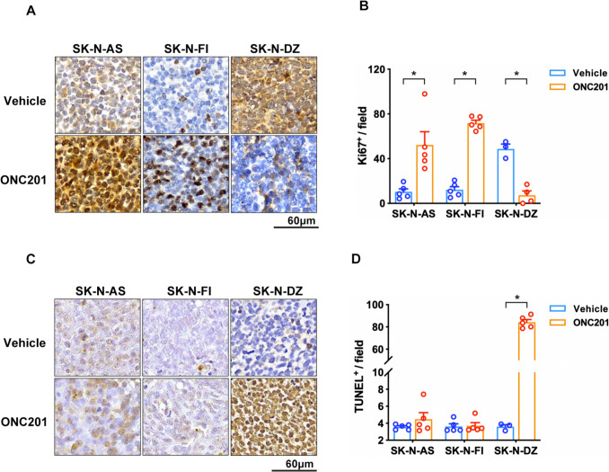
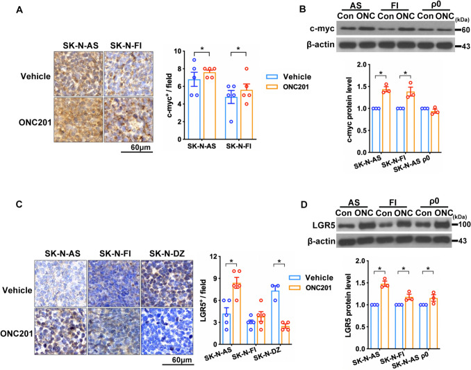
Neuroblastoma (NB) is a formidable challenge in pediatric oncology due to its intricate molecular landscape, necessitating multifaceted therapeutic approaches. ONC201 is an imipridone antibiotic compound with a promising drug candidate leveraging its potent anticancer properties against the mitochondrial proteases ClpP and ClpX. Despite demonstrating early clinical promise, particularly in MYCN-amplified NB, its efficacy in non-MYCN-amplified NB remains a subject worthy of investigation. In this study, we extended the coverage of ONC201 to treat non-MYCN-amplified NB, and our data implicated ONC201's inability to reduce tumor growth in animal models harboring SK-N-AS or SK-N-FI cell lines. Interestingly, ONC201 induced the expression of oncogenic markers c-Myc and LGR5 while downregulating the tumor suppressor ATRX. While it fails to attenuate tumor neovascularization in non-MYCN-amplified NB xenografts, its effectiveness differs from that of its MYCN-amplified counterpart. Rho zero (ρ0)-SK-N-AS cells treated with ONC201 showed comparable observed trends in parental SK-N-AS cells, including LGR5 upregulation and ATRX downregulation, suggesting that ONC201's multifaceted actions extend beyond mitochondrial targets. Our elucidation highlights the need to discern molecular signatures when deploying ONC201 monotherapy against NB, which lacks MYCN-amplification.

Keywords: ATRX; LGR5; Neuroblastoma; Non-MYCN-amplified cell line; ONC201.


Conflict of interest statement

Declarations. Ethics approval: The study adhered to the Declaration of Helsinki and received approval from the Institutional Review Board (IRB) of Chang Gung Medical Foundation (protocol code 201901723B0, dated 2 December 2019). The protocol for animal studies was also sanctioned by the Institutional Animal Care and Use Committee of Chang Gung Memorial Hospital (protocol code 2019091805, dated 1 January 2021, and 2023031303, dated 24 July 2023). Informed consent: The IRB of Chang Gung Medical Foundation approved the waiver of patient consent for protocol code 201901723B0, dated 2 December 2019, allowing the use of de-identified data. Conflict of interest: The authors declare no competing interests. Declaration of generative AI and AI-assisted technologies in the writing process: During the preparation of this work, the authors used chatGPT in order to improve language and readability. After using this tool/service, the authors reviewed and edited the content as needed and took full responsibility for the content of the publication.

Figures

Fig. 1
Fig. 1
Effects of ONC201 and 2DG on NB xenografts in NOD/SCID mice. A Experimental schematic showing the experimental design, treatment schedules, and analysis process. B Measurement of tumor occurrence after subcutaneous injection of NB cells with or without ONC201 and/or 2DG treatment. C Quantification of tumor weight and volume in SK-N-AS cell group. D Quantification of tumor weight and volume in SK-N-FI cell group. Dots represent the numbers in each group, and colors indicate different groups. Tumor growth and volume quantifications are represented as mean ± SEM and are considered significant at *P < 0.05 and **P < 0.01, calculated using ordinary one-way ANOVA and Tukey’s multiple comparison test
Fig. 2
Fig. 2
ONC201 induced tumorigenesis. A Immunohistochemical staining to examine the expression of Ki- 67 and C TUNEL staining in tissue sections. B Ki- 67 expression was significantly upregulated by ONC201, whereas D apoptosis was unaffected. Dots represent the numbers in each group, and colors indicate different groups. Data are presented as means ± SEM, and differences are considered significant at *P < 0.05. A two-group comparison was conducted utilizing an unpaired two-tailed t-test
Fig. 3
Fig. 3
ONC201 increased c-Myc and LGR5 expression in non-MYCN-amplified cells. A c-Myc expression was increased in ONC201-treated (ONC) SK-N-AS and SK-N-FI xenograft tissues. B Immunoblots showing c-Myc expression in SK-N-AS and SK-N-FI cells after treatment with ONC201. C LGR5 expression was increased in ONC201-treated SK-N-AS xenograft tissue. D Immunoblots showing LGR5 expression in SK-N-AS, SK-N-FI, and SK-N-AS ρ0 cells after treatment with ONC201. Dots represent the numbers in each group, and colors indicate different groups. Data are presented as means ± SEM, and differences are considered significant at *P < 0.05. A two-group comparison was conducted utilizing an unpaired two-tailed t-test
Fig. 4
Fig. 4
Analysis of ATRX expression in NB cells and tissues. A Images of immunohistochemical staining for ATRX on tissue sections from SK-N-AS or SK-N-FI xenografts with or without ONC201 treatment. B Immunoblots show ATRX expression after ONC201 treatment in SK-N-AS, SK-N-FI, and SK-N-AS ρ0 cells. Data are presented as means ± SEM, and differences are considered significant at *P < 0.05. A two-group comparison was conducted utilizing an unpaired two-tailed t-test. C Representative profiles of ATRX immunostaining in human NB tissues. D Histological scoring of ATRX intensity in NB tissues. Dots represent the numbers in each group, and colors indicate different groups
Fig. 5
Fig. 5
Effects of ONC201 in murine endothelial cells. A and B NB xenograft tissue sections were stained to identify IB4 and then counterstained with hematoxylin. IB4 staining areas were examined in ImageJ to quantify the percentage of endothelial coverage. Data are presented as means ± SEM, and differences are considered significant at *P < 0.05. A two-group comparison was conducted utilizing an unpaired two-tailed t-test. SVEC4 - 10 cells were treated with 15 µM ONC201 for 24–96 h, and flow cytometry was performed to determine the proportion of cell death via Trypan blue assay (C and D) and staining with annexin V (fluorescein isothiocyanate) and propidium iodide (PI) (E and F). Dots represent the numbers in each group, and colors indicate different groups. For (D) and (F), time course analysis is presented as mean ± SEM, considered significant at **P < 0.01, and calculated by ordinary two-way ANOVA and Tukey’s multiple comparison test

References

    1. Matthay KK, Maris JM, Schleiermacher G, Nakagawara A, Mackall CL, Diller L, Weiss WA (2016) Neuroblastoma. Nat Rev Dis Primers 2:16078. 10.1038/nrdp.2016.78 - PubMed
    1. Kushner BH, Ostrovnaya I, Cheung IY, Kuk D, Kramer K, Modak S, Yataghene K, Cheung NK (2015) Prolonged progression-free survival after consolidating second or later remissions of neuroblastoma with Anti-G(D2) immunotherapy and isotretinoin: a prospective Phase II study. Oncoimmunology 4:e1016704. 10.1080/2162402x.2015.1016704 - PMC - PubMed
    1. Prabhu VV, Morrow S, Rahman Kawakibi A, Zhou L, Ralff M, Ray J, Jhaveri A, Ferrarini I, Lee Y, Parker C et al (2020) ONC201 and imipridones: anti-cancer compounds with clinical efficacy. Neoplasia 22:725–744. 10.1016/j.neo.2020.09.005 - PMC - PubMed
    1. Gardner SL, Tarapore RS, Allen J, McGovern SL, Zaky W, Odia Y, Daghistani D, Diaz Z, Hall MD, Khatib Z et al (2022) Phase I dose escalation and expansion trial of single agent ONC201 in pediatric diffuse midline gliomas following radiotherapy. Neurooncol Adv 4:vdac143. 10.1093/noajnl/vdac143 - PMC - PubMed
    1. Wu JC, Huang CC, Wang PW, Chen TY, Hsu WM, Chuang JH, Chuang HC (2023) ONC201 suppresses neuroblastoma growth by interrupting mitochondrial function and reactivating nuclear ATRX expression while decreasing MYCN. f 10.3390/ijms24021649 - PMC - PubMed

MeSH terms