. 2025 Apr 10;15(1):12272.
doi: 10.1038/s41598-025-96910-x.

The usefulness of different imaging modalities in mandibular osteonecrosis and osteomyelitis diagnosis

Affiliations

The usefulness of different imaging modalities in mandibular osteonecrosis and osteomyelitis diagnosis

Masaya Kawasaki et al. Sci Rep. .

Abstract

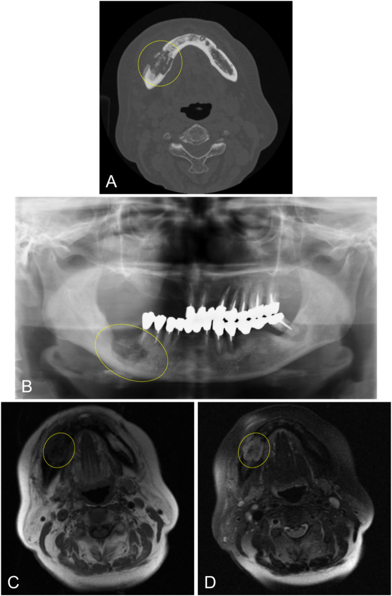
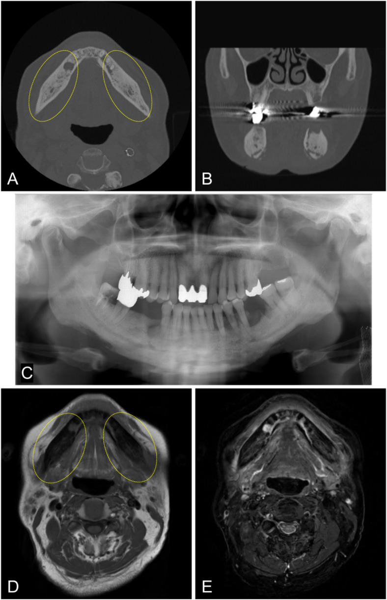
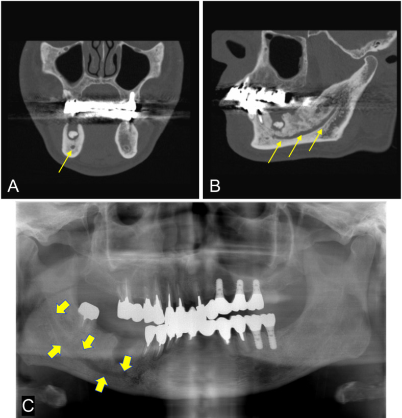
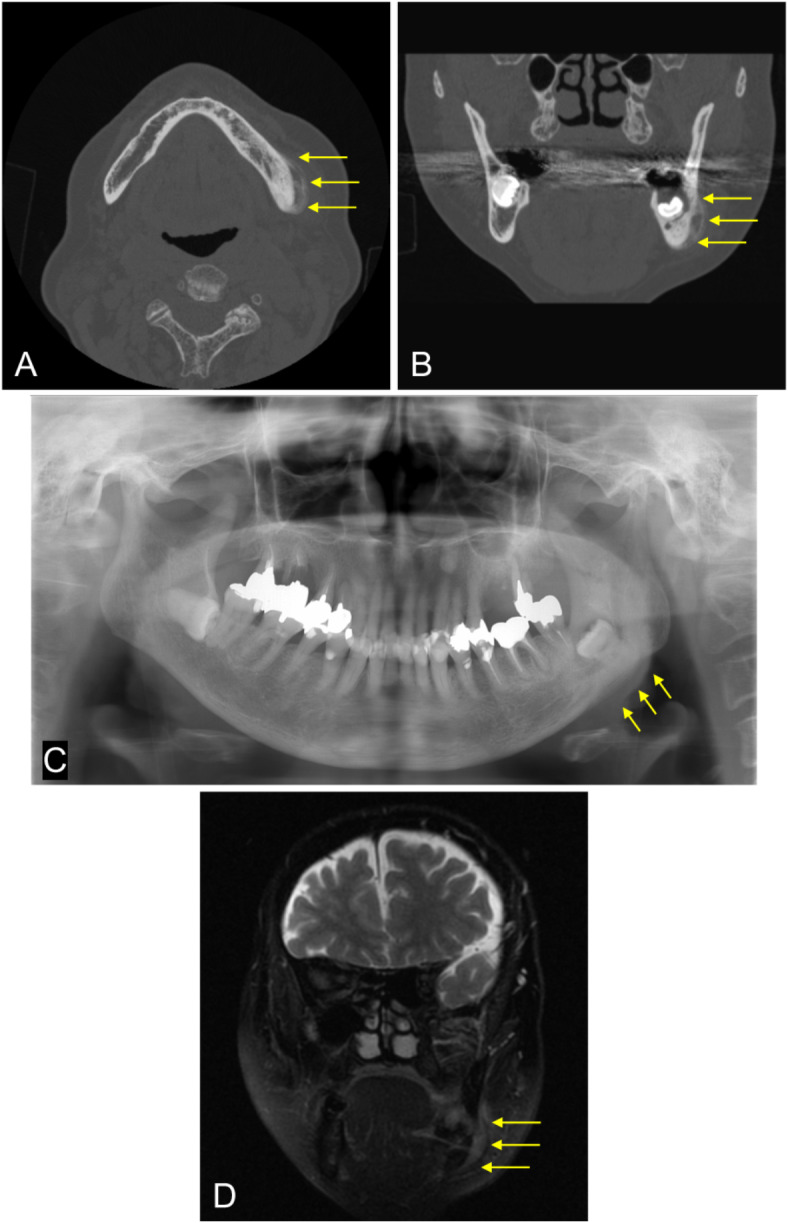
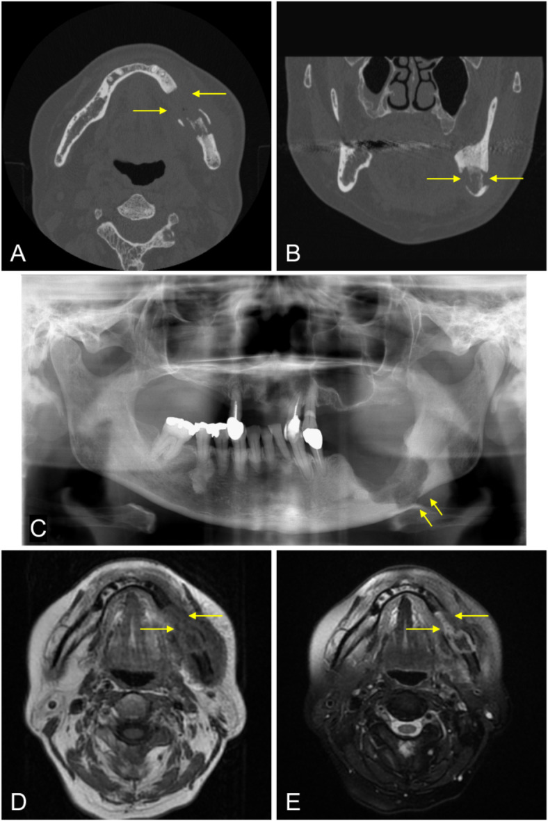
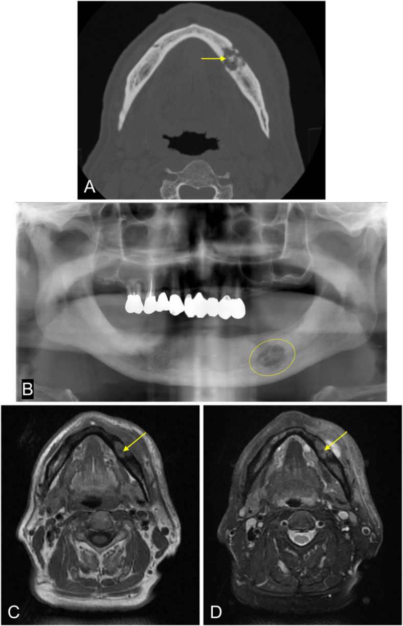
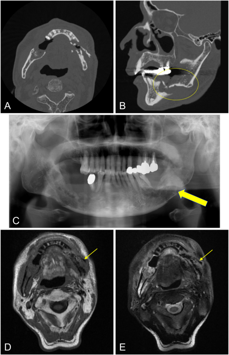
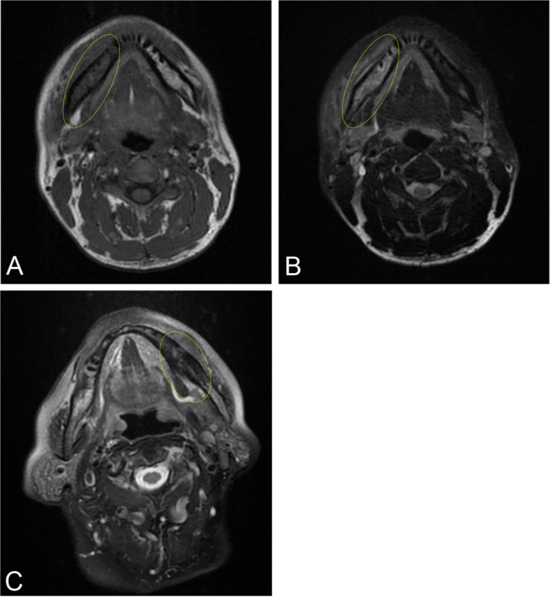
To examine the CT-imaging features of subjects with bacterial osteomyelitis (OM), osteoradionecrosis (ORN), and medication-related osteonecrosis of the jaw (MRONJ) with histopathological confirmation, and to examine the diagnostic efficacy of panoramic radiography and MRI in detecting these disease features. 150 cases with preoperative CT data were selected: 61 bacterial OM, 19 ORN, and 70 MRONJ. 143 cases underwent panoramic X-ray examination, and 47 underwent MRI. The assessment criteria for imaging findings included: (1) bone resorption, (2) osteosclerosis, (3) clarity of the mandibular canal, (4) periosteal reactions, (5) cortical bone perforation, (6) sequestrum, and (7) pathological fractures. CT was considered the gold standard for assessing these features. Compared with CT, all panoramic radiographs were detectable for diagnostic features of the disease. Bone resorption was detected in 123 cases (sensitivity 91.1%), and osteosclerosis was detected in 131 cases (sensitivity 98.5%). With panoramic radiography, most changes to clarity of the mandibular canal and pathological fractures were detected (sensitivities of 87.8% and 68.8%, respectively). However, the sensitivities for detection of periosteal reactions, cortical bone perforation and sequestration were low (19.6%, 17.8% and 19.4%, respectively). Sensitivity of MRI for detecting periosteal reactions, cortical bone perforation, sequestration, and pathological fractures (27.3%, 73.5%, 35.7%, and 60.0%, respectively) was equivalent or superior to panoramic imaging. MR-specific characteristics of bone marrow edema were depicted on almost all examinations. Panoramic radiography may be adequate for identifying bone resorption and osteosclerosis. However, MRI provides more value than panoramic radiography in detecting periosteal reactions, cortical bone perforation, sequestration, and bone marrow edema.

Keywords: CT; MRI; Osteomyelitis; Panoramic radiography.


Conflict of interest statement

Declarations. Competing interests: The authors declare no competing interests.

Figures

Fig. 1
Fig. 1
Case with bone resorption. A 79-year-old female underwent extraction of the right mandibular first molar at a dental clinic. Upon investigation of medication due to incomplete healing of the extraction socket, it was revealed that she had been taking risedronate for osteoporosis for the past 3 years and alendronate for the past 1.5 years. She was then referred to our hospital for further treatment. (A) CT image (bone mode) showing a low CT value area in the trabecular bone of the right mandibular molar region (ellipse). (B) Panoramic X-ray image revealing radiolucency in the same area with surrounding sclerotic changes (ellipse). (C) T1-weighted MR image demonstrating the bone resorption area in the same region, with low signal intensity in the bone marrow compared with the contralateral side (ellipse). (D) T2-weighted fat-suppressed MR image demonstrating the bone resorption area in the same region, with high signal intensity in the bone marrow compared with the contralateral side (ellipse).
Fig. 2
Fig. 2
Case with osteosclerosis. A 72-year-old male with a history of denosumab use for bone metastasis from bile duct cancer. Although he underwent extraction of the right mandibular second premolar at a dental clinic after a 2-month hiatus from medication, he was referred to our hospital due to treatment for bone exposure. (A, B) CT images (bone mode) showing an increase in CT values in the mandibular trabecular bone on both sides as well as the extraction socket of the right mandibular second premolar (ellipses). (C) Panoramic X-ray image revealing increased radiopacity in the mandibular trabecular bone on both sides. (D, E) T1-weighted MR image and T2-weighted fat-suppressed MR image demonstrate a sclerotic area in the same region, and the bone marrow signal in that area shows low signal intensity (ellipses).
Fig. 3
Fig. 3
Case with clarification of the mandibular canal. A 60-year-old male underwent extraction of the lower right first molar at a dental clinic. Pain and suppuration occurred in the same area, leading to a referral to our hospital. He was diagnosed with bacterial OM because of exclusion from the ORN or MRONJ criteria. (A, B) CT images (bone mode) showing clarification of the right inferior alveolar canal (arrows). (C) Panoramic X-ray image revealing similar findings (arrows). However, the results of MRI were inconclusive.
Fig. 4
Fig. 4
Case with periosteal reaction. A 70-year-old female experienced discomfort in the lower left molar region and sought care at a dental clinic. Initially, she was informed about the impacted lower left third molar but was left untreated. Later, swelling in the area developed, leading to a referral to our hospital. She was diagnosed with bacterial OM because of exclusion from the ORN or MRONJ criteria. (A, B) The CT images (bone mode) showing an area of increased CT values continuous with the outer cortical bone (arrows). (C) Panoramic X-ray image revealing increased radiopacity (new bone formation) along the lower border of the mandible (arrows). (D) T2-weighted fat-suppressed MR image displaying a high signal intensity area continuous with the outer cortical bone (arrows).
Fig. 5
Fig. 5
Case with cortical bone perforation. A 64-year-old female had a history of using zoledronate for 3 years and pamidronate for 2 years to prevent bone metastasis from breast cancer. She was referred to our hospital due to swelling on the left side of the cheek and limited mouth opening. (A, B) CT images (bone mode) not showing continuity of the cortical bone on the buccal and lingual sides (arrows). (C) Panoramic X-ray image not revealing continuity of the cortical bone along the lower border of the mandible (arrows). (D, E) T1-weighted MR image and T2-weighted fat-suppressed MR image not showing continuity of the cortical bone on the buccal and lingual sides (arrows).
Fig. 6
Fig. 6
Case with a sequestrum. An 81-year-old male underwent extraction of the left lower second premolar at a dental clinic. Nonhealing extraction socket complications persisted, with periods of improvement and worsening. CBCT imaging revealed sequestrum in the left mandibular molar region, leading to referral to our hospital. He was diagnosed with bacterial OM because of exclusion from the ORN or MRONJ criteria. (A) CT image (bone mode) showing islands of high CT values within the low CT value area (arrow). (B) Panoramic X-ray image revealing radiopaque areas within the radiolucent area (ellipse). (C, D) T1-weighted MR image and T2-weighted fat-suppressed MR image demonstrating no signal intensity region suspected to be sequestrum (arrows).
Fig. 7
Fig. 7
Case with a pathological fracture. A 75-year-old male received chemoradiotherapy with 70 Gy for nasopharyngeal cancer. He subsequently developed radiation-induced osteomyelitis on both sides of the mandible. (A, B) CT images (bone mode) showing separation and displacement of the mandible in the region of the left lower molar (ellipse). (C, D, E) Panoramic X-ray images, T1-weighted MR images, and T2-weighted fat-suppressed MR images revealing separation and displacement of the mandible in the region of the left lower molar (arrows).
Fig. 8
Fig. 8
Examples of MR signal intensities of lesions compared with those of the contralateral side. A 39-year-old male presented at a local clinic with pain and swelling on the right side of the mandible. Root canal treatment was performed on the right lower first molar, but the swelling did not improve. He also developed abnormal sensation of the mental nerve and was referred to our hospital. (A) T1-weighted image showing low signal intensity compared with the healthy side (ellipse). (B) T2-weighted fat-suppressed image showing high signal intensity compared with the healthy side (ellipse). An 86-year-old female presented at a local clinic with swelling in the left lower molar region of the mandible. She was treated with alendronate for osteoporosis and was referred to our clinic. (C) T2-weighted fat-suppressed image showing low signal intensity compared with the healthy side (ellipse).

Similar articles

References

    1. Schmitt, S. K. Osteomyelitis. Infect. Dis. Clin. North. Am.31, 325–338 (2017). - PubMed
    1. Krakowiak, P. A. Alveolar osteitis and osteomyelitis of the jaws. Oral Maxillofac. Surg. Clin. North. Am.23, 401–413 (2011). - PubMed
    1. Otto, S. et al. Infection as an important factor in medication-related osteonecrosis of the jaw (MRONJ). Medicina (Kaunas)57, 1–12 (2021). - PMC - PubMed
    1. Schuknecht, B. & Valavanis, A. Osteomyelitis of the mandible. Neuroimaging Clin. N. Am.13, 605–618 (2003). - PubMed
    1. Chronopoulos, A., Zarra, T., Ehrenfeld, M. & Otto, S. Osteoradionecrosis of the jaws: definition, epidemiology, staging and clinical and radiological findings. A concise review. Int. Dent. J.68, 22–30 (2018). - PMC - PubMed

LinkOut - more resources