

Epithelial Remodelling in Myopia After Keratorefractive Lenticule Extraction
- PMID: 40210829
- PMCID: PMC12069778
- DOI: 10.1007/s40123-025-01138-7
Epithelial Remodelling in Myopia After Keratorefractive Lenticule Extraction
Abstract
Introduction: We analyzed longitudinal epithelial changes after the treatment of myopia with keratorefractive lenticule extraction (KLEx) and the zonal change in epithelial thickness up to 12 months after SmartSight for myopic astigmatism with the SCHWIND ATOS femtosecond laser.
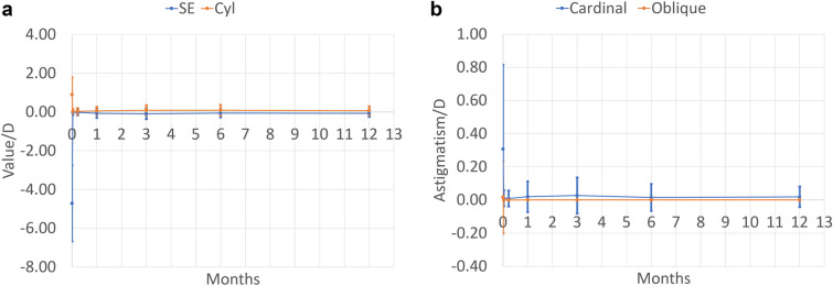
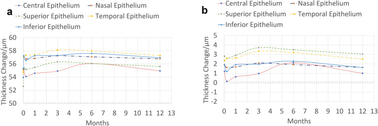
Methods: We used anterior segment optical coherence tomography (AS-OCT) data and analyzed changes in the epithelium after treatment to ascertain how much epithelium hyperplasia occurred after KLEx. Data from 80 eyes treated with SmartSight, with a complete follow-up from postoperative day 1 (POD1) to 12 months postoperative, were used. The mean age of the patients was 29 ± 6 years with a mean spherical equivalent (SEQ) of - 4.72 ± 1.97 diopters (D) (- 1.25 to - 9.88 D) and a mean magnitude of refractive astigmatism of 0.90 ± 0.89 D. Preoperative central epithelial thickness was from 46 to 67 µm.
Results: Postoperative central epithelial thickness at 12-month follow-up was 3 ± 5 µm thicker than preoperatively. The other epithelial zones (nasal, superior, temporal, inferior) thickened by + 4 ± 4 µm. The epithelial change showed larger variability at POD1 and stabilized from 1 week onwards. Postoperatively, the change in epithelium was not different for the different zones, and it did not correlate with the achieved refractive changes for any zone at any time point.
Conclusions: The changes in epithelial thickness after KLEx for moderate myopia with SmartSight were minimal, indicating a low level of epithelial hyperplasia without resembling a regression-inducing lentoid. Findings suggest that KLEx with SCHWIND ATOS has a subtle impact on the epithelial thickness (with postoperative epithelium becoming slightly thicker). However, the differences remain below any clinical relevance.
Keywords: Corneal laser refractive surgery; Epithelial remodelling; Femtosecond laser; KLEx; Lenticule; Myopic astigmatism.
© 2025. The Author(s).
Conflict of interest statement
Declarations. Conflict of Interest: Samuel Arba Mosquera is an employee at and inventor in several patents owned by SCHWIND eye-tech-solutions. None of the other authors have financial or proprietary interests in materials or methods presented herein. Ethical Approval: All patients provided written informed consent (ICF) in accordance with the Declaration of Helsinki for both treatment and the use of de-identified clinical data for research purposes. The study was evaluated under the Medical Research Involving Human Subjects Act by the Specialty Eye Hospital Svjetlost and was deemed exempt from ethics approval due to its retrospective chart review nature. The purpose of this clinical research does not represent a clinical investigation. The medical device was used within its intended purpose without any additional invasive or patient burdensome procedures used.
Figures
Similar articles
-
Three-Month Clinical Outcomes to Correct Myopia or Myopic Astigmatism Using a Femtosecond Laser for Lenticule Creation With Automated Centration and Cyclotorsion Compensation.J Refract Surg. 2024 Jan;40(1):e30-e41. doi: 10.3928/1081597X-20231212-03. Epub 2024 Jan 1. J Refract Surg. 2024. PMID: 38190561
-
Comparing high and low energy outcomes on day one for SmartSight myopic-astigmatism treatments with the SCHWIND ATOS: a retrospective case series.BMC Ophthalmol. 2023 Jul 18;23(1):328. doi: 10.1186/s12886-023-03076-z. BMC Ophthalmol. 2023. PMID: 37464345 Free PMC article.
-
Predictability of the Achieved Lenticule Thickness in Keratorefractive Lenticule Extraction for Myopia Correction.J Refract Surg. 2023 Nov;39(11):728-735. doi: 10.3928/1081597X-20230925-02. Epub 2023 Nov 1. J Refract Surg. 2023. PMID: 37937760
-
[Comparison of corneal epithelial remodeling after small incision lenticule extraction and femtosecond laser-assisted LASIK].Zhonghua Yan Ke Za Zhi. 2020 Feb 11;56(2):93-102. doi: 10.3760/cma.j.issn.0412-4081.2020.02.004. Zhonghua Yan Ke Za Zhi. 2020. PMID: 32074819 Chinese.
-
Differences in ocular high order aberrations before and after small incision lenticule extraction for correction of myopia: a systematic review and meta-analysis.Front Med (Lausanne). 2024 Mar 27;11:1274101. doi: 10.3389/fmed.2024.1274101. eCollection 2024. Front Med (Lausanne). 2024. PMID: 38601117 Free PMC article.
References
-
- Munnerlyn CR, Koons SJ, Marshall J. Photorefractive keratectomy: a technique for laser refractive surgery. J Cataract Refract Surg. 1988;14(1):46–52. 10.1016/s0886-3350(88)80063-4. - PubMed
-
- Buratto L, Ferrari M. Excimer laser intrastromal keratomileusis: case reports. J Cataract Refract Surg. 1992;18(1):37–41. 10.1016/s0886-3350(13)80381-1. - PubMed
-
- Shah R, Shah S, Sengupta S. Results of small incision lenticule extraction: all-in-one femtosecond laser refractive surgery. J Cataract Refract Surg. 2011;37(1):127–37. 10.1016/j.jcrs.2010.07.033. - PubMed
-
- Ficker LA, Bates AK, Steele AD, et al. Excimer laser photorefractive keratectomy for myopia: 12 month follow-up. Eye (Lond). 1993;7(Pt 5):617–24. 10.1038/eye.1993.143. - PubMed
LinkOut - more resources
Full Text Sources
Research Materials
