Early diagnosis of acute lymphoblastic leukemia utilizing clinical, radiographic, and dental age indicators
- PMID: 40210878
- PMCID: PMC11986116
- DOI: 10.1038/s41598-025-95014-w
Early diagnosis of acute lymphoblastic leukemia utilizing clinical, radiographic, and dental age indicators
Abstract
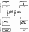
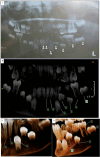
Leukemic patients often display clinical signs like anemia, thrombocytopenia, and hepatosplenomegaly. Early diagnosis is crucial for intervention and improved prognosis. Dentists can help identify these signs through oral masses, gingival bleeding, and oral ulceration, with radiographical features like bone osteolysis, moth-eating appearance, and abnormal tooth chronology. This study aimed to achieve early diagnosis of leukemic child patients (LCP) by the dentist based on their clinical, age estimation, and radiographical oral signs. Twenty-three children suffer from leukemia, selected after an initial diagnosis based on their clinical signs with an abnormal CBC or abnormal WBCs. These patients were accessed clinically for oral signs and radiographically using panoramic radiographs and cone beam computed tomography (CBCT) to evaluate chronology and bone density. LCP were compared with systematically free control child cases (SFC) who went for a panoramic image and CBCT scan needed for their orthodontic problems. Clinical results for LCP revealed (100%) of cases showed gingival bleeding, (87%) of cases showed gingival masses, (83%) of cases revealed aphthous-like ulceration, and (100%) of cases had different grades of mobility related to the lower first permanent molar used as markers for tooth affection. Radiographical results revealed a statistically significant decrease (P value ≤ 0.05) in LCP age revealed by panoramic and CBCT images in comparison with their actual age. Also, there was a statistically significant decrease in bone density shown by LCP regarding selected regions. LCP could be early diagnosed by the dentist through clinical and radiographical indicators. Diagnosing acute lymphoblastic leukemia (ALL) in its early stages remains a significant challenge due to the nonspecific and often subtle nature of initial symptoms. Dental practitioners can bridge the gap between routine dental care and early systemic disease detection, potentially expediting medical intervention and improving outcomes for children with acute lymphoblastic leukemia.
Keywords: Age Estimation; Bone density; Cone beam computed tomography (CBCT); Leukemic child patient.
© 2025. The Author(s).
Conflict of interest statement
Declarations. Consent for publication: Not applicable. Competing interests: The authors declare no competing interests. Ethical approval and consent to participate: . The Helsinki Declaration of 1964 and its later amendments were complied with, and the ethical committee46 of Tanta University’s Faculty of Dentistry granted ethical approval for this study under code (#R-OMPDR-1-20-2). The patients’ guardians were told about the goal of the study and an informed consent form was signed before clinical procedures.
Figures


Similar articles
-
A comparison of estimated age based on pulp volume from cone beam computed tomography (CT) images and panoramic radiography data with chronological age.J Clin Pediatr Dent. 2024 Mar;48(2):149-162. doi: 10.22514/jocpd.2024.043. Epub 2024 Mar 3. J Clin Pediatr Dent. 2024. PMID: 38548645
-
[Comparison of mesiodistal tooth angulations determined through traditional panoramic radiographs and cone beam CT panoramic images].Hua Xi Kou Qiang Yi Xue Za Zhi. 2014 Aug;32(4):331-5. doi: 10.7518/hxkq.2014.04.004. Hua Xi Kou Qiang Yi Xue Za Zhi. 2014. PMID: 25241531 Free PMC article. Chinese.
-
Inferior alveolar nerve injury: Correlation between indicators of risk on panoramic radiographs and the incidence of tooth and mandibular canal contact on cone-beam computed tomography scans in a Western Australian population.J Investig Clin Dent. 2018 Aug;9(3):e12323. doi: 10.1111/jicd.12323. Epub 2018 Feb 4. J Investig Clin Dent. 2018. PMID: 29399983
-
Is cone-beam computed tomography (CBCT) an alternative to plain radiography in assessments of dental disease? A study of method agreement in a medically compromised patient population.Clin Oral Investig. 2024 Jan 30;28(2):127. doi: 10.1007/s00784-024-05527-3. Clin Oral Investig. 2024. PMID: 38289447 Free PMC article.
-
Imaging modalities to inform the detection and diagnosis of early caries.Cochrane Database Syst Rev. 2021 Mar 15;3(3):CD014545. doi: 10.1002/14651858.CD014545. Cochrane Database Syst Rev. 2021. PMID: 33720395 Free PMC article.
References
-
- Mitchell, C., Hall, G. & Clarke, R. T. Acute leukaemia in children: diagnosis and management. BMJ ;338. (2009).
-
- Kaplan, J. A. Leukemia in children. Pediatr. Rev.40 (7), 319–331. 10.1542/pir.2018-0192 (2019). - PubMed
-
- Greaves, M. Aetiology of acute leukaemia. Lancet349 (9048), 344–349. 10.1016/S0140-6736(96)09412-3 (1997). - PubMed
-
- Redaelli, A., Laskin, B. L., Stephens, J. M., Botteman, M. F. & Pashos, C. L. A systematic literature review of the clinical and epidemiological burden of acute lymphoblastic leukaemia (ALL). Eur. J. Cancer Care (Engl). 14 (1), 53–62. 10.1111/j.1365-2354.2005.00513.x (2005). - PubMed
MeSH terms
LinkOut - more resources
Full Text Sources
Miscellaneous

