. 2025 Apr 10;15(1):12351.
doi: 10.1038/s41598-025-96868-w.

CircSCD1 inhibits ferroptosis in breast Cancer through stabilizing SCD1 protein via deubiquitinase OTUB1

Affiliations

CircSCD1 inhibits ferroptosis in breast Cancer through stabilizing SCD1 protein via deubiquitinase OTUB1

Zhiwei Wu et al. Sci Rep. .

Abstract

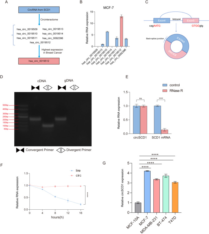
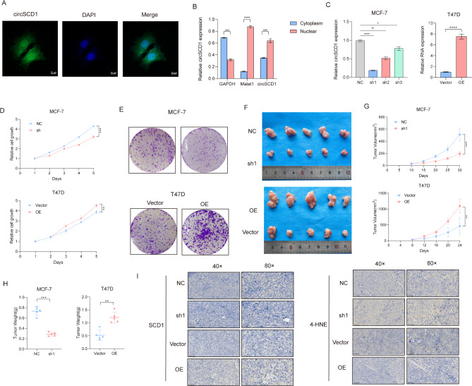
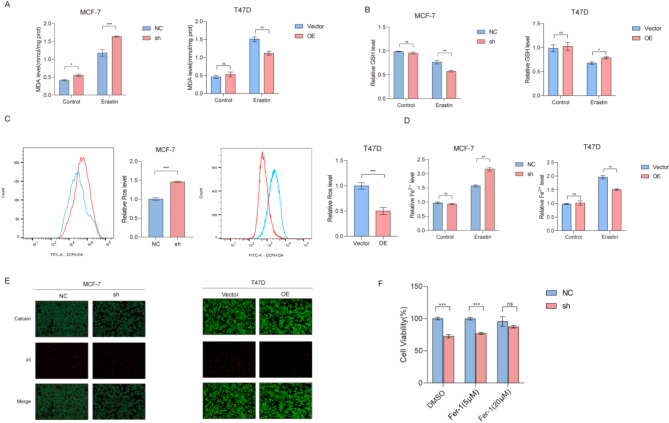
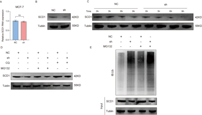
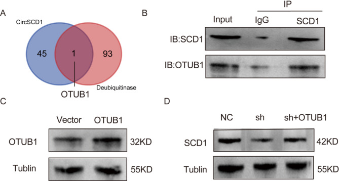
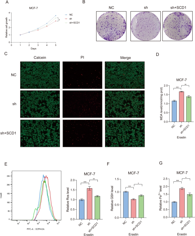
Breast cancer is the leading cause of cancer-related death in women worldwide, and its developmental mechanisms involve complex factors. Recent studies have shown that ferroptosis is closely related to the occurrence and progression of breast cancer. However, the role of circular RNAs (circRNAs) in regulating ferroptosis in breast cancer remains unclear. In this study, we investigated the regulatory role of circSCD1 (hsa_circ_0019512) in breast cancer. We examined the expression of circSCD1 in breast cancer cell lines and explored its impact on cell viability and colony formation. We also evaluated the involvement of circSCD1 in ferroptosis by measuring the levels of malondialdehyde (MDA), glutathione (GSH), reactive oxygen species (ROS), and intracellular iron. In vivo xenograft experiments were performed to confirm the role of circSCD1 in promoting tumor growth and inhibiting ferroptosis.Furthermore, we investigated the mechanism by which circSCD1 regulates SCD1 protein stability through ubiquitination and identified the interaction between circSCD1 and the deubiquitinase OTUB1. Our results showed that circSCD1 was upregulated in breast cancer cell lines and promoted cell viability and colony formation. Knockdown of circSCD1 increased MDA and ROS levels, decreased GSH levels, and enhanced ferroptosis in breast cancer cells. In vivo, circSCD1 knockdown significantly reduced tumor size and weight, while its overexpression enhanced tumor growth. Mechanistically, circSCD1 interacted with OTUB1 to inhibit the ubiquitination and degradation of SCD1 protein, thereby stabilizing its expression. Rescue experiments demonstrated that SCD1 overexpression partially reversed the effects of circSCD1 knockdown on cell proliferation and ferroptosis. Our findings suggest that circSCD1 plays a crucial role in promoting breast cancer cell growth and inhibiting ferroptosis by regulating SCD1 protein stability. Targeting the circSCD1/OTUB1/SCD1 axis may provide a potential therapeutic strategy for breast cancer treatment.


Conflict of interest statement

Declarations. Competing interest: The authors declare no competing interests. Ethical approval: All animal experiments were conducted in compliance with the ARRIVE guidelines ( https://arriveguidelines.org ). The experimental protocols were approved by the Medical Ethics Committee of Xiangya Hospital, Central South University (Approval No. XY20240521005). Every effort was made to minimize animal suffering during the study, including the implementation of humane endpoints and appropriate anesthesia protocols.

Figures

Fig. 1
Fig. 1
Expression and validation of CircSCD1 in breast cancer cell lines. (A) The diagram of screening SCD1-derived circRNAs in the MCF-7. (B) Detection of SCD1-derived circRNA expression levels in MCF-7 cells. (C) Schematic representation of CircSCD1, showing its circularization via splicing of the 4th exon of SCD1, resulting in a mature sequence length of 436 nucleotides. (D) The reverse transcription–PCR (RT-PCR) products from different primers of CircSCD1 were detected by agarose gel electrophoresis. (E) Comparison of RNase R digestion resistance between CircSCD1 and the linear form of SCD1 mRNA. (F) Degradation rates of CircSCD1 and the linear form of SCD1 mRNA after actinomycin treatment, shown over time. (G) Expression levels of CircSCD1 in normal human mammary epithelial cell line MCF-10 A and breast cancer cell lines MCF-7, MDA-MB-231, BT-474 and T47D. All data from three independent experiments shown as mean ± SD. ns p > 0.05; **** p < 0.0001.
Fig. 2
Fig. 2
Functional investigation and cellular localization of CircSCD1 in MCF-7 and T47D breast cancer cells. (A) Cellular localization of CircSCD1 in MCF-7 cells visualized using RNA Fluorescence In Situ Hybridization (FISH). (B) Distribution of CircSCD1 in the cytoplasm and nucleus of MCF-7 cells, as determined by nucleolar fractionation experiments. (C) Relative expression levels of CircSCD1 in MCF-7 and T47D cells after knockdown (sh1, sh2, sh3) and overexpression (OE) compared to control (NC and Vector) cells. (D) Cell viability assessed by CCK-8 assay in MCF-7 and T47D cells with CircSCD1 knockdown (sh1) and overexpression (OE). (E) Colony formation ability of CircSCD1 knockdown and overexpression cells in MCF-7 and T47D cells, demonstrated by colony formation assays. (F) Tumor images from MCF-7 and T47D cell xenograft mouse models with CircSCD1 knockdown (sh1) and overexpression (OE). (G) Tumor volume measurements from MCF-7 and T47D xenograft models with CircSCD1 knockdown and overexpression. (H) Tumor weight measurements from MCF-7 and T47D xenograft models with CircSCD1 knockdown and overexpression. (I) Immunohistochemistry analysis of SCD1 and 4-HNE in tumor tissues from MCF-7 xenograft models with CircSCD1 knockdown and overexpression. All data are from three independent experiments, shown as mean ± SD. *p < 0.05; **p < 0.01; ***p < 0.001; ****p < 0.0001.
Fig. 3
Fig. 3
(A) Measurement of malondialdehyde (MDA) levels in MCF-7 and T47D cells with circSCD1 knockdown (sh) and overexpression (OE), respectively, under control or Erastin treatment conditions. (B) Measurement of glutathione (GSH) levels in MCF-7 and T47D cells with circSCD1 knockdown (sh) and overexpression (OE), respectively, under control or Erastin treatment conditions. (C) Measurement of reactive oxygen species (ROS) levels in MCF-7 cells with circSCD1 knockdown (sh) and T47D cells with circSCD1 overexpression (OE) using flow cytometry. (D) Measurement of intracellular Fe²⁺ levels in MCF-7 cells with circSCD1 knockdown (sh) and T47D cells with circSCD1 overexpression (OE), under control or Erastin treatment conditions. (E) Propidium iodide (PI) and Calcein-AM staining to assess cell death in MCF-7 cells with circSCD1 knockdown (sh) and T47D cells with circSCD1 overexpression (OE). (F) Measurement of cell viability in MCF-7 cells with circSCD1 knockdown (sh) treated with ferrostatin-1 (5 µM and 20 µM) after ferroptosis induction. All data are from three independent experiments and are presented as mean ± SD. ns p > 0.05; * p < 0.05; ** p < 0.01; *** p < 0.001; **** p < 0.0001.
Fig. 4
Fig. 4
CircSCD1 enhances SCD1 protein stability by inhibiting its ubiquitination. (A) Measurement of SCD1 mRNA levels in MCF-7 cells with circSCD1 knockdown. (B) Measurement of SCD1 protein levels in MCF-7 cells with circSCD1 knockdown. (C) Time-dependent degradation of SCD1 protein in MCF-7 cells treated with an cycloheximide (CHX), comparing control and circSCD1 knockdown groups. (D) Effect of Chloroquine(CQ) and MG132 treatment on SCD1 protein levels in MCF-7 cells with Control/circSCD1 knockdown. (E) Measurement of SCD1 ubiquitination levels in MCF-7 cells with circSCD1 knockdown. All data from three independent experiments shown as mean ± SD. ns p > 0.05.
Fig. 5
Fig. 5
The interplay of the circSCD1/OTUB1/SCD1 axis in the post-transcriptional regulation of SCD1 protein stability. (A) The intersection of circSCD1 interacting proteins enriched through CO-IP combined with mass spectrometry and the list of deubiquitinases. (B) CO-IP experiment demonstrating the interaction between SCD and OTUB1. (C) Verification of overexpression efficiency of OTUB1 using synthesized OTUB1 overexpression plasmid. (D) Analysis of SCD1 protein expression in MCF-7 cells with circSCD1 knockdown and OTUB1 overexpression.
Fig. 6
Fig. 6
The pivotal role of circSCD1-mediated SCD1 upregulation in promoting Breast Cancer. (A) Cell viability assessed by CCK-8 assay in MCF-7 cells with circSCD1 knockdown and SCD1 overexpression. (B) Colony formation assays for MCF-7 cells with circSCD1 knockdown and SCD1 overexpression. (C) PI staining to evaluate cell death in MCF-7 cells with circSCD1 knockdown and SCD1 overexpression. (D) Measurement of MDA levels after circSCD1 knockdown and SCD1 overexpression in MCF-7 cells. (E) Measurement of ROS levels after circSCD1 knockdown and SCD1 overexpression in MCF-7 cells after ferroptosis induction. (F) Measurement of lipid peroxidation levels after circSCD1 knockdown and SCD1 overexpression in MCF-7 cells. Measurement of Fe2+ levels after circSCD1 knockdown and SCD1 overexpression in MCF-7 cells after ferroptosis induction. All data from three independent experiments shown as mean ± SD. * p < 0.05; ** p < 0.01; *** p < 0.001.

Similar articles

References

    1. Dixon, S. J. et al. Ferroptosis: An iron-dependent form of nonapoptotic cell death. Cell149 (5), 1060–1072. (2012). - PMC - PubMed
    1. Tang, D. et al. Ferroptosis: Molecular mechanisms and health implications. Cell. Res.31 (2), 107–125. 10.1038/s41422-020-00441-1 (2021). - PMC - PubMed
    1. Zhao, Y. et al. Neutrophils resist ferroptosis and promote breast cancer metastasis through aconitate decarboxylase 1. Cell. Metab.35 (10), 1688–1703. 10.1016/j.cmet.2023.09.004 (2023). - PMC - PubMed
    1. LI, J. et al. Obacunone alleviates ferroptosis during lipopolysaccharide-induced acute lung injury by upregulating Nrf2-dependent antioxidant responses. Cell. Mol. Biol. Lett.27 (1), 29. 10.1186/s11658-022-00318-8 (2022). - PMC - PubMed
    1. Liang, D. et al. Ferroptosis surveillance independent of GPX4 and differentially regulated by sex hormones. Cell186 (13), 2748–2764. 10.1016/j.cell.2023.05.003 (2023). - PMC - PubMed

MeSH terms