. 2025 Apr 10;15(1):12225.
doi: 10.1038/s41598-025-97122-z.

Oroxylin A may promote cell apoptosis and inhibit epithelial-mesenchymal transition in endometrial cancer, associated with the ERβ/PI3K/AKT pathway

Affiliations

Oroxylin A may promote cell apoptosis and inhibit epithelial-mesenchymal transition in endometrial cancer, associated with the ERβ/PI3K/AKT pathway

Xue Fan et al. Sci Rep. .

Abstract

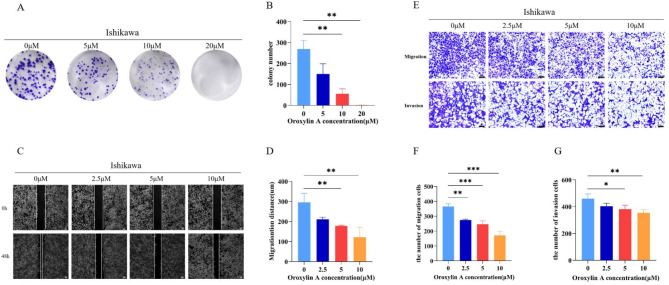
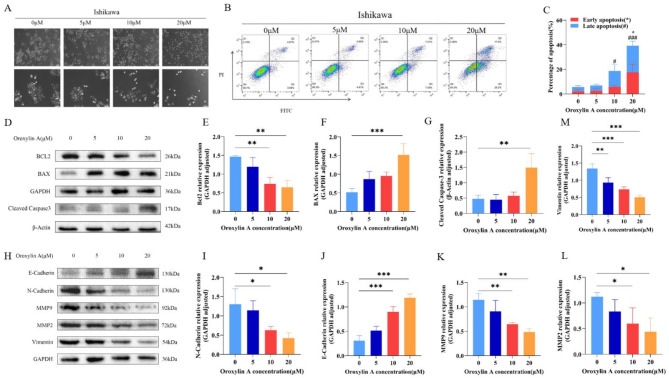
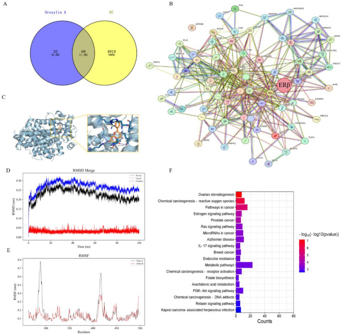
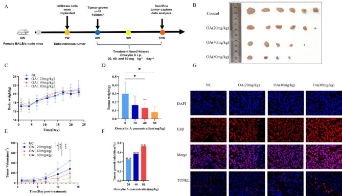
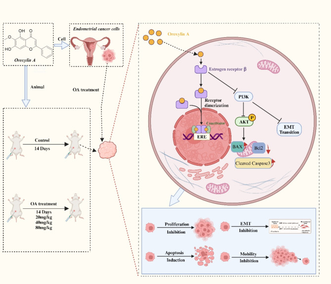
Endometrial cancer (EC) is a prevalent gynecological cancer worldwide, often associated with poor prognosis after recurrence or metastasis. Oroxylin A (OA) is an active flavonoid compound with a strong anti-tumor function. However, the effects of OA on EC remain unknown. In this study, we planned to investigate the anti-EC effects of OA and explore its mechanisms. Five cell lines were used for in vitro experiments, and female BALB/c nude mice were applied for xenograft experiments. The cytotoxicity and experimental concentration of OA were detected by CCK-8. Wound healing, transwell, and colony formation assays were used to evaluate the anti-metastatic and anti-proliferative activities of OA on EC cells. TUNEL assay and flow cytometry were applied for the evaluation of apoptosis. Network pharmacology was used to explore potential targets, and molecular dynamics simulations and dockings were applied for the quantification of binding energy, and stability of OA. RT-qPCR, WB, and immunofluorescence were applied for the detection of localization and expression of correlated markers. The results showed that OA notably inhibited the proliferation, migration, and invasion of Ishikawa cells. Meanwhile, in vivo Ishikawa xenograft assays demonstrated that OA notably inhibited growth and promoted apoptosis of EC. Mechanistically, after treatment with OA, the expressions of Cleaved Caspase-3, BAX, E-cadherin, and ERβ were increased, while the expressions of Bcl-2, Vimentin, N-cadherin, MMP2, MMP9, PI3K and phospho-AKT (Ser473) were decreased. Therefore, OA may exhibit significant anti-EC effects by regulating the ERβ/PI3K/AKT pathway to promote apoptosis and inhibit epithelial-mesenchymal transition (EMT).

Keywords: Apoptosis; EMT; ERβ; Endometrial cancer; Oroxylin A.


Conflict of interest statement

Declarations. Competing interests: The authors declare no competing interests. Ethical approval: We promise that all animal studies received approval from First Hospital of Lanzhou University Ethical Committee on Experimental Animal Care and Use (No. LDYYLL2024-491) and were conducted in accordance with ARRIVE guidelines. The mice were humanely sacrificed after anesthesia with sodium pentobarbital.

Figures

Fig. 1
Fig. 1
Chemical structure of OA and its effects on cell viability in a range of cell lines. (A) Chemical structure of OA. (B) Cell viability of Ishikawa, RL95-2, HEC-1 A, HEC-1B, and HEK293 cells after treatment with OA for 48 h. (C) Viability of Ishikawa cells after exposure to OA for 24 h, 48 h, and 72 h. (D) Viability of Ishikawa cells after 5-FU treatment for 48 h.
Fig. 2
Fig. 2
Inhibitory effects of OA on the proliferation, migration, and invasion of Ishikawa cells. (A) Cell colony formation and (B) the analysis of colony formation number. (C) Wound healing assay (Scale bar is 100 μm) and (D) the quantification of the wound healing. (E) Transwell migration and invasion assay (Scale bar is 100 μm), and the quantification of (F) migration cells and (G) invasion cells. *p < 0.05, **p < 0.01, ***p < 0.001 vs. OA 0 µM group.
Fig. 3
Fig. 3
Effects of OA on apoptosis induction and EMT inhibition in Ishikawa cells. (A) Morphological changes of Ishikawa cells after OA intervention (Scale bar is 100 μm). (BC) Evaluation of apoptosis rate and the quantification of apoptosis in Ishikawa cells after treatment with OA for 48 h. (DG) Effect of OA on the expression of apoptosis-related proteins Bcl2 (E), Bax (F), and Cleaved Caspase3 (G). (HM) Effects of different concentrations of OA on the expressions of EMT-related proteins N-Cadherin (I), E-Cadherin (J), MMP9 (K), MMP2 (L), and Vimentin (M). *p < 0.05, **p < 0.01, ***p < 0.001 vs. OA 0 μM group. Late apoptosis: #p < 0.05, ##p < 0.01, ###p < 0.001 vs. OA 0 µM group.
Fig. 4
Fig. 4
Target screening of OA for its anti-EC effect. (A) Cross-protein targets of OA and EC. (B) Protein–protein interaction (PPI) network of 68 proteins. (C) Optimal docking mode of OA and ERβ. (DE) Molecular dynamics simulation between OA and ERβ, RMSD (D), RMSF (E), (chain A: protein, chain B: ligand). (F) KEGG pathway analysis of 68 proteins.
Fig. 5
Fig. 5
OA exerted anti-EC effects by regulating the ERβ/PI3K/AKT pathway. (A) ERβ mRNA expression level. (B) Protein expression levels of ERβ, AKT, p-AKT(Ser473) and PI3K. (C) Relative protein expression of ERβ. (D) Relative protein expression of AKT. (E) Relative protein expression of p-AKT(Ser473). (F) Relative protein expression of PI3K. (G) CCK-8 used for Ishikawa cells rescue experiment showed that OA inhibited cell proliferation, but the mixture of OA and PHTPP reversed this effect. (H) Protein expression level of ERβ when treated with DMSO, OA (10 µM), PHTPP(1 pM) and OA (10 µM) + PHTPP(1 pM). (I) Relative protein expression of ERβ when treated with DMSO, OA (10 µM), PHTPP(1 pM) and OA (10 µM) + PHTPP(1 pM). (J) Immunofluorescence analysis of subcellular localization and protein expression level of ERβ (red). Cell nuclei were stained with DAPI (blue) (Scale is 20 μm). (K) Average fluorescence of ERβ. *p < 0.05, **p < 0.01, ***p < 0.001 vs. OA 0 µM group.
Fig. 6
Fig. 6
OA inhibited tumor growth and induced apoptosis in Ishikawa xenograft mice. (A) Schematic diagram of xenograft tumors and drug administration. (B) Photos of Ishikawa xenograft tumors in all mice at the end of the experiment. (C) Changes in mouse body weight during the experimental period. (D) Tumor weight of mice after OA treatment. (E) Tumor volume of mice after OA treatment. (F) Tumor growth inhibition rate after OA treatment. (G) Representative images of ERβ and TUNEL immunofluorescence staining in tumor tissues of Ishikawa xenograft mice after OA treatment (Scale bar is 10 μm). *p < 0.05, **p < 0.01, ***p < 0.001 vs. OA 0 µM group.
Fig. 7
Fig. 7
Potential mechanisms of action of OA in EC (BioRender.com is used for the creation of Fig. 7).

References

    1. Crosbie, E. J. et al. Endometrial cancer. Lancet399, 1412–1428. 10.1016/s0140-6736(22)00323-3 (2022). - PubMed
    1. Emons, G. & Gründker, C. The role of gonadotropin-releasing hormone (GnRH) in endometrial cancer. Cells1010.3390/cells10020292 (2021). - PMC - PubMed
    1. He, S., Fang, X., Xia, X., Hou, T. & Zhang, T. Targeting CDK9: A novel biomarker in the treatment of endometrial cancer. Oncol. Rep.10.3892/or.2020.7746 (2020). - PMC - PubMed
    1. Wu, M. et al. Adjuvant chemotherapy versus radiotherapy in high-risk, early-stage endometrioid endometrial carcinoma. Curr. Med. Sci.42, 185–191. 10.1007/s11596-021-2437-8 (2021). - PubMed
    1. Yao, L., Chen, S. & Li, W. Fatostatin inhibits the development of endometrial carcinoma in endometrial carcinoma cells and a xenograft model by targeting lipid metabolism. Arch. Biochem. Biophys.684, 108327. 10.1016/j.abb.2020.108327 (2020). - PubMed

MeSH terms

Substances

LinkOut - more resources