. 2025 Apr 11;31(1):134.
doi: 10.1186/s10020-025-01178-6.

D-chiro-inositol effectively counteracts endometriosis in a mouse model

Affiliations

D-chiro-inositol effectively counteracts endometriosis in a mouse model

Martina Placidi et al. Mol Med. .

Abstract

Background: Endometriosis, a common condition affecting 5-10% of women of reproductive age, is the growth of endometrial-like tissue outside the uterus, leading to pain and infertility. Current treatments, such as surgery and hormonal therapy, offer limited long-term benefits. This study investigated the potential of D-chiro inositol (DCI), a natural compound that influences ovarian steroidogenesis, to treat endometriosis and compared its efficacy with a progestin drug such as Dienogest (DG).

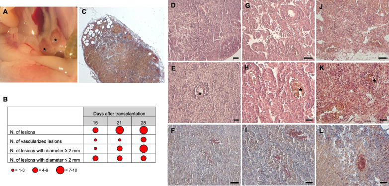
Methods: We established a non-surgical mouse model of endometriosis in CD1 mice. Uterine horns were removed from donor mice, cut into fragments and inoculated in recipient mice by intraperitoneal injection. Endometriosis progression was assessed at 15, 21 and 28 days after transplantation, with the 28-day window being the most effective. The mice were then randomly assigned to four experimental groups, which received for 28 days: water (EMS); DCI 0.4 mg/die (DCI); DCI 0.2 mg/die and Dienogest 0.33 ng/die (DCI + DG); DG 0.67 ng/die (DG). At the end of the treatments, endometriotic lesions, ovaries and circulating estradiol levels were analyzed.

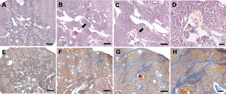
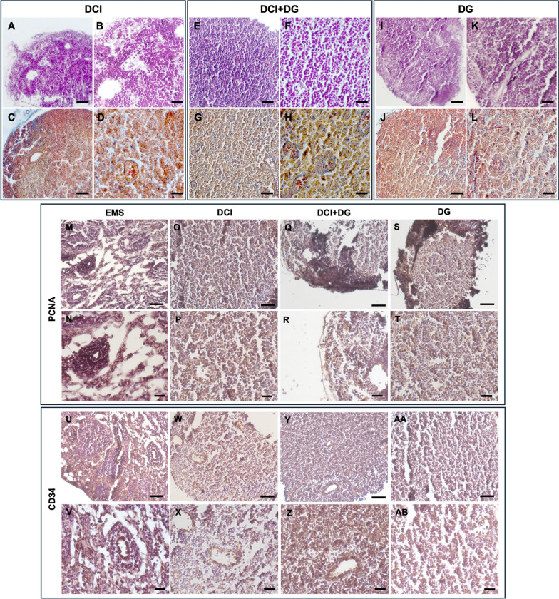
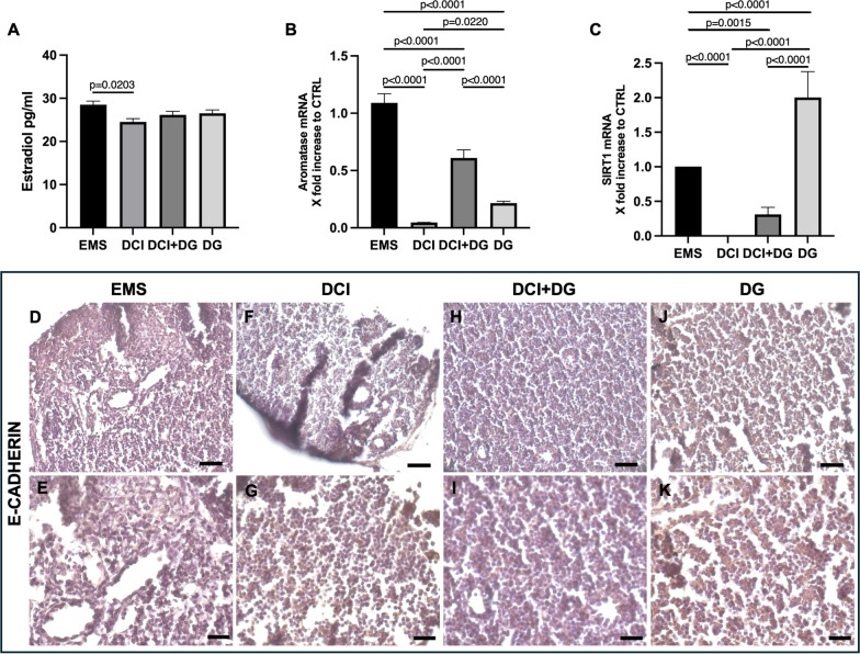
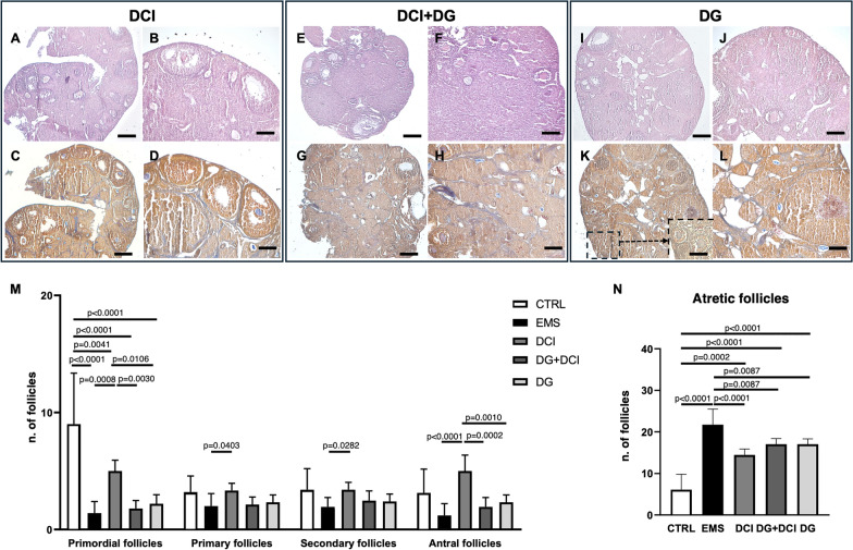
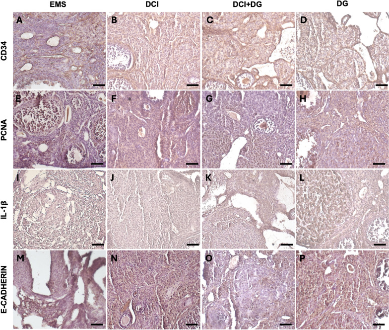
Results: The results showed that treatment with DCI, both alone and in combination with DG, significantly reduced the number, size and vascularization of endometriotic lesions compared to the EMS control group. Histological analysis confirmed a decrease in endometriotic foci across all treatment groups, with the most pronounced effects in the DCI group. To investigate the underlying molecular mechanisms, we found that DCI led to a significant reduction in the expression of Sirt1 and an increase in E-Cadherin, indicating a reduction in EMT transition relevant for lesion development. In addition, DCI decreased cell proliferation and,blood vessel formation, as evaluated by PCNA and CD34, respectively. Futhermore, in the ovary, DCI treatment downregulated the expression of aromatase (Cyp19a1), the enzyme critical for estrogen biosynthesis, and increased the number of primordial to antral follicles, suggesting a beneficial effect on ovarian folliculogenesis.

Conclusions: By modulating proliferation, EMT transition and aromatase activity, DCI emerges as a promising compound for endometriosis treatment.

Keywords: Aromatase; DCI; DG; E-Cadherin; EMT; Endometriosis; Sirt1.


Conflict of interest statement

Declarations. Ethics approval and consent to participate: The project was approved by the Italian Ministry of Health and the internal Committee of the University of L’Aquila (authorization no. 917/2023-PR). Consent for publication: Not applicable. Competing interests: The authors declare no competing interests.

Figures

Fig. 1
Fig. 1
A Representative images of lesions isolated from the abdominal cavity of mice. Asterisks indicate lesions. B Mean number of lesions, vascularization and size at 15, 21 and 28 after the induction of endometriosis. Five-six mice per experimental group were employed. C H&E of a representative endometriotic lesion showing the external connective capsule. LM, mag: 5X. DL H&E (D, E, G, H, J, K) and Trichrome AZAN (F, I, L) stainings of a representative endometriotic lesions from 15-days p.t. (DF), 21 days p.t. (GI) and 28 days p.t. (JL) mice by LM. Asterisks indicate endometriotic foci: D, G, J LM, mag. 20X; bar: 50 µm. E, F, H, I, K, L LM, mag. 40X; bar: 20 µm
Fig. 2
Fig. 2
H&E (AD) and Trichrome AZAN (EH) stainings of endometriotic ovaries from 15 days p.t. mice by LM. A, B, E, F low magnification pictures of a representative endometriotic ovary. C, D, G, H high magnification showing the presence of endometriotic foci (arrows) and an encapsulated cyst (asterisk). A, E mag. 5X; bar: 200 µm; B, F mag. 10X; bar: 100 µm. C, G mag. 20X; bar: 50 µm. D, H mag. 40X; bar: 20 µm
Fig. 3
Fig. 3
H&E (AB) and Trichrome AZAN (CD) stainings of representative endometriotic lesions in DCI-treated mice 28 days p.t. by LM. A, D mag. 10X; bar: 100 µm. B, E mag. 20X; bar: 50 µm. C, F mag. 40X; bar: 20 µm (EF) and Trichrome AZAN (G, H) stainings of representative endometriotic lesions in DG + DCI-treated mice 28 days p.t. by LM. E, G mag. 20X; bar: 50 µm. F, H mag. 40X; bar: 20 µm. H&E (IK) and Trichrome AZAN (JL) stainings of representative endometriotic lesions in DG-treated mice 28 days p.t. by LM. I, J mag. 20X; bar: 50 µm. K, L mag. 40X; bar: 20 µm. MT PCNA immunoreactivity in endometriotic lesions 28 days p.t. of control EMS (M, N), DCI-treated (O, P), DG + DCI (Q, R) and DG-treated (S, T)-treated mice by LM. M, O, Q, S LM, mag. 20X; bar: 50 µm. N, P, R, T mag. 40X; bar: 20 µm. UAB CD34 immunoreactivity in endometriotic lesions 28 days p.t. of control EMS (U, V), DCI-treated (W, X), DG + DCI (Y, Z) and DG-treated (AA, AB)-treated mice by LM. U, W, Y, AA mag. 20X; bar: 50 µm. V, X, Z, AB mag. 40X; bar: 20 µm
Fig. 4
Fig. 4
A Serum estradiol levels in the different experimental classes. Three mice per experimental group were employed. Experiments were done in triplicate. One-way ANOVA: p = 0.0322, followed by Tukey HSD multiple comparisons. B Aromatase gene expression in ovaries. Three mice per experimental group were employed. Experiments were done in triplicate. One-way ANOVA: p < 0.0001, followed by Tukey HSD multiple comparisons. C SIRT1 gene expression in lesions. Three mice per experimental group were employed. Experiments were done in triplicate. One-way ANOVA p < 0.0001, followed by Tukey HSD multiple comparisons. DK E-Cadherin immunoreactivity in endometriotic lesions 28 days p.t. of control EMS (D, E), DCI-treated (F, G), DG + DCI (H, I) and DG-treated (J, K)-treated mice by LM. D, F, H, J mag. 20X; bar: 50 µm. E, G, I, K LM, mag. 40X; bar: 20 µm
Fig. 5
Fig. 5
H&E (AB) and Trichrome AZAN (CD) stainings of a representative endometriotic ovary 28 days p.t. from DCI-treated mice by LM. A, C mag. 5X; bar: 200 µm; B, D mag. 10X; bar: 100 µm. H&E (E, F) and Trichrome AZAN (G, H) stainings of a representative endometriotic ovary 28 days p.t. from DG + DCI-treated mice by LM. E, G mag. 5X; bar: 200 µm; F, H mag. 10X; bar: 100 µm. H&E (I, J) and Trichrome AZAN (K, L) stainings of a representative endometriotic ovary 28 days p.t. from DG-treated mice by LM. I, K mag. 5X; bar: 200 µm; J, L mag. 10X; bar: 100 µm. M Ovarian follicle count. Values are expressed as mean ± SD. Differences were evaluated via ANOVA (p < 0.05) followed by Tukey HSD post-hoc test: * p < 0.05; ** p < 0.01. N Total atretic follicles. Values are expressed as mean ± SD. Differences were evaluated via ANOVA (p < 0.05) followed by Tukey HSD post-hoc test: * p < 0.05; ** p < 0.01
Fig. 6
Fig. 6
AD CD34 immunoreactivity in endometriotic ovary 28 days p.t. of control EMS (A), DCI (B), DG + DCI (C) and DG (D)-treated mice by LM. mag. 20X; bar: 50 µm; EH) PCNA immunoreactivity in endometriotic ovary 28 days p.t. of control EMS (E), DCI (F), DG + DCI (G) and DG (H)-treated mice by LM. mag. 20X; bar: 50 µm. IL IL-1β immunoreactivity in endometriotic ovary 28 days p.t. of control EMS (I), DCI (J), DG + DCI (K) and DG-treated (L)-treated mice by LM. mag. 20X; bar: 50 µm. MP E-Cadherin immunoreactivity in endometriotic ovary 28 days p.t. of control EMS (M), DCI (N), DG + DCI (O) and DG (P)-treated mice by LM. mag. 20X; bar: 50 µm

Similar articles

References

    1. Ahmed I, Balestrieri E, Tudosa I, Lamonaca F. Morphometric measurements of blood cell. Meas Sens. 2021;18:100294. 10.1016/j.measen.2021.100294.
    1. Ahn SH, Monsanto SP, Miller C, et al. Pathophysiology and immune dysfunction in endometriosis. Biomed Res Int. 2015;2015:795976. 10.1155/2015/795976. - PMC - PubMed
    1. Akhmetkaliyev A, Alibrahim N, Shafiee D, Tulchinsky E. EMT/MET plasticity in cancer and go-or-grow decisions in quiescence: the two sides of the same coin? Mol Cancer. 2023;22:90. 10.1186/s12943-023-01793-z. - PMC - PubMed
    1. Amaral C, Augusto TV, Almada M, et al. The potential clinical benefit of targeting androgen receptor (AR) in estrogen-receptor positive breast cancer cells treated with Exemestane. Biochim Biophys Acta Mol Basis Dis. 2020;1866:165661. 10.1016/j.bbadis.2019.165661. - PubMed
    1. Ansieau S, Bastid J, Doreau A, et al. Induction of EMT by twist proteins as a collateral effect of tumor-promoting inactivation of premature senescence. Cancer Cell. 2008;14:79–89. 10.1016/j.ccr.2008.06.005. - PubMed

LinkOut - more resources