. 2025 Apr 10;30(1):44.
doi: 10.1186/s11658-025-00705-x.

Repression of ZNFX1 by LncRNA ZFAS1 mediates tobacco-induced pulmonary carcinogenesis

Affiliations

Repression of ZNFX1 by LncRNA ZFAS1 mediates tobacco-induced pulmonary carcinogenesis

Sichuan Xi et al. Cell Mol Biol Lett. .

Abstract

Background: Despite exhaustive research efforts, integrated genetic and epigenetic mechanisms contributing to tobacco-induced initiation and progression of lung cancers have yet to be fully elucidated. In particular, limited information is available regarding dysregulation of noncoding RNAs during pulmonary carcinogenesis.

Methods: We examined correlations and interactions of long noncoding (lnc) RNAs and protein-coding genes in normal respiratory epithelial cells (NREC) and pulmonary tumor cells following exposure to cigarette smoke condensate (CSC) using gene expression arrays, qRT-PCR, western blot, growth assays, transwell assays, and murine xenograft models, as well as methylated DNA immunoprecipitation, RNA cross-link immunoprecipitation, and quantitative chromatin immunoprecipitation techniques with bioinformatics analyses.

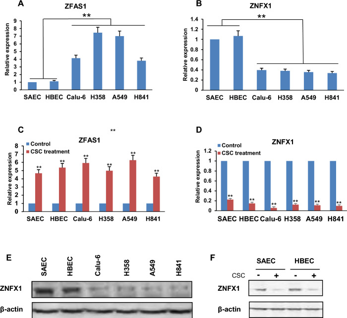
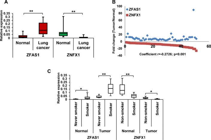
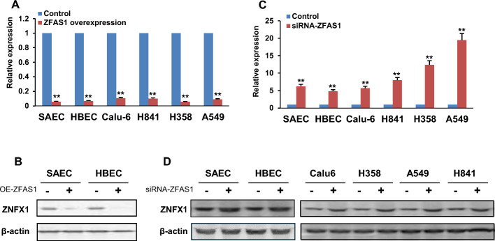
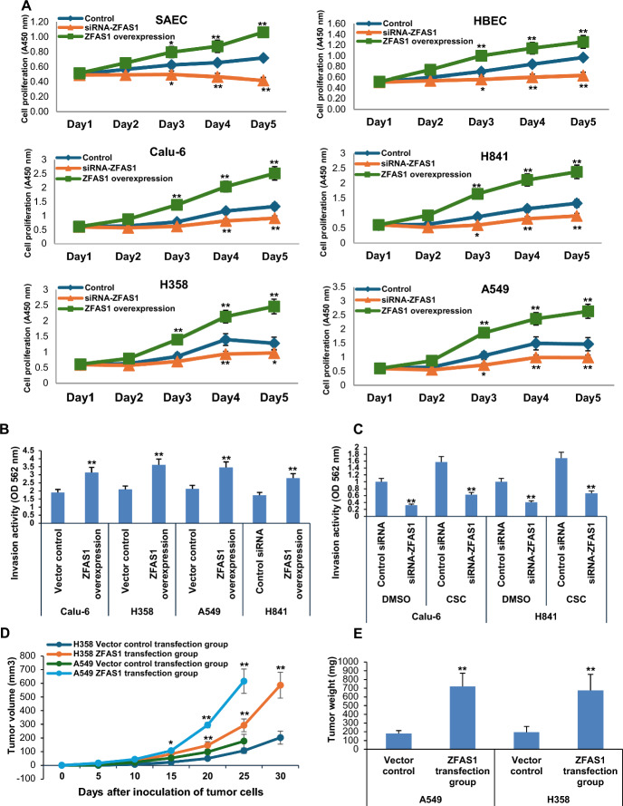
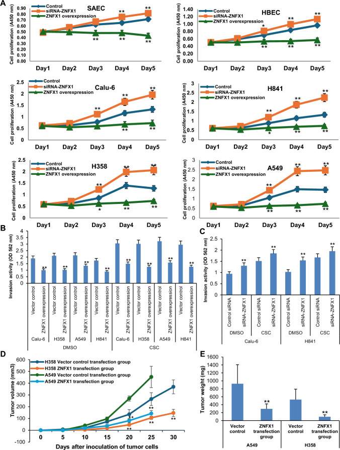
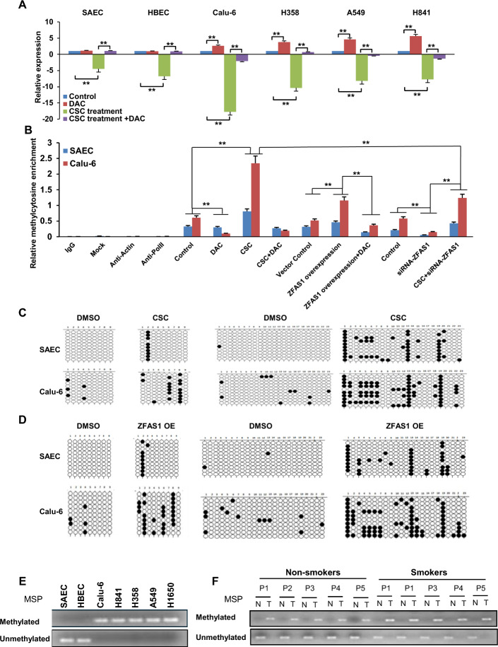
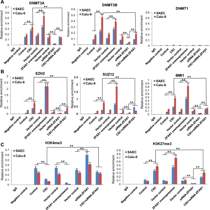
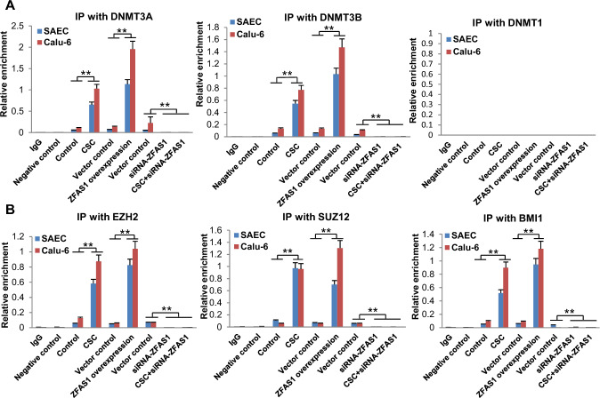
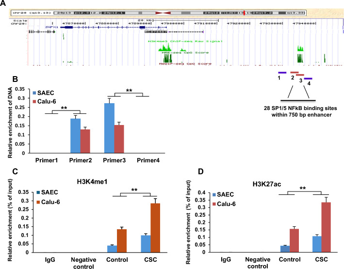
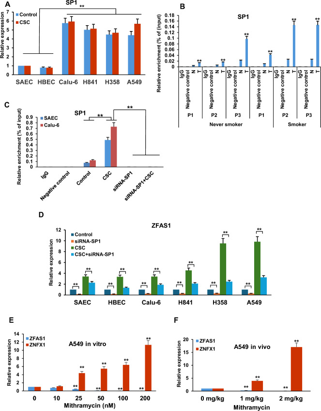
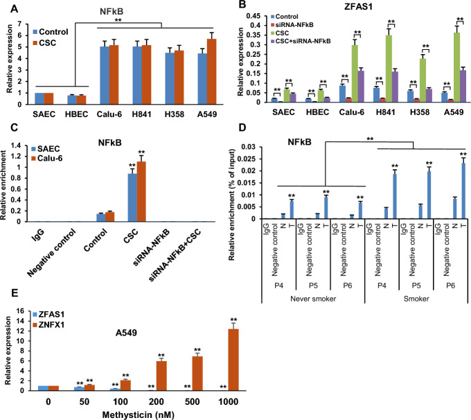
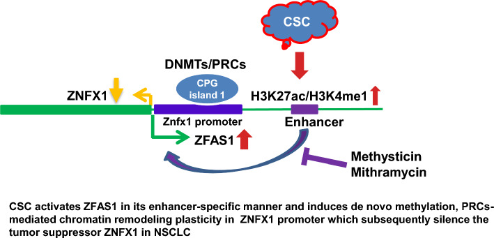
Results: Among diverse alterations of lncRNA and coding gene expression profiles in NREC exposed to CSC, we observed upregulation of lncRNA ZFAS1 and repression of an adjacent protein-coding gene, ZNFX1, and confirmed these findings in primary lung cancers. Phenotypic experiments indicated that ZFAS1 is an oncogene, whereas ZNFX1 functions as a tumor suppressor in lung cancer cells. Mechanistically, CSC induces ZFAS1 expression via SP1 and NFĸB-associated activation of an enhancer linked to ZFAS1. Subsequently, ZFAS1 interacts with DNA methyltransferases and polycomb group proteins to silence ZNFX1. Mithramycin and methysticin repress ZFAS1 and upregulate ZNFX1 in lung cancer cells in vitro and in vivo.

Conclusion: These studies reveal a novel feedforward lncRNA circuit contributing to pulmonary carcinogenesis and suggest that pharmacologic targeting of SP1 and/or NFĸB may be useful strategies for restoring ZNFX1 expression for lung tumor therapy.

Keywords: ZFAS1; ZNFX1; BMI1; Cigarette smoke; DNMT; EZH2; Epigenetics; Lung cancer; NFĸB; Noncoding RNA; SP1; SUZ12.


Conflict of interest statement

Declarations. Ethics approval and consent to participate: The study protocol was approved by the NIH internal review board (approval no. 06C0014; date: 02/28/2023) based on the Helsinki Declaration. All animal experiments were approved by the National Cancer Institute Animal Care and Use Committee (approval no. SB-200; date: 07/03/2024) and were performed according to international guidelines and the Basel Declaration. Competing interests: The authors declare that they have no competing interests.

Figures

Fig. 1
Fig. 1
CSC modulates reciprocal transcriptional activities of ZFAS1 and ZNFX1 in normal respiratory epithelia and lung cancer cells. A, B qRT-PCR analysis demonstrating endogenous ZFAS1 (A) and ZNFX1 (B) mRNA levels are significantly higher or lower, respectively, in SAEC and HBEC cells relative to Calu-6, H358, A549, and H841 cells. C, D qRT-PCR analysis demonstrating upregulation of ZFAS1 (C) with concomitant downregulation of ZNFX1 (D) in SAEC, HBEC, Calu-6, H358, A549, and H841 cells following 5-day CSC exposure. * p < 0.05; **p < 0.01. E Immunoblot analysis demonstrates that endogenous ZNFX1 protein levels are higher in SAEC and HBEC cells compared with Calu-6, H358, A549, and H841 cells. F Immunoblot demonstrating decreased ZNFX1 protein levels in SAEC and HBEC following 5-day CSC exposure. Data are mean ± SEM; T-test; n = 3; * p < 0.05; **p < 0.01
Fig. 2
Fig. 2
Reverse correlation between expression of ZNFX1 and  ZFAS1 in lung cancer specimens. A qRT-PCR analysis of ZFAS1 and ZNFX1 mRNA levels in 51 lung cancer specimens and their paired adjacent normal lung tissues. B Correlation patterns and corresponding correlation coefficient values between ZFAS1 and ZNFX1 in 51 lung cancers. Coefficient r = −0.2726, p < 0.001. C qRT-PCR analysis of ZFAS1 and ZNFX1 expression in lung cancers from smokers/former smokers versus never smokers. * p < 0.05; **p < 0.01
Fig. 3
Fig. 3
ZFAS1 directly antagonizes transcriptional activity of ZNFX1 in normal respiratory epithelia and lung cancer cells. A qRT-PCR analysis demonstrating that overexpression of ZFAS1 downregulates ZNFX1 in normal respiratory epithelia and lung cancer cells. B Immunoblotting analysis demonstrating that overexpression of ZFAS1 reduces ZNFX1 protein levels in SAEC and HBEC. C qRT-PCR analysis demonstrating that knockdown of ZFAS1 upregulates ZNFX1 in normal respiratory epithelia and lung cancer cells. D Immunoblot demonstrating that depletion of ZFAS1 increases ZNFX1 protein levels in normal respiratory epithelia and lung cancer cells. Data are mean ± SEM; t-Test; n = 3; * p < 0.05; **p < 0.01
Fig. 4
Fig. 4
ZFAS1 functions as an oncogene in lung cancer cells. A Effects of ZFAS1 expression on in vitro proliferation of SAEC and HBEC, as well as Calu-6, H841, H358, and A549 lung cancer cells. ZFAS1 promotes cell growth in normal respiratory epithelia and lung tumor cells. B, C Matrigel invasion assays demonstrate that overexpression of ZFAS1 enhances invasion of Calu-6, H358, A549, and H841 lung cancer cells, whereas siRNA knockdown of ZFAS1 inhibits invasion of control lung cancer cells and attenuates CSC-mediated invasion potential in these cells. *p < 0.05; **p < 0.01. D Growth of H358 and A549 subcutaneous xenografts in nude mice. Volumes of xenografts derived from H358 and A549 cells overexpressing ZFAS1 are significantly larger than control xenografts. E Tumor masses from H358 and A549 xenografts. ZFAS1 overexpression significantly increases the average mass of tumor xenografts. Data are mean ± SEM; T-test; n = 3; * p < 0.05; **p < 0.01
Fig. 5
Fig. 5
ZNFX1 functions as a tumor suppressor in lung cancer cells. A Effects of ZNFX1 expression on in vitro proliferation in SAEC and HBEC as well as Calu-6, H841, H358, and A549 cells. ZNFX1 promotes cell growth in both normal respiratory epithelia and lung tumor cells. B, C Matrigel invasion assays demonstrating that overexpression of ZNFX1 inhibits, whereas knockdown of ZNFX1 increases invasion of Calu-6 and H841 lung cancer cells. Knockdown of ZNFX1 enhances CSC-induced invasion of lung cancer cells. * p < 0.05; ** p < 0.01. D Growth of H358 and A549 subcutaneous xenografts in nude mice. Volumes of xenografts derived from H358 and A549 cells overexpressing ZNFX1 are significantly smaller than control xenografts. E Tumor masses from H358 and A549 xenografts. ZNFX1 overexpression significantly decreases the average mass of tumor xenografts. Data are mean ± SEM; T-test; n = 3; * p < 0.05; **p < 0.01
Fig. 6
Fig. 6
Promoter CpG island-specific methylation alterations coincide with the repression of ZNFX1. A qRT-PCR analysis demonstrating that DAC increases ZNFX1 expression in lung cancer cells not in normal lung epithelia. DAC abrogates CSC-mediated repression of ZNFX1 in normal lung epithelia and lung cancer cells. B MeDIP analysis of DNA methylation profiles in the first CpG island proximal to the TSS of ZNFX1 in SAEC and Calu-6 cells; DAC decreases CSC- or ZFAS1-mediated DNA hypermethylation. C, D Bisulfite sequencing of two genomic regions in the first CpG island of the ZNFX1 promoter, demonstrating that CSC or ZFAS1 overexpression enhances DNA methylation in this CpG island in SAEC and Calu-6 cells. E MSP demonstrating site-specific DNA methylation in the ZNFX1 promoter in lung cancer cells but not in NREC. F MSP analysis demonstrating DNA methylation in the first CpG island of the ZNFX1 promoter in lung cancer tissues from both smokers and nonsmokers relative to normal adjacent lung tissues. Data are mean ± SEM; T-test; n = 3; * p < 0.05; **p < 0.01
Fig. 7
Fig. 7
CSC-mediated activation of ZFAS1 coincides with epigenetic alterations in the promoter of ZNFX1. A qChIP analysis of DNMT3A, DNMT3B, and DNMT1 levels within the first CpG island of the ZNFX1 promoter in SAEC and Calu-6 cells exposed to NM or CSC, following overexpression or knockdown of ZFAS1. B qChIP analysis of EZH2, SUZ12, and BMI1 levels within the promoter of ZNFX1 in SAEC and Calu-6 cells either exposed to NM or CSC following overexpression or knockdown of ZFAS1. C qChIP analysis of H3K4me3 or H3K27me3 within the promoter of ZNFX1 in SAECs and Calu-6 cells exposed to NM or CSC following overexpression or knockdown of ZFAS1. CSC and ZFAS1 decrease levels of H3K4me3 while increasing H3K27me3 levels in the proximal promoter region (0 to −1 kb) of ZNFX1 in SAEC and Calu-6 cells; depletion of ZFAS1 partially abrogates CSC-induced alterations in these histone marks within this region. Data are mean ± SEM; T-test; n = 3; * p < 0.05; **p < 0.01
Fig. 8
Fig. 8
Interactions of ZFAS1 transcripts with DNA methylation and polycomb group proteins. A, B qCLIP analyses of interactions of ZFAS1 with DNMT3A or DNMT3B and DNMT1 as well as EZH2, SUZ12, BMI1 in SAEC and Calu-6 cells exposed to NM or CSC, with or overexpression or knockdown of ZFAS1. CSC and ZFAS1 over-expression induce binding of DNMT3A, DNMT3B, EZH2, SUZ12, and BMI1 but not DNMT1 to ZFAS1 transcripts; knockdown of ZFAS1 diminishes these interactions. Data are mean ± SEM; T-test; n = 3; * p < 0.05; ** p < 0.01
Fig. 9
Fig. 9
Enhancer-specific modulation of ZFAS1 by CSC. A Schematic depiction demonstrating putative enhancer-like regulatory element region for ZFAS1. The numbers 1, 2, 3, and 4 indicate the primer positions for the formaldehyde-assisted isolation of regulatory elements (FAIRE) assay to characterize upstream regulatory elements of ZFAS1. B Quantitative FAIRE analysis for mapping the regulatory elements of ZFAS1 identified significant specific amplification signals in primer 2 and 3 positions compared with negative signals from primer 1 and 4 sites, demonstrating that the narrow genetic region (40–41 kb upstream from TSS of ZFAS1) is the potential enhancer for ZFAS1. C, D qChIP analysis demonstrating increased levels of H3K4me1 and H3K27ac (histone activation marks) in the putative enhancer of ZFAS1 in SAEC and Calu-6 cells following CSC exposure. Data are mean ± SEM; T-test; n = 3; * p < 0.05; ** p < 0.01
Fig. 10
Fig. 10
Role of SP1 in CSC-mediated regulation of ZFAS1. A qRT-PCR analysis of S1  SP1 expression in NREC and lung cancer cells. SP1 expression is much higher in lung cancer cells compared with NREC. Short term CSC exposure does not appear to increase SP1 expression in these cells. B qChIP analysis of SP1 occupancy in the ZFAS1 enhancer in lung cancers and paired normal adjacent lung tissues from smokers and nonsmokers. C qChIP analysis demonstrating that CSC exposure induces recruitment of SP1 to the putative enhancer of ZFAS1 in SAEC and Calu-6 cells. D qChIP analysis demonstrating that knockdown of SP1 decreases endogenous ZFAS1 expression and markedly attenuates CSC-mediated upregulation of ZFAS1 in NREC and lung cancer cells. E, F qRT-PCR analysis demonstrating that mithramycin mediates dose-dependent reduction of ZFAS1 with a concomitant increase in ZNFX1 expression in A549 lung cancer cells in vitro (E) and in vivo (F). Data are mean ± SEM; T-test; n = 3; * p < 0.05; ** p < 0.01
Fig. 11
Fig. 11
CSC upregulates ZFAS1 via selective interaction of NFkB with the ZFAS1 enhancer. A qRT-PCR analysis demonstrating expression of NFkB-p65 in normal respiratory epithelia and lung cancer cells cultured in the presence or absence of CSC. B qRT-PCR analysis demonstrating that siRNA knockdown of NFkB-p65 significantly decreased ZFAS1 expression in normal respiratory epithelia and lung cancer cells treated with or without exposure to CSC. C/D) Quantitative ChIP analysis demonstrating increased levels of NFkB in the enhancer region of ZFAS1 in SAEC and Calu-6 cells following CSC exposure (C) or in lung cancer tissues from both smokers and nonsmokers (D). E qRT-PCR analysis demonstrating ZFAS1 and ZNFX1 expression in A549 lung cancer cells treated with or without mythysticin at 0 to 1000 nM concentration for 48 h. Methysticin dose-dependently enhanced expression of ZNFX1 while dose-dependently inhibiting expression of ZFAS1 in normal respiratory epithelia and lung cancer cells. Data are mean ± SEM; T-test; n = 3; * p < 0.05; **p < 0.01
Fig. 12
Fig. 12
Mechanistic diagram summarizing mechanisms by which repression of ZNFX1 by ZFAS1 mediates tobacco-induced pulmonary carcinogenesis. CSC activates ZFAS1 in an enhancer-specific manner and induces de novo DNA methylation and polycomb-mediated chromatin remodeling within the ZNFX1 promoter, which subsequently silences this tumor suppressor in NSCLC.

Similar articles

References

    1. Leiter A, Veluswamy RR, Wisnivesky JP. The global burden of lung cancer: current status and future trends. Nature Rev Clin Oncol. 2023;20:624–39. - PubMed
    1. Bray F, Laversanne M, Sung H, Ferlay J, Siegel RL, Soerjomataram I, et al. Global cancer statistics 2022: GLOBOCAN estimates of incidence and mortality worldwide for 36 cancers in 185 countries. CA Cancer J Clin. 2024;74:229–63. - PubMed
    1. Thai AA, Solomon BJ, Sequist LV, Gainor JF, Heist RS. Lung cancer. Lancet. 2021;398:535–54. - PubMed
    1. Rudin CM, Brambilla E, Faivre-Finn C, Sage J. Small-cell lung cancer. Nature Rev Dis Primers. 2021;7:3. - PMC - PubMed
    1. He S, Li H, Cao M, Sun D, Yang F, Yan X, et al. Survival of 7,311 lung cancer patients by pathological stage and histological classification: a multicenter hospital-based study in China. Transl Lung Cancer Res. 2022;11:1591–605. - PMC - PubMed

Substances