. 2025 Apr 10;25(1):153.
doi: 10.1186/s12883-025-04171-y.

Resveratrol attenuates the CoCl2-induced hypoxia damage by regulation of lysine β-hydroxybutyrylation in PC12 cells

Affiliations

Resveratrol attenuates the CoCl2-induced hypoxia damage by regulation of lysine β-hydroxybutyrylation in PC12 cells

Yamei Wang et al. BMC Neurol. .

Abstract

Background: Stroke is a cerebrovascular disease that is the main cause of death and disability worldwide. Hypoxia is a major factor that causes neuronal damage and even cellular death. However, the mechanism and therapeutic drugs for hypoxia are not completely understood.

Methods: In this study, PC12 cells (a rat adrenal pheochromocytoma cell line) were exposed to Cobalt chloride (CoCl2) to induce hypoxia. Using this cell model, the impacts of hypoxia on cell viability, proliferation, reactive oxygen species (ROS), and the levels of lysine β-hydroxybutyrylation (Kbhb) and the inflammatory signaling factor P65 were examined. In addition, we explored the ability of resveratrol (RES) to alleviate CoCl2-induced hypoxia damage.

Results: RES attenuated CoCl2-induced decreases of cell viability and cell proliferation and increase of ROS production in PC12 cells. CoCl2 downregulated Kbhb in PC12 cells, but RES alleviated this effect. In addition, upregulated Kbhb by 3-hydroxybutyric acid sodium could partially recover the CoCl2-induced hypoxia damage to PC12 cells, including cell viability, cell proliferation, oxidative stress, and the protein level of the inflammatory signaling factor P65.

Conclusion: Our results indicate that RES protects against CoCl2-induced hypoxia damage in PC12 cells by modulating Kbhb, a novel post-translational modification.

Keywords: CoCl2; Hypoxia; Lysine β-hydroxybutyrylation; PC12 cells; Reactive oxygen species; Resveratrol.


Conflict of interest statement

Declarations. Ethics approval and consent to participate: Not applicable. Consent for publication: Not applicable. Competing interests: The authors declare no competing interests.

Figures

Fig. 1
Fig. 1
Effect of CoCl2 on the viability of PC12 cells. (a-c) PC12 cells were exposed to 5 µM, 50 µM, or 100 µM CoCl2 for assessment of cell viability at 24 h (a), 48 h (b) and 72 h (c) using the CCK-8 assay. n = 3. The data conformed to normal distribution analyzed by Shapiro-Wilk test. The data are presented as the mean ± SD. Differences between groups were assessed by one-way ANOVA. **p < 0.01 and ***p < 0.001
Fig. 2
Fig. 2
Effect of CoCl2 combined with resveratrol (RES) on the viability of PC12 cells. PC12 cells were exposed to 100 µM CoCl2, 50 μm RES, and 100 µM CoCl2 plus 50 μm RES for 48 h. (a) Morphological characterization of PC12 cells was performed using an optical microscope (the red arrows for abnormal morphology); scale bars in the figures indicate 100 μm. (b) The percentage of abnormal cells in different groups have been quantified. (c) Cell viability was examined by CCK-8 assay. The data are presented as the mean ± SD. n = 3. The data conformed to normal distribution analyzed by Shapiro-Wilk test. Differences between groups were assessed by one-way ANOVA. *p < 0.05, **p < 0.01
Fig. 3
Fig. 3
Effect of CoCl2 combined with RES on PC12 cell proliferation. (a) EdU staining (red) and Hoechst staining (blue) of PC12 cells treated with 100 µM CoCl2 combined with 50 µM RES. Scale bar: 100 μm. (b) Statistical result of CoCl2 combined with RES on PC12 cell proliferation. EdU positive cells (%) were calculated as the ratio of EdU staining cells to Hoechst staining cells and multiplied by 100 (%). The data are presented as the mean ± SD. n = 4. The data conformed to normal distribution analyzed by Shapiro-Wilk test. Differences between groups were assessed by one-way ANOVA. ns: not significant, **p < 0.01, ***p < 0.001
Fig. 4
Fig. 4
Effect of RES on CoCl2-induced reactive oxygen species (ROS) production in PC12 cells. PC12 cells were exposed to 100 µM CoCl2, 50 µM RES, and 100 µM CoCl2 plus 50 µM RES for 48 h. ROS production was measured with the ROS fluorescent dye DCFH-DA. The data are presented as the mean ± SD. n = 6. The data conformed to normal distribution analyzed by Shapiro-Wilk test. Differences between groups were assessed by one-way ANOVA. ns: not significant, **p < 0.01, ***p < 0.001
Fig. 5
Fig. 5
Effect of CoCl2, RES, and CoCl2 plus RES on the level of Kbhb in PC12 cells. (a) PC12 cells were exposed to CoCl2 (5, 50 and 100 µM) for 48 h, and the level of Kbhb was analyzed via western blotting. (b) PC12 cells were exposed to 50 µM RES, 100 µM CoCl2, and 50 µM RES plus 100 µM CoCl2 for 48 h, and the level of Kbhb was analyzed via western blotting. The data are presented as the mean ± SD. n = 3. The data conformed to normal distribution analyzed by Shapiro-Wilk test. Differences between groups were assessed by one-way ANOVA. *p < 0.05, **p < 0.01
Fig. 6
Fig. 6
Effect of 3-hydroxybutyric acid sodium (3-HBA·Na) on the level of Kbhb and viability of PC12 cells. PC12 cells were exposed to 100 µM CoCl2, 20 mM 3-HBA·Na, and 20 mM 3-HBA·Na plus 100 µM CoCl2 for 48 h. (a and b) The level of Kbhb was examined by western blotting. (c) Cell viability was examined via a CCK-8 assay. The data are presented as the mean ± SD. n = 3 or 6. The data conformed to normal distribution analyzed by Shapiro-Wilk test. Differences between groups were assessed by one-way ANOVA. **p < 0.01, ***p < 0.001
Fig. 7
Fig. 7
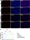
Effect of 3-hydroxybutyric acid sodium (3-HBA·Na) on the proliferation of PC12 cells. EdU staining (red) and Hoechst staining (blue) of PC12 cells treated with 100 µM CoCl2, 20 mM 3-HBA·Na, and 20 mM 3-HBA·Na plus 100 µM CoCl2. Scale bar: 100 μm. (b) Statistical result of 3-HBA·Na combined with RES on PC12 cell proliferation. The data are presented as the mean ± SD. n = 3. The data conformed to normal distribution analyzed by Shapiro-Wilk test. Differences between groups were assessed by one-way ANOVA. ns: not significant, *p < 0.05
Fig. 8
Fig. 8
Effect of 3-hydroxybutyric acid sodium (3-HBA·Na) on the expression of P65. PC12 cells were exposed to 100 µM CoCl2, 20 mM 3-HBA·Na, and 20 mM 3-HBA·Na plus 100 µM CoCl2 for 48 h. The protein level of P65 in the nuclear fraction was analyzed via western blotting (a and b)

Similar articles

References

    1. Malik P, Patel UK, Kaul S, Singla R, Kavi T, Arumaithurai K, et al. Risk factors and outcomes of intravenous tissue plasminogen activator and endovascular thrombectomy utilization amongst pediatrics acute ischemic stroke. Int J Stroke. 2021;16(2):172–83. 10.1177/1747493020904915. - PubMed
    1. Chen R, Zhang X, Gu L, Zhu H, Zhong Y, Ye Y, et al. New insight into neutrophils: A potential therapeutic target for cerebral ischemia. Front Immunol. 2021;12. 10.3389/fimmu.2021.692061. - PMC - PubMed
    1. Hermann DM, Xin W, Bähr M, Giebel B, Doeppner TR. Emerging roles of extracellular vesicle-associated non-coding RNAs in hypoxia: insights from cancer, myocardial infarction and ischemic stroke. Theranostics. 2022;12 13:5776–802. 10.7150/thno.73931. - PMC - PubMed
    1. Chou TT, Trojanowski JQ, Lee VM-Y. Neurotrophin signal transduction in Medulloblastoma. J Neurosci Res. 1997;49 5:522–7. 10.1002/(SICI)1097-4547(19970901)49:5%3C522::AID-JNR2%3E3.0.CO;2-D. - PubMed
    1. Wang Y, Qin X, Han Y, Li B. VGF: A prospective biomarker and therapeutic target for neuroendocrine and nervous system disorders. Biomed Pharmacother. 2022;151:113099. 10.1016/j.biopha.2022.113099. - PubMed

LinkOut - more resources