

Age-specific normative values of sacral development and fusion in children and adolescents: a cross-sectional study utilizing multiplanar reconstruction computed tomography imaging
- PMID: 40211180
- PMCID: PMC11983754
- DOI: 10.1186/s12891-025-08597-w
Age-specific normative values of sacral development and fusion in children and adolescents: a cross-sectional study utilizing multiplanar reconstruction computed tomography imaging
Abstract
Background: This study aimed to determine the index of the sacral vertebrae fusion period in children and adolescents to diagnose the lesion around the sacral spine accurately.
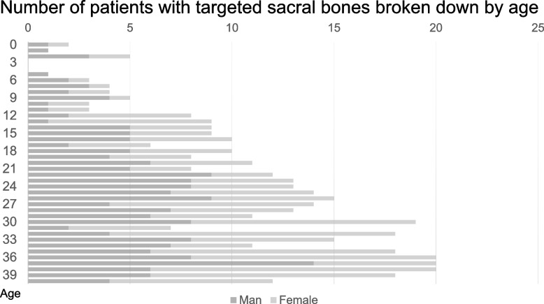
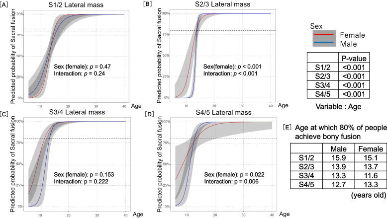
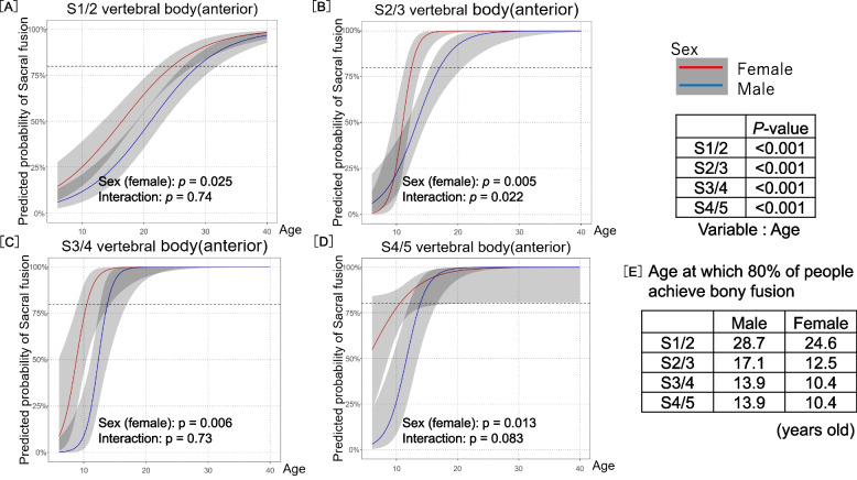
Methods: Patients aged 0-40 years who underwent computed tomography (CT), including the normal sacrum for screening abdominal disorders and pan-scan in trauma between 2019 and 2022 were retrospectively examined. There were 402 eligible sacra (385 patients: 206 women and 179 men). We evaluated bony fusion at six parts of the sacral vertebrae (anterior or posterior of each intervertebral and both side lateral masses). The predicted probability of bony fusion obtained from the logistic regression model is depicted graphically by sex.
Results: The association between bony fusion in each vertebral segment and age was evaluated using a logistic regression model with a Huber-White robust sandwich estimator, including the patient as a clustering variable. Bony fusion of the sacral bodies of S1/S2 was slowest, with 80% of patients achieving bony fusion at 28.7 and 24.6 years of age for men and women, respectively. Compared to men, women exhibited earlier fusion of the intervertebral segments of the sacral vertebrae; however, no significant difference between the sexes in terms of eventual bony fusion at the lateral mass was observed, while the initiation of bony fusion occurred earlier in women.
Conclusion: The predicted probability of bony fusion could aid pediatricians, orthopedists, radiologists, and other physicians in understanding the normal development of the sacral spine and accurately differentiating the lesion around the sacral spine.
Keywords: Adolescent; Bony fusion; Sacral spine development; Sacrum.
© 2025. The Author(s).
Conflict of interest statement
Declarations. Ethics approval and consent to participate: The Gifu University Graduate School of Medicine Institutional Review Board has accepted this retrospective study under study number #2022–175. Informed consent was obtained using an opt-out method disclosed on the Gifu University website. For any participants under the age of 16, the consent was obtained using same method from the parents or legal guardians. The research was conducted following the tenets of the Declaration of Helsinki and its later amendments. Consent for publication: Not applicable. Competing interests: The authors declare no competing interests.
Figures


References
-
- Jaremko JL, Siminoski K, Firth GB, Matzinger MA, Shenouda N, Konji VN, Roth J, Sbrocchi AM, Reed MH, O’Brien MK, et al. Common normal variants of pediatric vertebral development that mimic fractures: a pictorial review from a national longitudinal bone health study. Pediatr Radiol. 2015;45(4):593–605. - DOI - PMC - PubMed
-
- Grant’s Atlas of Anatomy, 16e. Agur AMR, Dalley AFI. agur. Lippincott Williams & Wilkins, a Wolters Kluwer business. 2025. https://premiumbasicsciences.lwwhealthlibrary.com/book.aspx?bookid=3319&.... Accessed 04 Apr 2025.
-
- Rios L, Weisensee K, Rissech C: Sacral fusion as an aid in age estimation. Forensic Sci Int 2008, 180(2–3):111 e111–117. - PubMed
-
- Mahon TJ, Friedling LJ, Gordon GM: The use of ventral fusion between sacral elements S1 and S2 as an additional age-at-death indicator in a black South African skeletal sample. Forensic Sci Int 2018, 286:267 e261–267 e266. - PubMed
MeSH terms
LinkOut - more resources
Full Text Sources
Medical
