. 2025 Apr 10;45(1):9.
doi: 10.1186/s41232-025-00373-6.

Critical roles of IL-6 signaling in myoblast differentiation of human adipose-derived mesenchymal stem cells

Affiliations

Critical roles of IL-6 signaling in myoblast differentiation of human adipose-derived mesenchymal stem cells

Takashi Otsuka et al. Inflamm Regen. .

Abstract

Background: Ectopic fat is also formed in muscles as well as the liver, where adipose-derived mesenchymal stem cells (ADSCs) promote adipogenesis. On the other hand, after muscle injury, muscle satellite cells (SCs) contribute to muscle repair through myodifferentiation. Human ADSCs are multipotent stem cells, but it remains unclear whether they are involved in myoblast differentiation. The aim is to find a novel myogenic cytokine and its signaling pathway that promotes the differentiation of human ADSCs-a potential source of new muscle precursor cells-into myoblasts.

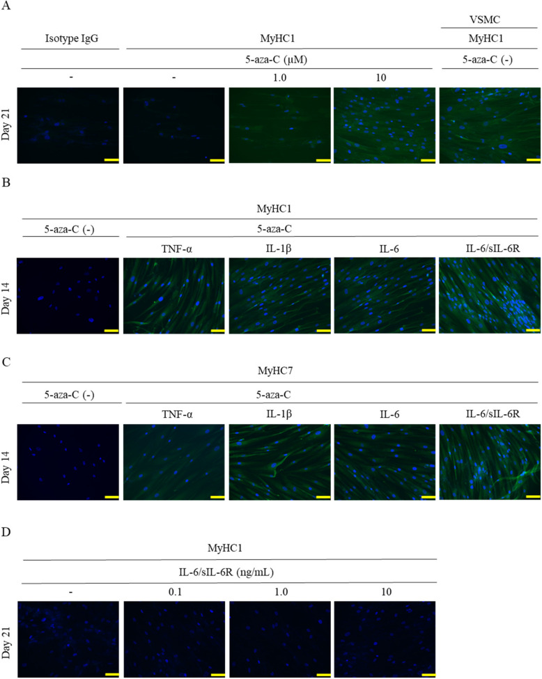
Methods: An array kit was used to detect cytokines produced by ADSCs. After treating ADSCs with the DNA methyltransferase inhibitor 5-Aza-2'-deoxycytidine (5-aza-C) and different JAK inhibitors, MyHC1, a myodifferentiation marker, was detected by immunofluorescence staining and reverse transcription-quantitative polymerase chain reaction (RT-qPCR). The expression status of signaling molecules was determined by Western blotting and the recruitment of transcription factors to the MYOG promoter by chromatin immunoprecipitation (ChIP).

Results: IL-6 was detected at high concentrations in the culture supernatant of ADSCs. ADSCs stimulated with 5-aza-C became strongly positive for MyHC1 on day 21 post-stimulation. When co-stimulated with 5-aza-C and IL-6/sIL-6R, ADSCs became positive for MyHC1 protein and upregulated MYOG mRNA as early as day 14 post-stimulation. Co-stimulation with 5-aza-C and IL-6/sIL-6R resulted in phosphorylation of STAT1 and STAT3. The addition of a JAK2 inhibitor, but not JAK1/3 inhibitors, abolished the MyHC1 positivity and phosphorylation of STAT1 and STAT3. Co-stimulation with 5-aza-C and IL-6/sIL-6R during the myogenesis process resulted in the recruitment of STAT1, but not STAT3, to the MYOG promoter. Myoblast differentiation induced by stimulation with 5-aza-C was enhanced by activation of the IL-6/JAK2/STAT1/MYOG pathway.

Conclusions: Therefore, sustained IL-6/JAK2/STAT1 activation may serve as an important driver of human ADSC differentiation into myoblast, suggesting an important candidate signaling pathway for ameliorating muscle atrophy.

Keywords: 5-aza-C; ADSCs; IL-6; Myoblast; STAT1.


Conflict of interest statement

Declarations. Ethics approval and consent to participate: Not applicable. Consent for publication: Not applicable. Competing interests: S. Nakayamada has received consulting fees, speaking fees, lecture fees, and/or honoraria from AstraZeneca, GlaxoSmithKline, Pfizer, Bristol-Myers, Astellas, Asahi-kasei, AbbVie, Chugai, Sanofi, Eisai, Gilead Sciences, Mitsubishi-Tanabe, Janssen, Eli Lilly, Boehringer Ingelheim, and Ayumi. Y. Okada has received lecture fees from AstraZeneca, MSD, Ono, Mitsubishi-Tanabe, Bayer, Novo Nordisk, Eli Lilly, and Boehringer Ingelheim and research funds from Kowa and Mitsubishi-Tanabe. Y. Tanaka has received speaking fees and/or honoraria from Abbvie, Eisai, Chugai, Eli-Lilly, Behringer-Ingelheim, GlaxoSmithKline, Taisho, AstraZeneca, Daiichi-Sankyo, Gilead, Pfizer, UCB, Asahi-kasei, and Astellas and received research grants from Behringer-Ingelheim, Taisho, and Chugai. The other authors declare that they have no competing interests.

Figures

Fig. 1
Fig. 1
Human ADSCs produce IL-6 at high levels. A Cytokine array with ADSC culture supernatant. The one on the left was with MSCGM alone, and the three on the right were with ADSC culture supernatant. The solid and dashed frames indicate the positive and negative internal controls, respectively. Cytokines produced at significant levels were numbered 1 to 7. Three independent lots of ADSCs were used. B The top 7 factors with high secretion levels. DPI, dots per inch. C A panel of cytokines was measured in the culture supernatant of ADSCs using the CBA method. D The production of IL-6, IL-1β, and TNF-α from human breast cancer cell line (MCF-7), normal human dermal fibroblasts (NHDF), and three lots of ADSCs was quantified by RT-qPCR. C, D Three independent lots of ADSCs (C, D) and NHDF (D) were used. Data are expressed as mean ± standard deviation. Student’s unpaired two-tailed t-test was used for comparisons between two groups
Fig. 2
Fig. 2
IL-6/sIL-6R promotes myoblast differentiation induced by 5-aza-C. AD ADSCs were stimulated with 5-aza-C alone (1.0, 10 μM) (A) or co-stimulated with 5-aza-C (10 μM) and TNF-α (10 ng/mL), IL-1β (10 ng/mL), IL-6 (10 ng/mL), or IL-6/sIL-6R (10 ng/mL) (B, C), or varying concentrations of IL-6/sIL-6R (0.1, 1.0, 10 ng/mL) (D), followed by fluorescence staining with anti-MyHC1 antibody on days 14 (B) and 21 (A, D). Cells were also fluorescence-stained with anti-MyHC7 antibody on day 21 (C). Human vascular smooth muscle cells (VSMCs) were used as the positive control. Three independent lots of ADSCs were used. Data from a representative lot are presented. The images were captured at 200 × magnification. Scale bar indicates 100 μM
Fig. 3
Fig. 3
Co-stimulation of human ADSCs with 5-aza-C and IL-6/sIL-6R induces expression of skeletal muscle lineage cell markers. A, B ADSCs were stimulated with 5-aza-C alone (A) or co-stimulated with 5-aza-C and different concentrations of IL-6/sIL-6R (A, B). The expression of MYOG, DESMIN, LGALS, PPARγ, and RUNX2 was quantified by RT-qPCR. Human skeletal muscle myoblasts (HSMMs) were used as the positive control. Five independent lots of ADSCs were used. Data are expressed as mean ± standard deviation. Student’s unpaired two-tailed t-test was used for comparisons between two groups
Fig. 4
Fig. 4
Stimulation of human ADSCs with IL-6/sIL-6R induces phosphorylation of STAT1/3, which is inhibited by a JAK2 inhibitor. AC The whole-cell extract of ADSCs (lot #1, 2, 3, 4, and 5) was subjected to Western blotting (WB). A The expression of JAK1, JAK2, and JAK3 molecules was examined. BJAB and LNCaP cells were used as controls. B Following co-stimulation with 5-aza-C and IL-6, IL-6/sIL-6R, TNF-α, or IL-1β (30 min), expression of STAT1 and STAT3 was determined. C Cells were pre-treated with a JAK1, JAK2, or JAK3 inhibitor for 1 h, stimulated with IL-6/sIL-6R for 30 min, and then measured for STAT1, STAT3, and STAT5 expressions. Five independent lots of ADSCs were used. Data from a representative lot are presented
Fig. 5
Fig. 5
Stimulation with a JAK2 inhibitor inhibits myogenesis of human ADSCs. A, B When co-stimulated with 5-aza-C and IL-6/sIL-6R, ADSCs became strongly positive for MyHC1 on day 21 post-stimulation. A Cells were pre-treated with a SFK, JAK1, JAK2, or JAK3 inhibitor (10 μM) for 1 h. B Cells pre-treated with a JAK2 inhibitor at concentrations of 0.1, 1, and 10 μM were stained for MyHC1. Representative photographs from three independent experiments that showed similar findings are shown. The images were captured at 200 × magnification. Scale bar indicates 100 μM
Fig. 6
Fig. 6
Co-stimulation of ADSCs with IL-6/sIL-6R induced recruitment of STAT1 to the MYOG gene promoter. A Expected STAT binding site of the MYOG gene promoter. B ADSCs incubated with or without 5-azaC and IL-6/sIL-6R (30 min) were subjected to ChIP assay using a series of antibodies

References

    1. Inoue T, Maeda K, Nagano A, et al. Related factors and clinical outcomes of osteosarcopenia: a narrative review. Nutrients. 2021;13(2):291. 10.3390/nu13020291. - PMC - PubMed
    1. Rosenberg IH. Sarcopenia: origins and clinical relevance. J Nutr. 1997;127:990S–991S. 10.1093/jn/127.5.990S. - PubMed
    1. Prentice AM, Jebb SA. Beyond body mass index. Obes Rev. 2001;2:141–7. 10.1046/j.1467-789x.2001.00031.x. - PubMed
    1. Olmos Martínez JM, Hernández Martínez P, González Macías J. Frailty, sarcopenia and osteoporosis. Med Clin (Barc). 2024;163(2):e17–23. 10.1016/j.medcli.2024.03.004. - PubMed
    1. Yin H, Price F, Rudnicki MA. Satellite cells and the muscle stem cell niche. Physiol Rev. 2013;93(1):23–67. 10.1152/physrev.00043.2011. - PMC - PubMed

LinkOut - more resources