Altered immune signatures in breast cancer lymph nodes with metastases revealed by spatial proteome analyses
- PMID: 40211433
- PMCID: PMC11987258
- DOI: 10.1186/s12967-025-06415-4
Altered immune signatures in breast cancer lymph nodes with metastases revealed by spatial proteome analyses
Abstract
Background: Metastasis to lymph nodes is strongly associated with reduced survival in breast cancer patients. To increase the understanding on how lymph node metastasis impairs the local immune response in affected lymph nodes, we here studied spatial proteomic changes of critical lymph node immune populations in uninvolved lymph nodes (UnLN) and paired lymph nodes with metastases (LNM) from five breast cancer patients.
Methods: The proteome was analyzed for cortical lymphocyte compartments, subcapsular sinus (SCS) and medullary sinus (MS) CD169+ macrophages, using the Digital Spatial Profiling (DSP) platform from NanoString.
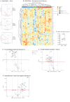
Results: Our results identified a stable proteome of SCS CD169+ macrophages in LNM, with the exception for downregulation of the anti-apoptotic protein Bcl-xL and FAPα, but a clear reduction in numbers of SCS CD169+ macrophages in LNM. In contrast, the proteome of MS CD169+ macrophages, B-cell compartments and interfollicular T-cells showed altered immune signatures in LNM, indicating that the decline in SCS CD169+ macrophages coincide with a malfunction in the local, anti-tumor immune responses.
Conclusions: The findings from our study support the notion that metastasis to lymph nodes in breast cancer patients modifies local immune responses. These changes may contribute to explain unsuccessful therapeutic responses, and thereby worsened prognosis, for breast cancer patients with LNM.
Keywords: B-cell follicles; Breast cancer; CD169+ macrophages; Germinal centers; Interfollicular T-cells; Lymph node metastasis.
© 2025. The Author(s).
Conflict of interest statement
Declarations. Ethical approval and consent to participate: The study was conducted in accordance with the Declaration of Helsinki. Ethical approval was obtained by the Swedish Ethical Review Authority (Dnr 2021–04869). Consent for publication: All authors read, revised and approved the final manuscript. Competing interests: Authors declare no competing interest.
Figures


Similar articles
-
Difference between sentinel and non-sentinel lymph nodes in the distribution of dendritic cells and macrophages: An immunohistochemical and morphometric study using gastric regional nodes obtained in sentinel node navigation surgery for early gastric cancer.J Anat. 2025 Feb;246(2):272-287. doi: 10.1111/joa.14147. Epub 2024 Oct 5. J Anat. 2025. PMID: 39367691 Free PMC article.
-
Topohistology of dendritic cells and macrophages in the distal and proximal nodes along the lymph flow from the lung.J Anat. 2025 Aug;247(2):393-407. doi: 10.1111/joa.14251. Epub 2025 Mar 28. J Anat. 2025. PMID: 40151162 Free PMC article.
-
Positron emission tomography (PET) and magnetic resonance imaging (MRI) for the assessment of axillary lymph node metastases in early breast cancer: systematic review and economic evaluation.Health Technol Assess. 2011 Jan;15(4):iii-iv, 1-134. doi: 10.3310/hta15040. Health Technol Assess. 2011. PMID: 21276372 Free PMC article.
-
Intra-patient spatial comparison of non-metastatic and metastatic lymph nodes reveals the reduction of CD169+ macrophages by metastatic breast cancers.EBioMedicine. 2024 Sep;107:105271. doi: 10.1016/j.ebiom.2024.105271. Epub 2024 Aug 21. EBioMedicine. 2024. PMID: 39173531 Free PMC article.
-
Post-mastectomy radiotherapy for women with early breast cancer and one to three positive lymph nodes.Cochrane Database Syst Rev. 2023 Jun 16;6(6):CD014463. doi: 10.1002/14651858.CD014463.pub2. Cochrane Database Syst Rev. 2023. PMID: 37327075 Free PMC article.
References
-
- Ping J, Liu W, Chen Z, Li C. Lymph node metastases in breast cancer: mechanisms and molecular imaging. Clin Imaging. 2023;103. - PubMed
MeSH terms
Substances
Grants and funding
- 2020-01842/Vetenskapsrådet
- 21 1392 Pj 01 H/Cancerfonden
- Allmänna Sjukhusets i Malmö Stiftelse för Bekämpande av Cancer/Allmänna Sjukhusets i Malmö Stiftelse för Bekämpande av Cancer
- Gyllenstiernska Krapperupsstiftelsen/Gyllenstiernska Krapperupsstiftelsen
- Kungliga Fysiografiska Sällskapet i Lund/Kungliga Fysiografiska Sällskapet i Lund
LinkOut - more resources
Full Text Sources
Medical
Research Materials
Miscellaneous

