. 2025 May;14(13):e2403944.
doi: 10.1002/adhm.202403944. Epub 2025 Apr 10.

Ovarian Function Restoration with Biomimetic Scaffold Incorporating Angiogenic Molecules and Antioxidant in Chemotherapy-Induced Perimenopausal Model

Affiliations

Ovarian Function Restoration with Biomimetic Scaffold Incorporating Angiogenic Molecules and Antioxidant in Chemotherapy-Induced Perimenopausal Model

Da-Seul Kim et al. Adv Healthc Mater. 2025 May.

Abstract

Chemotherapy-induced premature ovarian insufficiency (POI) is a major cause of infertility and hormonal imbalance in young female cancer survivors. In this study, developed a biomimetic scaffold is developed that incorporates polydeoxyribonucleotide (PDRN) and melatonin to restore ovarian function. The scaffold is designed to mimic the ovarian extracellular matrix (ECM), enhancing angiogenesis, promoting antioxidant effects, and reducing reactive oxygen species (ROS). Human embryonic stem cell-derived mesenchymal progenitor cells (hESC-MPCs) are also incorporated to further support tissue regeneration. The scaffold demonstrated strong efficacy in improving cell survival, promoting folliculogenesis, and restoring ovarian function in a chemotherapy-induced perimenopausal mouse model. Results showed that the scaffold enhanced vascularization, reduced fibrosis, and normalized hormone levels, including estrogen (E2) and anti-Müllerian hormone (Amh). Additionally, the transplantation of the scaffold restored fertility rates and increased the number of offspring in treated mice. This approach presents a promising solution for improving ovarian recovery and fertility preservation in patients with chemotherapy-induced POI, offering a novel therapeutic strategy for reproductive health.

Keywords: ROS scavenging; angiogenesis; anti‐inflammation; biomimetic scaffold; chemotherapy‐induced premature ovarian insufficiency; embryonic stem cell‐derived mesenchymal progenitor cells; ovarian function restoration.


Conflict of interest statement

The authors declare no conflict of interest.

Figures

Figure 1
Figure 1
The schematic illustration of hESC‐MPCs laden biomimetic scaffold incorporating antioxidant and angiogenic molecules restorates the chemotherapy‐induced premature ovarian insufficiency in mice. The biomimetic scaffold containing PDRN and melatonin provides higher hESC‐MPCs adhsion, immune moduation, and angiogenesis for ovarian function restoation.
Figure 2
Figure 2
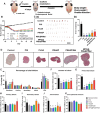
The characterization of biomimetic scaffold for restoration of ovarian function in chemotherapy‐induced perimenopause. A) Optical images of the porcine‐derived oECM fabrication process (scale bar indicates 1000 µm). B) Quantification of decellularization efficiency using DNA content of the native porcine ovary and the oECM (n = 5). C) Representative images with DAPI staining of the native porcine ovary and the oECM. D) Western blot analysis of the native porcine ovary and the oECM. E) Representative images of scanning electron microscopy (SEM, scale bars indicate 200 µm). F) Changes in the pH of scaffolds during in vitro degradation in PBS solution at 37 °C (n = 3). (G) Thermogravimetric analysis of the each scaffolds (n = 3). (H) The porosity of the each scaffolds. Cumulative release profiles of PDRN (I) and Melatonin (J) during in vitro degradation in PBS solution at 37 °C for 28 days (n = 3).
Figure 3
Figure 3
Biofunctional Abilities of Biomimetic Scaffold in ROS Scavenging and Angiogenesis for Ovarian Function Restoration. A) Representative image of tubule‐forming assay: calcein AM‐stained images (scale bar, 100 µm). Quantification of total tube length and number of branch points (n = 3). B) Fluorescence microscopy images of 2′,7′‐dichlorofluorescin diacetate (DCFDA)‐treated. And 2,2‐Diphenyl‐1‐(2,4,6‐trinitrophenyl)‐hydrazyl (DPPH) radical scavenging activity (n = 3). C) Gene expression levels of macrophage markers (iNOS and CD206) and inflammation‐related genes (TNF‐α, and IL‐10) by quantitative PCR with reverse transcription (RT‐qPCR; n = 3). **** p < 0.0001, ***p < 0.001, **p < 0.01, and *p < 0.05 indicate statistically significant differences, respectively (One‐way ANOVA with Tukey's multiple comparisons test).
Figure 4
Figure 4
Chemotherapy‐induced Perimenopausal Model. A) Schematic illustration of in vivo experimental design. B) Body weights were assessed daily during cisplatin injection, followed by measurements every two days post‐transplantation of the scaffold. C) Gross morphology of ovaires at 4 weeks after scaffold implantation (scale ber, 500 µm). D) H&E staining (scale bar, 200 µm). E) Body weights at 1, 2, 3, and 4 weeks following scaffold implantation. E) Percentage of total follicles of each groups in different stages per ovary. F–K) The number of follicles at each stage in primordial (G), primary (H), secondary (I), antral (J), and zona pellucida remnant (K). Data represent mean ± standard error of the mean. Different superscript letters indicate a significant difference (p < 0.05, n ≥ 10) (One‐way ANOVA with Tukey's multiple comparisons test).
Figure 5
Figure 5
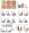
Chemotherapy‐induced Perimenopausal Model. A) Represent of PicroSirius Red (PSR)‐stained ovarian section (scale bar, 100 µm) and quantification of PSR stained ovarian stromal fibrosis area. B) Immunofluorsence of Amh (green), Ki‐67 (Red), DAPI (Blue) (scale bar, 50 and 200 µm). And the quantification of Amh positive area. C) Expression of Amh and b‐actin in ovary tissue were evaluated by Western blot. And normalized result to β‐actin. D) Plasma levels of E2 and E) FSH. Data represent mean ± standard error of the mean. Different superscript letters indicate a significant difference (p < 0.05, n ≥ 5) (One‐way ANOVA with Tukey's multiple comparisons test).
Figure 6
Figure 6
Angiogenic and antioxidantic abilities of biomimetic scaffold in mouse chemotherapy‐induced perimenopausal model. A) Blood vessle formation and quantification using H&E staining (scale bar, 100 µm). B) Gene expression levels of inflammation markers (TNF‐α, IL‐6, and IFN‐γ), C) macrophage makers (IL‐4 and CD206), D) apoptosis markers (Bcl‐2 and Bax), and E) senescence markers (p16 and p21) by RT‐qPCR. F) Expression of apoptosis markers and β‐actin in ovary tissue were evaluated by Western blot. And normalized the result to β‐actin. G) Pregnancy rate and H) number of offspring. Data represent mean ± standard error of the mean. Different superscript letters indicate a significant difference (p < 0.05, n ≥ 5) (One‐way ANOVA with Tukey's multiple comparisons test).

References

    1. a) Reynolds A. C., McKenzie L. J., J. Clin. Oncol. 2023, 41, 2281; - PMC - PubMed
    2. b) Zhao P., Guo C., Du H., Xiao Y., Su J., Wang X., Yeung W. S. B., Li G., Wang T., Human Fertility 2023, 26, 887. - PubMed
    1. a) Buigues A., Marchante M., Herraiz S., Pellicer A., Reprod. Sci. 2020, 27, 1609; - PubMed
    2. b) Moustaki M., Kontogeorgi A., Tsangkalova G., Tzoupis H., Makrigiannakis A., Vryonidou A., Kalantaridou S. N., Frontiers in Reproductive Health 2023, 5, 1194575. - PMC - PubMed
    1. a) Ke H., Tang S., Guo T., Hou D., Jiao X., Li S., Luo W., Xu B., Zhao S., Li G., Zhang X., Xu S., Wang L., Wu Y., Wang J., Zhang F., Qin Y., Jin L., Chen Z.‐J., Nat. Med. 2023, 29, 483; - PMC - PubMed
    2. b) Lie Fong S., Lugtenburg P. J., Schipper I., Themmen A. P. N., de Jong F. H., Sonneveld P., Laven J. S. E., Hum. Reprod. 2008, 23, 674. - PubMed
    1. a) Unuane D., Tournaye H., Velkeniers B., Poppe K., Best Practice & Research Clinical Endocrinology & Metabolism 2011, 25, 861; - PubMed
    2. b) Tsiligiannis S., Panay N., Stevenson C. J., Curr. Vasc. Pharmacol. 2019, 17, 604. - PubMed
    1. a) Perin E. C., Borow K. M., Henry T. D., Mendelsohn F. O., Miller L. W., Swiggum E., Adler E. D., Chang D. H., Fish R. D., Bouchard A., Jenkins M., Yaroshinsky A., Hayes J., Rutman O., James C. W., Rose E., Itescu S., Greenberg B., J. Am. Coll. Cardiol. 2023, 81, 849; - PubMed
    2. b) Amirdelfan K., Bae H., McJunkin T., DePalma M., Kim K., Beckworth W. J., Ghiselli G., Bainbridge J. S., Dryer R., Deer T. R., Brown R. D., The Spine Journal 2021, 21, 212. - PubMed

MeSH terms