Review
. 2025 Sep;45(5):1407-1422.
doi: 10.1002/med.22112. Epub 2025 Apr 10.

Molecular Tools to Study and Control Dopaminergic Neurotransmission With Light

Affiliations
Review

Molecular Tools to Study and Control Dopaminergic Neurotransmission With Light

Galyna Maleeva et al. Med Res Rev. 2025 Sep.

Abstract

Dopaminergic neurotransmission is involved in several important brain functions, such as motor control, learning, reward-motivated behavior, and emotions. Dysfunctions of dopaminergic system may lead to the development of various neurological and psychiatric disorders, like Parkinson's disease, schizophrenia, depression, and addictions. Despite years of sustained research, it is not fully established how dopaminergic neurotransmission governs these important functions through a relatively small number of neurons that release dopamine. Light-driven neurotechnologies, based on the use of small light-regulated molecules or overexpression of light-regulated proteins in neurons, have greatly contributed to the advancement of our understanding of dopaminergic circuits and our ability to control them selectively. Here, we overview the current state-of-the-art of light-driven control of dopaminergic neurotransmission. While we provide a concise guideline for the readers interested in pharmacological, pharmacogenetic, and optogenetic approaches to modulate dopaminergic neurotransmission, our primary focus is on the usage of photocaged and photo-switchable small dopaminergic molecules. We argue that photopharmacology, photoswitchable molecules of varied modalities, can be employed in a wide range of experimental paradigms, providing unprecedent insights into the principles of dopaminergic control, and represent the most promising light-based therapeutic approach for spatiotemporally precise correction of dopamine-related neural functions and pathologies.

Keywords: azobenzene; caged ligands; catecholamine; dopamine; neuromodulation; neuronal circuits; optogenetics; optopharmacology; photoisomerization; photopharmacology; photoswitch.


Figures

Figure 1
Figure 1
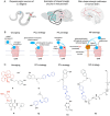
Endogenous DAergic circuits and strategies to control them with light. (A). Examples of DAergic pathways from the nervous systems of C. elegans, mice, and humans. Left panel. C. elegans has 4 pairs of DAergic neurons – CEPV, CEPD, ADE, and PDE – that release DA synaptically and extrasynaptically to mediate different behaviors of the nematode, such as memory, locomotion, and learning. Middle panel. Examples of DAergic pathways in the nervous system of mice: (i) projections from substantia nigra (SN) to dorsal striatum (DS); (ii) projections from ventral tegmental area (VTA) to nucleus accumbens (NAcc); (iii) projections from VTA to prefrontal cortex (PFC). Right panel. Main DAergic circuits of human brain: nigrostriatal pathway – from substantia nigra pars compacta to dorsal striatum; mesocortical pathway – from ventral tegmental area to prefrontal cortex; mesolimbic pathway – from ventral tegmental area to limbic system (amygdala, nucleus accumbens, hippocampus); tuberoinfundibular pathway – from hypothalamus to pituitary gland. (B). An overview of various strategies of light‐dependent activation of DARs. Left panel: Caged DAergic compounds are freely diffusible; the cage undergoes photolysis, and the caged molecule can interact with DAR and activate or inhibit it. Left middle panel: PCL (photochromic ligand) strategy is based on freely diffusible photoswitchable molecules that can be reversibly toggled between two conformations (e.g., based on cis‐ and trans‐azobenzene, one of them being more active than the other) by illuminating with light of two specific wavelengths. Right middle panel: PTL (photochromic tethered ligand) strategy is based on the ability of photoswitchable molecules carrying specific linker to tether to endogenous or introduced cysteine residues (like in the case of DARs). When illuminated with a specific wavelength, the photoswitchable linker changes its conformation and enables the interaction between the pharmacologically active moiety with the receptor binding site. Right panel: Another approach combines (1) a genetically overexpressed membrane anchor protein and (2) the application of a photoswitchable orthogonal remotely tethered ligand compound (MP strategy). This compound first conjugates to the cysteine introduced in the membrane anchor and then illumination is used to change the compound conformation between ligand‐obstructed to ligand‐exposed to allow the interaction with the receptor. (C). Examples of DAergic light‐dependent modulators reported in literature. Uncaging strategy: 2, RuBi‐Dopa, DA caged in [Ru(bpy)2(PMe3) (Dopa)] (PF6)2 (bpy = 2,2′ bipyridine, PMe3 = trimethylphosphine). 8, Caged inverse agonist of D2R/D3R (dechloroeticlopride‐based). PCL strategy: 11, PCL cis‐on DAergic agonist. 12, Azodopa, PCL trans‐on DAergic agonist. PTL strategy: 13, MAP, PTL trans‐on inverse DAergic agonist or neutral antagonist. MP strategy: 15, MP‐D2ago, PTL membrane anchored D2R cis‐on agonist. [Color figure can be viewed at wileyonlinelibrary.com]
Figure 2
Figure 2
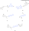
Literature reported caged‐DAergic modulators. [Color figure can be viewed at wileyonlinelibrary.com]
Figure 3
Figure 3
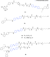
Literature reported PCL and PTL DAergic modulators. [Color figure can be viewed at wileyonlinelibrary.com]
Figure 4
Figure 4
Literature reported PCL and MP DAergic modulators. [Color figure can be viewed at wileyonlinelibrary.com]

References

    1. Odoemelam C. S., Percival B., Wallis H., et al., “G‐Protein Coupled Receptors: Structure and Function in Drug Discovery,” RSC Advances 10 (2020): 36337–36348. - PMC - PubMed
    1. Yang D., Zhou Q., Labroska V., et al., “G Protein‐Coupled Receptors: Structure‐And Function‐Based Drug Discovery,” Signal Transduction and Targeted Therapy 6 (2021): 7. - PMC - PubMed
    1. Beaulieu J.‐M. and Gainetdinov R. R., “The Physiology, Signaling, and Pharmacology of Dopamine Receptors,” Pharmacological Reviews 63 (2011): 182–217. - PubMed
    1. Missale C., Nash S. R., Robinson S. W., Jaber M., and Caron M. G., “Dopamine Receptors: From Structure to Function,” Physiological Reviews 78 (1998): 189–225. - PubMed
    1. Jaber M., Robinson S. W., Missale C., and Caron M. G., “Dopamine Receptors and Brain Function,” Neuropharmacology 35 (1996): 1503–1519. - PubMed