. 2025 Apr;15(4):e70289.
doi: 10.1002/ctm2.70289.

ATG16L1 restrains macrophage NLRP3 activation and alveolar epithelial cell injury during septic lung injury

Affiliations

ATG16L1 restrains macrophage NLRP3 activation and alveolar epithelial cell injury during septic lung injury

Yan Bai et al. Clin Transl Med. 2025 Apr.

Abstract

Background: The lung is the organ most commonly affected by sepsis. Additionally, acute lung injury (ALI) resulting from sepsis is a major cause of death in intensive care units. Macrophages are essential for maintaining normal lung physiological functions and are implicated in various pulmonary diseases. An essential autophagy protein, autophagy-related protein 16-like 1 (ATG16L1), is crucial for the inflammatory activation of macrophages.

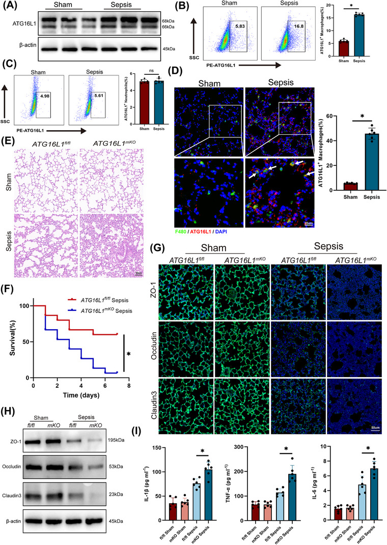
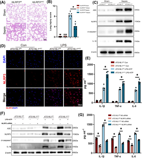
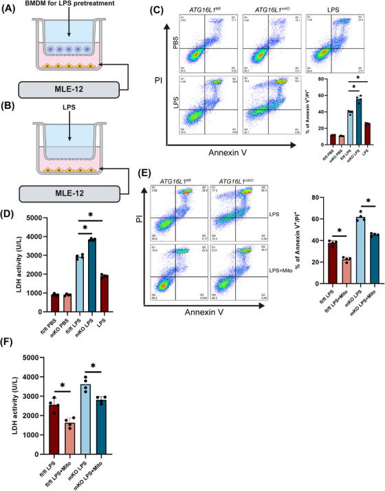
Methods: ATG16L1 expression was measured in lung from mice with sepsis. ALI was induced in myeloid ATG16L1-, NLRP3- and STING-deficient mice by intraperitoneal injection of lipopolysaccharide (LPS, 10 mg/kg). Using immunofluorescence and flow cytometry to assess the inflammatory status of LPS-treated bone marrow-derived macrophages (BMDMs). A co-culture system of BMDMs and MLE-12 cells was established in vitro.

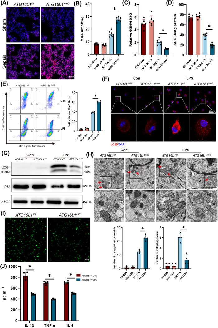
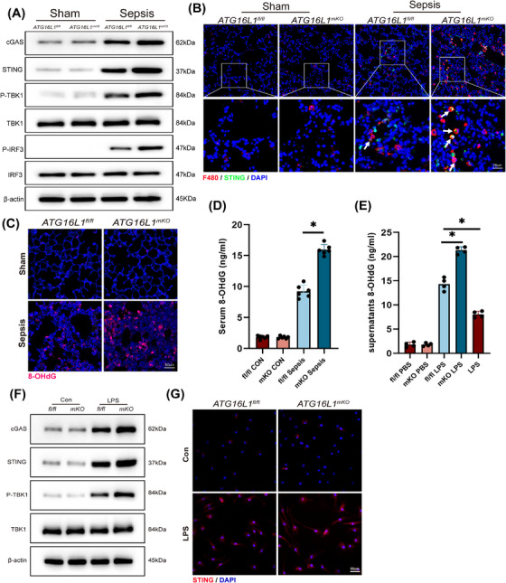
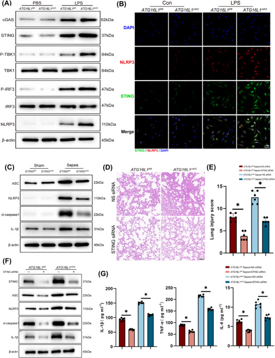
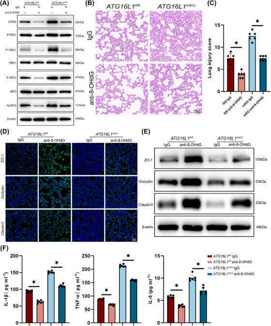
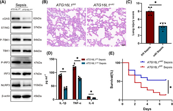
Results: Myeloid ATG16L1-deficient mice exhibited exacerbated septic lung injury and a more intense inflammatory response following LPS treatment. Mechanistically, ATG16L1-deficient macrophages exhibited impaired LC3B lipidation, damaged mitochondria and reactive oxygen species (ROS) accumulation. These abnormalities led to the activation of NOD-like receptor family pyrin domain-containing protein 3 (NLRP3), subsequently enhancing proinflammatory response. Overactivated ATG16L1-deficient macrophages aggravated the damage to alveolar epithelial cells and enhanced the release of double-stranded DNA (dsDNA), thereby promoting STING activation and subsequent NLRP3 activation in macrophages, leading to positive feedback activation of macrophage NLRP3 signalling. Scavenging mitochondrial ROS or inhibiting STING activation effectively suppresses NLRP3 activation in macrophages and alleviates ALI. Furthermore, overexpression of myeloid ATG16L1 limits NLRP3 activation and reduces the severity of ALI.

Conclusions: Our findings reveal a new role for ATG16L1 in regulating macrophage NLRP3 feedback activation during sepsis, suggesting it as a potential therapeutic target for treating sepsis-induced ALI.

Key points: Myeloid-specific ATG16L1 deficiency exacerbates sepsis-induced lung injury. ATG16L1-deficient macrophages exhibit impaired LC3B lipidation and ROS accumulation, leading to NLRP3 inflammasome activation. Uncontrolled inflammatory responses in ATG16L1-deficient macrophages aggravate alveolar epithelial cell damage. Alveolar epithelial cells release dsDNA, activating the cGAS-STING-NLRP3 signaling pathway, which subsequently triggers a positive feedback activation of NLRP3. Overexpression of ATG16L1 helps mitigate lung tissue inflammation, offering a novel therapeutic direction for sepsis-induced lung injury.

Keywords: ATG16L; NLRP3 inflammasome; STING; acute lung injury; macrophages.


Conflict of interest statement

The authors declare they have no conflicts of interest.

Figures

FIGURE 1
FIGURE 1
Myeloid‐specific ATG16L1 deficiency exacerbates sepsis‐induced lung injury. ATG16L1 fl/fl and ATG16L1 mKO mice were intraperitoneally injected with lipopolysaccharide (LPS; 10 mg/kg) or PBS. Lung samples and BALF were collected 24 h after the treatment. (A) Protein levels of ATG16L1 in lung tissues of Sham and sepsis groups (n = 6/group). (B) Flow cytometry analysis the expression levels of ATG16L1 in alveolar macrophages isolated from BALF. (C) Flow cytometry analysis the expression levels of ATG16L1 in neutrophils isolated from BALF (n = 6/group). (D) Representative immunofluorescence images of F4/80 (green) and ATG16L1 (red) in lung tissues. Blue indicates nuclei stained with 4',6‐diamidino‐2‐phenylindole(DAPI). White arrows indicate macrophages with elevated ATG16L1 levels (n = 6/group; scale bar: 25 µm). (E) Haematoxylin and eosin (H&E) staining of lung tissue sections (n = 6/group; scale bar: 50 µm). (F) Survival analysis of ATG16L1fl/fl and ATG16L1mKO mice after LPS (20 mg/kg) treatment (n = 15/group). (G) Representative images of immunofluorescence staining of ZO‐1 (green), Occludin (green) and Claudin 3 (green) in lung tissues. Blue indicates nuclei stained with DAPI (n = 6/group; scale bar: 50 µm). (H) Western blot analysis of ZO‐1, Occludin, Claudin 3 and β‐actin in lung tissues (n = 6/group). (I) Concentration of inflammatory cytokines IL‐1β, TNF‐α and IL‐6 in murine BALF (n = 6/group). Data are presented as the mean ± SEM. * p < .05.
FIGURE 2
FIGURE 2
ATG16L1 deficiency promotes macrophage NLRP3 inflammasome activation to aggravate acute lung injury (ALI). (A and B) NLRP3fl/fl and NLRP3mKO mice were intraperitoneally injected with lipopolysaccharide (LPS; 10 mg/kg) or PBS. Lung samples and BALF were collected 24 h after the treatment. Haematoxylin and eosin (H&E) staining of lung tissue sections and lung tissues injury scores (n = 6/group; scale bar: 50 µm). (C) Western blot analysis of apoptosis‐associated speck‐like protein containing a CARD (ASC), NLRP3, cl‐caspase1, IL‐1β and β‐actin in lung tissues from LPS‐treated ATG16L1fl/fl and ATG16L1mKO mice (n = 6/group). (D) Representative images of immunofluorescence staining of NLRP3 (red) immunofluorescence staining. Blue indicates nuclei stained with DAPI (n = 4/group; scale bar: 50 µm). (E) Levels of inflammatory cytokines IL‐1β, TNF‐α and IL‐6 in the supernatants of bone marrow‐derived macrophages (BMDMs) (n = 4/group). (F) BMDMs were transfected with NLRP3 siRNA or non‐specific siRNA (control) and then stimulated with 100 ng/mL of LPS. Protein levels of ASC, NLRP3, cl‐caspase1, IL‐1β and β‐actin were measured using Western blotting (n = 4/group). (G) Levels of inflammatory cytokines IL‐1β, TNF‐α and IL‐6 in the supernatants of BMDMs with NLRP3 siRNA treatment (n = 4/group). Data are presented as the mean ± SEM. * p < .05.
FIGURE 3
FIGURE 3
Deletion of ATG16L1 increases macrophage reactive oxygen species (ROS) accumulation by inhibiting autophagy. (A) Representative images of ROS fluorescence (red) detection in lung tissues. Blue indicates nuclei stained with DAPI (n = 6/group; scale bar: 50 µm). Levels of malondialdehyde (MDA) (B), GSH/GSSG (C) and superoxide dismutase (SOD) activity (D) in lung tissues from ATG16L1fl/fl and ATG16L1mKO mice treated with or without lipopolysaccharide (LPS) (n = 6/group). (E) Mitochondrial membrane potential detected by flow cytometry in ATG16L1fl/fl and ATG16L1mKO bone marrow‐derived macrophages (BMDMs) (n = 4/group). (F) Representative images of LC3B (red) immunofluorescence staining. Blue indicates nuclei stained with DAPI (n = 4/group; scale bar: 50 µm). (G) Western blot analysis of P62, LC3B‐I, LC3‐BII and β‐actin in BMDMs stimulated with LPS (n = 4/group). (H) Mitochondrial and phagosome ultrastructure observed using transmission electron microscopy in BMDMs with or without LPS treatment. Red arrows indicate mitochondria and black arrows indicate autophagosomes (n = 4/group; scale bar: 2 µm). (I) Representative immunofluorescence images of intracellular ROS in ATG16L1fl/fl and ATG16L1mKI BMDMs detected by DCFDA fluorescence probe (n = 4/group; scale bar: 100 µm). (J) Levels of inflammatory cytokines IL‐1β, TNF‐α and IL‐6 in the supernatants of ATG16L1fl/fl and ATG16L1mKI BMDMs (n = 4/group). Data are presented as the mean ± SEM. * p < .05.
FIGURE 4
FIGURE 4
ATG16L1‐deficient macrophages release inflammatory cytokines to damage alveolar epithelial cells after lipopolysaccharide (LPS) treatment. Established a co‐culture system of bone marrow‐derived macrophages (BMDMs) and MLE‐12 cells with or without LPS treatment (A). LPS stimulation of MLE‐12 cells alone served as a control (B). (C) MLE‐12 cells extent of cell death measured by Annexin V and propidium iodide(PI) staining. (D) Levels of lactate dehydrogenase (LDH) in the supernatants of MLE‐12 cells. (E and F) MLE‐12 cells extent of cell measured by Annexin V and PI staining and levels of LDH in the supernatants cultured with ATG16L1fl/fl and ATG16L1mKO BMDMs with or without Mitotempo pretreatment (n = 4/group). Data are presented as the mean ± SEM. * p < .05.
FIGURE 5
FIGURE 5
Inflammatory injury of alveolar epithelial cells enhances macrophage NLRP3 activation via dsDNA‒STING signalling. ATG16L1fl/fl and ATG16L1mKO mice were intraperitoneally injected with lipopolysaccharide (LPS; 10 mg/kg) or PBS. (A) Western blot analysis of cGAS, STING, P‐TBK1, TBK1, P‐IRF3, IRF3 and β‐actin in lung tissues (n = 6/group). (B) Representative immunofluorescence images of F4/80 (red) and STING (green) in lung tissues. Blue indicates nuclei stained with DAPI. White arrows indicate macrophages with elevated STING levels (n = 6/group; scale bar: 25 µm). (C and D) Representative immunofluorescence 8‐hydroxydeoxyguanosine (8‐OHdG) staining in ATG16L1fl/fl and ATG16L1mKO mice lung tissues (C) (scale bar: 50 µm) and serum 8‐OHdG levels (D) showing DNA damage (n = 6/group). (E) Levels of 8‐OHdG in the supernatants of MLE‐12 cells in the bone marrow‐derived macrophages (BMDMs) and MLE‐12 cells co‐culture system (n = 4/group). (F) Western blot analysis of cGAS, STING, P‐TBK1, TBK1 and β‐actin in BMDMs stimulated with MLE‐12 cells supernatants as conditioned media (n = 4/group). (G) Representative immunofluorescence images of STING (red) in BMDMs after stimulation with MLE‐12 cells conditioned media. Blue indicates nuclei stained with DAPI (n = 4/group; scale bar: 50 µm). Data are presented as the mean ± SEM. * p < .05.
FIGURE 6
FIGURE 6
Activation of the STING signalling pathway promotes the activation of the NLRP3 inflammasome. (A and B) Stimulating bone marrow‐derived macrophages (BMDMs) with MLE‐12 cells supernatants as conditioned media. Western blot analysis of cGAS, STING, P‐TBK1, TBK1, P‐IRF3, IRF3, NLRP3 and β‐actin in BMDMs (A). Immunofluorescence staining showing activation and co‐localisation of STING (green) and NLRP3 (red). Blue indicates nuclei stained with DAPI (B) (n = 4/group; scale bar: 50 µm). (C) STINGfl/fl and STINGmKO mice were intraperitoneally injected with lipopolysaccharide (LPS) (10 mg/kg) or PBS. Lung tissues were collected after 24 h of LPS treatment. Representative Western blot analysis of ASC, NLRP3, cl‐caspase1, IL‐1β and β‐actin in lung tissues (n = 6/group). (D and E) ATG16L1fl/fl and ATG16L1mKO mice were subjected to intraperitoneal LPS (10 mg/kg) injection after in vivo transfection with STING siRNA or non‐specific siRNA (control). Haematoxylin and eosin (H&E) staining of lung tissue sections and lung tissues injury scores (n = 6/group; scale bar: 50 µm). (F and G) Western blot analysis of STING, ASC, NLRP3, cl‐caspase1, IL‐1β and β‐actin in lung tissues after in vivo transfection with STING siRNA (n = 6/group). Levels of inflammatory cytokines IL‐1β, TNF‐α and IL‐6 in BALF (n = 6/group). Data are presented as the mean ± SEM. * p < .05.
FIGURE 7
FIGURE 7
Clearance of double‐stranded DNA (dsDNA) suppresses macrophage STING‒NLRP3 activation to protect lungs against septic injury. Immunoglobulin G (IgG) or anti‐8‐OHG antibody were pretreated 12 h before lipopolysaccharide (LPS)‐induced sepsis modelling. Lung tissues and BALF were collected after 24 h of LPS treatment. (A) Western blot analysis of cGAS, STING, P‐TBK1, TBK1, P‐IRF3, IRF3, NLRP3 and β‐actin in lung tissues (n = 6/group). (B and C) Haematoxylin and eosin (H&E) staining of lung tissue sections and lung tissues injury scores (n = 6/group; scale bar: 50 µm). (D) Representative immunofluorescence images of ZO‐1 (green), Occludin (green) and Claudin 3 (green) in lung tissues. Blue indicates nuclei stained with DAPI (n = 6/group; scale bar: 50 µm). (E) Western blot analysis of ZO‐1, Occludin, Claudin 3 and β‐actin in lung tissues (n = 6/group). (F) Levels of inflammatory cytokines IL‐1β, TNF‐α and IL‐6 in BALF (n = 6/group). Data are presented as the mean ± SEM. * p < .05.
FIGURE 8
FIGURE 8
Myeloid‐ATG16L1 overexpression alleviates septic lung injury. ATG16L1fl/fl and ATG16L1mKI mice were intraperitoneally injected with lipopolysaccharide (LPS) (10 mg/kg) or PBS. Lung samples and BALF were collected 24 h after the treatment. (A) Western blot analysis of cGAS, STING, P‐TBK1, TBK1, P‐IRF3, IRF3, NLRP3 and β‐actin proteins in lung tissues (n = 6/group). (B and C) Haematoxylin and eosin (H&E) staining of lung tissue sections and lung tissues injury scores (n = 6/group; scale bar: 50 µm). (D) Levels of inflammatory cytokines IL‐1β, TNF‐α and IL‐6 in the BALF (n = 6/group). (E) Survival analysis of ATG16L1fl/fl and ATG16L1mKI mice after LPS (20 mg/kg) treatment (n = 15/group). Data are presented as the mean ± SEM. * p < .05.

Similar articles

Cited by

References

    1. van der Poll T, van de Veerdonk FL, Scicluna BP, Netea MG. The immunopathology of sepsis and potential therapeutic targets. Nat Rev Immunol. 2017;17(7):407‐420. doi:10.1038/nri.2017.36 - DOI - PubMed
    1. Rudd KE, Johnson SC, Agesa KM, et al. Global, regional, and national sepsis incidence and mortality, 1990–2017: analysis for the Global Burden of Disease Study. Lancet. 2020;395(10219):200‐211. doi:10.1016/s0140-6736(19)32989-7 - DOI - PMC - PubMed
    1. Martin GS, Mannino DM, Eaton S, Moss M. The epidemiology of sepsis in the United States from 1979 through 2000. N Engl J Med. 2003;348(16):1546‐1554. doi:10.1056/NEJMoa022139 - DOI - PubMed
    1. Aziz M, Ode Y, Zhou M, et al. B‐1a cells protect mice from sepsis‐induced acute lung injury. Mol Med. 2018;24(1):26. doi:10.1186/s10020-018-0029-2 - DOI - PMC - PubMed
    1. Singer M, Deutschman CS, Seymour CW, et al. The Third International Consensus Definitions for Sepsis and Septic Shock (Sepsis‐3). JAMA. 2016;315(8):801‐810. doi:10.1001/jama.2016.0287 - DOI - PMC - PubMed

MeSH terms

Substances

LinkOut - more resources