. 2025 May 9;136(10):1113-1130.
doi: 10.1161/CIRCRESAHA.124.325550. Epub 2025 Apr 11.

The Heart Has Intrinsic Ketogenic Capacity that Mediates NAD+ Therapy in HFpEF

Affiliations

The Heart Has Intrinsic Ketogenic Capacity that Mediates NAD+ Therapy in HFpEF

Yen Chin Koay et al. Circ Res. .

Abstract

Background: Heart failure with preserved ejection fraction (HFpEF) has overtaken heart failure with reduced ejection fraction as the leading type of heart failure globally and is marked by high morbidity and mortality rates, yet with only a single approved pharmacotherapy: SGLT2i (sodium-glucose co-transporter 2 inhibitor). A prevailing theory for the mechanism underlying SGLT2i is nutrient deprivation signaling, of which ketogenesis is a hallmark. However, it is unclear whether the canonical ketogenic enzyme, HMGCS2 (3-hydroxy-3-methylglutaryl-coenzyme A synthase 2), plays any cardiac role in HFpEF pathogenesis or therapeutic response.

Methods: We used human myocardium, human HFpEF and heart failure with reduced ejection fraction transcardiac blood sampling, an established murine model of HFpEF, ex vivo Langendorff perfusion, stable isotope tracing in isolated cardiomyocytes, targeted metabolomics, proteomics, lipidomics, and a novel cardiomyocyte-specific conditional HMGCS2-deficient model that we generated.

Results: We demonstrate, for the first time, the intrinsic capacity of the human heart to produce ketones via HMGCS2. We found that increased acetylation of HMGCS2 led to a decrease in the enzyme's specific activity. However, this was overcome by an increase in the steady-state levels of protein. Oxidized form of nicotinamide adenine dinucleotide repletion restored HMGCS2 function via deacetylation, increased fatty acid oxidation, and rescued cardiac function in HFpEF. Critically, using a conditional, cardiomyocyte-specific HMGCS2 knockdown murine model, we revealed that the oxidized form of nicotinamide adenine dinucleotide is unable to rescue HFpEF in the absence of cardiomyocyte HMGCS2.

Conclusions: The canonical ketogenic enzyme, HMGCS2, mediates the therapeutic effects of the oxidized form of nicotinamide adenine dinucleotide repletion in HFpEF by restoring normal lipid metabolism and mitochondrial function.

Keywords: heart failure; ketone bodies; myocardium; oxygen consumption; stroke volume.


Conflict of interest statement

None.

Figures

Figure 1.
Figure 1.
Myocardial glycolytic metabolism. A, Heatmap showing decreased expression of glucose transporter and glycolytic enzymes in heart failure with preserved ejection fraction (HFpEF) myocardium. Protein label-free quantitation (LFQ) intensities were normalized and visualized using MetaboAnalyst 5.0. Each column in the figure represents data from an individual mouse. A color scale on the upper right side of the figure indicates normalized protein abundance (ranging from 2.0 to −2.0). B, Box and whisker plots with individual dot points in heart tissue from HFpEF (n=8) vs chow (n=8) illustrating significant reductions of several rate-limiting enzymes critical for the regulation of glycolysis and glucose oxidation, with upregulation of PDK4 (pyruvate dehydrogenase kinase 4) that inhibits the pyruvate dehydrogenase complex leading to metabolic inflexibility. P<0.05 is statistically significant, and exact values are specified in corresponding figures. Data were analyzed by the Mann-Whitney U test. C, Schematic summarizing the glycolytic pathway changes. GLUT4 indicates glucose transporter type 4; HK1, hexokinase 1; HK2, hexokinase 2; PDHA1, pyruvate dehydrogenase-A1; PDHB, pyruvate dehydrogenase B; PFKFB2, 6-phosphofructo-2-kinase/fructose-2,6-bisphosphatase 2; SLC2A4, solute carrier family 2, facilitated glucose transporter member 4; and TCA, tricarboxylic acid.
Figure 2.
Figure 2.
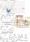
Proteomics pinpoints perturbed Hmgcs2 metabolism. A, Volcano plot depicting differentially abundant proteins in heart failure with preserved ejection fraction (HFpEF) vs control myocardium. The x axis displays the log2 fold change, and the y axis shows the −log10 of P values derived from a t test, adjusted using the Benjamini-Hochberg procedure. The dashed horizontal line shows the cutoff P value (<0.05), and the 2 vertical dashed lines represent the fold change cutoff of 1.5 (≈0.6 on log2 scale). Box plots illustrating changes in (B) proteins involved in fatty acid transport, ketogenesis, and ketone oxidation (n=8/group) and (C) substrates and products of the fatty acid transport, fatty acid oxidation, ketogenic, and ketone oxidation pathways in HFpEF group vs control myocardium (n=7/group). The y axis represents the log2-scaled abundance of each protein/metabolite. Statistical significance was assessed using the Mann-Whitney U test (B and C). D, Schematic summarizing the changes in ketogenic and lipid oxidation pathways with subcellular location indicated. HMGCS2 (3-hydroxy-3-methylglutaryl-coenzyme A synthase 2) is seen as an inflection point between increases and decreases in pathway intermediates; proteins indicated in yellow denote upregulation and blue downregulation in the HFpEF heart. Assessment of myocardial ketones using violin plots illustrated unchanged arterial and coronary sinus concentrations of (E) β-hydroxybutyrate (βHB), but lower arterial and coronary sinus concentrations of (F) acetoacetate (AcAc) in HFpEF subjects compared with HFrEF subjects. G, The percentage of myocardial extraction of ketones illustrated an increase in cardiac uptake of AcAc in HFpEF compared with heart failure with reduced ejection fraction (HFrEF) subjects (human transcardiac gradients: n=22 patients with HFpEF, and n=20 patients with HFrEF). The median of the violin plots is represented by a solid line, while the first and third quartiles are indicated by dashed lines. P<0.05 is statistically significant, and precise values are specified in corresponding figures. Data were analyzed by Kruskal-Wallis with the Dunn test (E), 2-way ANOVA followed by the Sidak test (F), or the Student t test (G). The percentage of myocardial extraction of ketones was calculated as arterial minus coronary sinus concentration divided by arterial concentrations multiplied by 100. A positive value indicates net uptake by the heart, whereas a negative value indicates net release. ACAA2 indicates acetyl-coenzyme A acyltransferase 2; ACAT1, acetyl-coenzyme A acetyltransferase 1; ACOT2, acyl-coenzyme A thioesterase 2; BDH1, beta-hydroxybutyrate dehydrogenase 1; CD36, cluster of differentiation 36; CER, ceramides; CPT1b, carnitine palmitoyltransferase I; CPT2, carnitine palmitoyltransferase 2; HMGCL, 3-hydroxymethyl-3-methylglutaryl-coenzyme A lyase; MCT1, monocarboxylate transporter 1; MPC, mitochondrial pyruvate carrier; OXCT1, 3-oxoacid coenzyme A-transferase 1; TCA, tricarboxylic acid; and βHB, β-hydroxybutyrate.
Figure 3.
Figure 3.
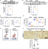
Oxidized form of nicotinamide adenine dinucleotide (NAD+) depletion and reduced myocardial ketogenic specific activity. A, Representative image of and Western Blot analysis of HMGCS2 (3-hydroxy-3-methylglutaryl-coenzyme A synthase 2) in chow and heart failure with preserved ejection fraction (HFpEF) groups (n=3 mice for chow; n=4 mice for HFpEF). B, Immunoprecipitated HMGCS2 from HFpEF myocardium revealed greater acetylation (n=3 mice for chow; n=4 mice for HFpEF). C, Total 13C6-labeled HMG-CoA (β-hydroxy β-methylglutaryl-coenzyme A) and 13C4-labeled acetoacetate from labeled [1,2-13C2]-acetyl-CoA was not significantly different in HFpEF vs control myocardium in mice (n=3 mice for chow; n=4 mice for HFpEF). D, The bar graph shows the abundance of SIRT3 (sirtuin 3) protein and ratios of NAD+/reduced form of nicotinamide adenine dinucleotide (NADH) in hearts from HFpEF mice over controls (n=8 mice/group). E, Schematic proposing depletion of NAD+ causing decreased SIRT3 that impairs deacetylation, potentially leading to decreased HMGCS2 activity. F, Upregulation of HMGCS2 in hypertrophic cardiomyopathy (HCM) samples. Representative immunohistochemistry (IHC) staining for HMGCS2 in the human myocardium (scale bar, 200 µmol/L). Semiquantitative analysis of the mean binding intensity of IHC staining. n=11 for control (healthy donors) and n=8 for HCM. Data represent the mean±SEM. Statistical significance was determined using the Mann-Whitney U test (AD and F), with P<0.05 considered significant. NAMPT indicates nicotinamide phosphoribosyltransferase.
Figure 4.
Figure 4.
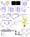
Dietary supplementation of nicotinamide riboside (NR) significantly improves diastolic dysfunction and exercise tolerance and increases cardiac ketogenesis and ketone oxidation in heart failure with preserved ejection fraction (HFpEF) mice. A, Schematic overview of NR, oxidized form of nicotinamide adenine dinucleotide (NAD+) precursor, and NR feeding protocol to HFpEF mice. B, Representative images of mitral pulsed Doppler echocardiography, showing ratio of peak velocity of mitral blood inflow in early diastole (E) to peak velocity of mitral blood inflow in late diastole (A); (C) ratio of peak velocity of mitral blood inflow in early diastole to peak velocity of mitral blood inflow in late diastole (E/A); (D) global longitudinal strain (GLS); (E) diastolic blood pressure (DBP) and (F) systolic blood pressure (SBP); and (G) exercise capacity as indicated by running distance in HFpEF (n=8) and NR-treated mice (n=7) at weeks 5 and 9. Data are presented as mean±SEM. Statistical significance was determined using the Wilcoxon matched-pair signed-rank test for paired comparisons between weeks 5 and 9 and the Mann-Whitney U test for unpaired comparisons between HFpEF and HFpEF+NR groups at each time point. Cardiac proteome profiling demonstrated increased expression of proteins involved in (H) deacetylation, ketogenesis, and ketone oxidation pathway (n=8 for HFpEF and n=7 for HFpEF+NR). I, Myocardial metabolite profile and (J) total 13C6-labeled HMG-CoA (β-hydroxy β-methylglutaryl-coenzyme A) from enzymatic activity assay in HFpEF (n=5) vs NR-treated (n=5) myocardium at week 9. Each data point was obtained from 2 to 3 pooled HFpEF and NR-supplemented mouse hearts. K, Overall schematic diagram highlighting the changes of cardiac ketogenesis and ketone oxidation in the post-NR therapy mice. L, Heart lysates from HFpEF (n=7) and NR-treated (n=7) mice were subjected to immunoprecipitation using an anti-acetyl-lysine antibody and analyzed using anti-HMGCS2 (3-hydroxy-3-methylglutaryl-coenzyme A synthase 2) antibody. M, The abundance percentages of isotopologues M+4 and M+6 of HMG-CoA, as well as M+2 isotopologues of acetyl-CoA (coenzyme A), and M+2 and M+4 isotopologues of alpha-ketoglutarate and fumarate, were determined from the metabolism of 13C16-labeled palmitate (n=5 mice in HFpEF and HFpEF+NR groups). Data represent the mean±SEM. In H through J, L, and M, statistical significance was assessed using the Mann-Whitney U test. P<0.05 is considered significant; exact values are provided in the corresponding figures. HFD indicates high-fat diet; L-NAME, L-NG-nitro arginine methyl ester; and ns indicates no significant.
Figure 5.
Figure 5.
Cardiomyocyte Hmgcs2 is critical for the therapeutic effects of NAD+ repletion. A, Schematic illustration of generation of cardiac-specific Hmgcs2 (3-hydroxy-3-methylglutaryl-coenzyme A synthase 2) knockdown mice. B, Metabolite HMG-CoA (β-hydroxy β-methylglutaryl-coenzyme A) levels; (C) systolic blood pressure (SBP) and (D) diastolic blood pressure (DBP); (E) ratio of peak velocity of mitral blood inflow in early diastole to peak velocity of mitral blood inflow in late diastole (E/A); (F) ratio between mitral E wave and E’ wave (E/E’); (G) global longitudinal strain (GLS); (H) lung water content-to-body weight ratio; and (I) oxygen consumption rate (OCR) in cardiac tissues utilizing palmitoylcarnitine as a substrate in Hmgcs2 (3-hydroxy-3-methylglutaryl-coenzyme A synthase 2) floxed mice (Hmgcs2fl/fl), αMHC (α-myosin heavy chain)-CreER (Cre-loxP recombination) (floxed mice) as controls and αMHC-CreER+, Hmgcs2fl/fl, knockdown mice fed by chow, high-fat diet (HFD)+L-NG-nitro arginine methyl ester (L-NAME), and HFD+L-NAME+NR for 12 weeks. The sample sizes were n=10/12/8 and n=10/6/6 for the chow/heart failure with preserved ejection fraction (HFpEF)/HFpEF+NR groups, respectively, in both control and Hmgcs2-deficient mice. J, Representative immunostaining images showing reduced expression of HMGCS2 in the cardiac tissue of Hmgcs2-deficient mice compared with control mice. Mice tissues were stained for HMGCS2 (red), wheat germ agglutinin (green), and 4′,6-diamidino-2-phenylindole (DAPI; blue) to delineate cell boundaries and nuclei, respectively (scale bar, 10 µm). K, Log2-fold change of specific 13C-metabolites derived from 13C16-labeled palmitate in cardiomyocytes isolated from flox-only control mice and HMGCS2-deficient mice; n=6 mice/group. Data are presented as mean±SEM. Statistical significance was assessed using 2-way ANOVA followed by the Tukey post hoc multiple comparisons test (BI) and the Mann-Whitney U test (K). P<0.05 was considered significant; exact values are provided in the corresponding figures. CreER indicates Cre-loxP recombination; Cre-LoxP, Cre recombinase–Locus of X-over P1; and Ns, not significant.

Comment in

References

    1. Wang Z, Ying Z, Bosy-Westphal A, Zhang J, Schautz B, Later W, Heymsfield SB, Müller MJ. Specific metabolic rates of major organs and tissues across adulthood: evaluation by mechanistic model of resting energy expenditure. Am J Clin Nutr. 2010;92:1369–1377. doi: 10.3945/ajcn.2010.29885 - PMC - PubMed
    1. Ashrafian H, Frenneaux MP, Opie LH. Metabolic mechanisms in heart failure. Circulation. 2007;116:434–448. doi: 10.1161/CIRCULATIONAHA.107.702795 - PubMed
    1. Ferrannini E, Mark M, Mayoux E. CV protection in the EMPA-REG outcome trial: a “thrifty substrate” hypothesis. Diabetes Care. 2016;39:1108–1114. doi: 10.2337/dc16-0330 - PubMed
    1. Veech RL. The therapeutic implications of ketone bodies: the effects of ketone bodies in pathological conditions: ketosis, ketogenic diet, redox states, insulin resistance, and mitochondrial metabolism. Prostaglandins Leukot Essent Fatty Acids. 2004;70:309–319. doi: 10.1016/j.plefa.2003.09.007 - PubMed
    1. Sato K, Kashiwaya Y, Keon CA, Tsuchiya N, King MT, Radda GK, Chance B, Clarke K, Veech RL. Insulin, ketone bodies, and mitochondrial energy transduction. FASEB J. 1995;9:651–658. doi: 10.1096/fasebj.9.8.7768357 - PubMed

MeSH terms