. 2024 Jun 30;4(9):2400061.
doi: 10.1002/smsc.202400061. eCollection 2024 Sep.

A Novel Piezo1 Agonist Promoting Mesenchymal Stem Cell Proliferation and Osteogenesis to Attenuate Disuse Osteoporosis

Affiliations

A Novel Piezo1 Agonist Promoting Mesenchymal Stem Cell Proliferation and Osteogenesis to Attenuate Disuse Osteoporosis

Ruihan Hao et al. Small Sci. .

Abstract

Disuse osteoporosis (OP) is a state of bone loss due to lack of mechanical stimuli, probably induced by prolonged bed rest, neurological diseases, as well as microgravity. Currently the precise treatment strategies of disuse OP remain largely unexplored. Piezo1, a mechanosensitive calcium (Ca2+) ion channel, is a key force sensor mediating mechanotransduction and it is demonstrated to regulate bone homeostasis and osteogenesis in response to mechanical forces. Using structure-based drug design, a novel small-molecule Piezo1 agonist, MCB-22-174, which can effectively activate Piezo1 and initiate Ca2+ influx, is developed and is more potent than the canonical Piezo1 agonist, Yoda1. Moreover, MCB-22-174 is found as a safe Piezo1 agonist without any signs of serious toxicity. Mechanistically, Piezo1 activation promotes the proliferation of bone marrow mesenchymal stem cells by activating the Ca2+-related extracellular signal-related kinases and calcium-calmodulin (CaM)-dependent protein kinase II (CaMKII) pathway. Importantly, MCB-22-174 could effectively promote osteogenesis and attenuate disuse OP in vivo. Overall, the findings provide a promising therapeutic strategy for disuse OP by chemical activation of Piezo1.

Keywords: Piezo1; disuse osteoporosis; mechenchymal stem cell proliferations; novel agonists; osteogenesis.


Conflict of interest statement

The authors declare no conflict of interest.

Figures
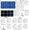
Figure 1
Figure 1
Piezo1 regulates MSC proliferation and osteogenesis as well as bone mass. a. μCT of rat distal femur from control and HU group. The left two lists of pictures are sagittal and coronal reconstruction images of the distal femur. The right two lists of pictures are the magnified trabecular bone structure from the most proximal growth plate of distal femur to the 200 slices underneath it. Scale bars equal to 2 mm. b) Statistic data of μCT (n = 3). BV/TV, bone volume/total volume. vBMD, volumetric bone mineral density. Tb.Th, trabecular thickness. Tb.Sp, trabecular separation. Tb.N. trabecular number. c) Immunofluorescent staining of Piezo1 and Lepr in the proximal right tibia of WB and HU14d groups (n = 3). Nuclei were counterstained with DAPI. Scale bars equal to 100 μm. d) Statistical analysis of costaining area in immunofluorescent staining (n = 3). e) Representative images of flow cytometry analysis of EdU+ rMSCs in control and 10% CMS groups. f) Statistical data of flow cytometry (n = 3). g) Relative expression of Col1a1 and Osx of C3H10T1/2 cell line in control and 10% CMS applicated groups h) Relative expression of Piezo1, Col1a1, Runx2, and Osx of rMSCs in control and 10% CMS applicated groups. i) Representative images of flow cytometry analysis of EdU+ rMSCs in control and Piezo1 siRNA transfected groups with 10% CMS application. j) Statistical data of flow cytometry (n = 3). k) Relative expression of Piezo1, Col1a1, Runx2, and Osx of rMSCs in control and Piezo1 siRNA transfected groups with 10% CMS. ns. No significance, *:p < 0.05, **:p < 0.01.
Figure 2
Figure 2
Development of Piezo1 agonist (MCB‐22‐174). a) The putative Yoda1 binding site in Piezo1 (PDB ID: 6B3R). Yoda1 is displayed in blue sticks, residues are depicted in white sticks. The allosteric binding pocket surrounded by residues Glu1688, Leu1689, Ala1718, Ala2091, Ala2094, Try2095, and Arg2098. b) Structure‐guided optimization of Yoda1. c) Docking mode of MCB‐22‐174 with Piezo1. Close‐up views of the hydrogen bonds and hydrophobic contacts formed between MCB‐22‐174 (yellow sticks) and Piezo1. Hydrogen bonds are shown in yellow dashed lines, and corresponding distances were labeled in angstrom. Interacting residues of Piezo1 are shown in the white stick representation. d) EC50 curve of Yoda1 in C3H10T1/2 cells. EC50 of Yoda1 equals to 42.84 μmol L−1 in C3H10T1/2 cell line. e) EC50 curve of MCB‐22‐174 in C3H10T1/2 cells. EC50 of MCB‐22‐174 equals 6.28 μmol L−1 in C3H10T1/2 cell line. f) Cell viability curve of Yoda1. g) Cell viability curve of MCB‐22‐174. h) Relative expression of Col1a1, Runx2, and Osx of rMSCs in control, 5 μmol L−1 Yoda1, 3, and 5 μmol·L−1 MCB‐22‐174‐treated groups for 72 h. (n = 3) ns. No significance, *:p < 0.05, compared with NC group.
Figure 3
Figure 3
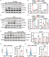
Piezo1 promoted the MSC proliferation through the CaMKII and ErK signaling pathway. a) Western blot images of CaMKII, p‐CaMKII, ERK, and p‐ERK in rMSCs after 0–60 min of 10% CMS stimulation. b) Statistical data of p‐ERK/ERK ratio and p‐CaMKII relative expression. c) Western blot images of CaMKII, p‐CaMKII, ERK, and p‐Eek in rMSCs after 0–60 min 5 μm Yoda1 treatment. d) Statistical data of p‐Erk/Erk ratio and p‐CaMKII relative expression. e) Western blot images of ERK and p‐Erk in control and Piezo1 siRNA transfected rMSCs after 0–15 min of 10% CMS treatment. f) Statistical data of p‐Erk/Erk ratio. g) Representative images and statistical data of flow cytometry analysis of EdU+ rMSCs in control and U1026‐treated groups. h) Representative images and statistical data of flow cytometry analysis of EdU+ rMSCs in control and KN93‐treated groups. For Western blot, GAPDH was employed as an internal reference (n = 3). *:p < 0.05, **:p < 0.01.
Figure 4
Figure 4
MCB‐22‐174 as a novel highly active agonist of Piezo1 could more effectively activate CaMKII/Erk signaling pathway than Yoda1 in vitro. a) Western blot images of CaMKII, p‐CaMKII, Erk, and p‐Erk in rMSCs after 0–15 min of 5 μmol L−1 MCB‐22‐174 treatment. b) Statistical data of p‐Erk/Erk ratio and p‐CaMKII relative expression. c) Western blot images of CaMKII, p‐CaMKII, Erk, and p‐Erk in rMSCs after 0–15 min treatment of 5 μmol L−1 Yoda1 or MCB‐22‐174. d) Statistical data of p‐Erk/Erk ratio and p‐CaMKII relative expression. e) Western blot images of CaMKII, p‐CaMKII, Erk, and p‐Erk in control and Piezo1 siRNA transfected rMSCs after 0–15 min of 5 μmol L−1 MCB‐22‐174 treatment. f) Statistical data of p‐Erk/Erk ratio and p‐CaMKII relative expression. For Western blot, GAPDH was chosen as internal reference (n = 3). *:p < 0.05, **:p < 0.01, ***p < 0.001.
Figure 5
Figure 5
In vivo, MCB‐22‐174 could significantly improve bone quality and MSC osteogenesis during HU. a) The procedure of in vivo application of Yoda1 and MCB‐22‐174. Created with BioRender.com. b) μCT of rat right femur from control, HU group, HU group treated with 5 μmol kg−1 Yoda1, and HU group treated with 5 μmol kg−1 MCB‐22‐174. The left two lists of pictures are sagittal and coronal reconstruction images of the distal femur. The right two lists of pictures are the magnified trabecular bone structure from the most proximal growth plate of distal femur to the 200 slices underneath it. Scale bars equal to 2 mm. c) Statistic data of μCT (n = 3). d) Immunofluorescent staining of ALP in the proximal right tibia of the four groups. Nuclei were counterstained with DAPI. Scale bars equal to 100 μm. e) Statistical analysis of the fluorescent staining area (n = 3). ns. No significance *p < 0.05, **p < 0.01 comparing with WB group.

References

    1. Rolvien T., Amling M., Calcif. Tissue Int. 2022, 110, 592. - PMC - PubMed
    1. Jepsen D. B., Ryg J., Hansen S., Jørgensen N. R., Gram J., Masud T., Osteoporosis Int. 2019, 30, 1827. - PMC - PubMed
    1. Delaine‐Smith R. M., Reilly G. C., Muscles Ligaments Tendons J. 2012, 2, 169. - PMC - PubMed
    1. Wang Y. K., Chen C. S., J. Cell. Mol. Med. 2013, 17, 823. - PMC - PubMed
    1. Watanabe‐Takano H., Ochi H., Chiba A., Matsuo A., Kanai Y., Fukuhara S., Ito N., Sako K., Miyazaki T., Tainaka K., Harada I., Sato S., Sawada Y., Minamino N., Takeda S., Ueda H. R., Yasoda A., Mochizuki N., Cell Rep. 2021, 36, 109380. - PubMed

LinkOut - more resources