

Comprehensive Management of Distal Femur Osteosarcoma in a 15-year-old Male: A Multidisciplinary Approach and Long-term Follow-up
- PMID: 40212505
- PMCID: PMC11981506
- DOI: 10.13107/jocr.2025.v15.i04.5450
Comprehensive Management of Distal Femur Osteosarcoma in a 15-year-old Male: A Multidisciplinary Approach and Long-term Follow-up
Abstract
Introduction: This comprehensive case report highlights the intricate diagnosis and multidisciplinary management of distal femur osteosarcoma in a 15-year-old male. Osteosarcoma is a rare and aggressive form of bone cancer, particularly affecting adolescents and young adults. The significance of this case lies in the detailed description of the diagnostic process, treatment strategy, and successful outcome, which may contribute valuable insights to the existing literature. While numerous case reports on osteosarcoma exist, this report offers unique insights into the management approach and outcome of distal femur osteosarcoma, potentially providing guidance for clinicians facing similar cases in the future.
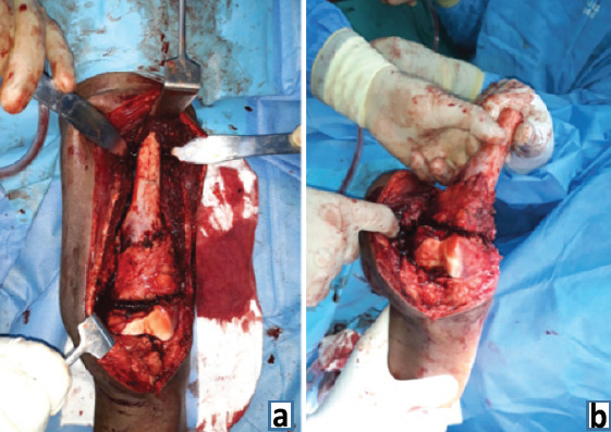
Case report: The patient in question is a 15-year-old male, whose ethnic background was not specified. He presented with symptoms suggestive of distal femur osteosarcoma, including localized bone pain, swelling, and limited range of motion in the affected knee joint. Radiological investigations confirmed the presence of a tumor in the distal femur, prompting further evaluation. Pathological markers and histopathological examination confirmed the diagnosis of osteosarcoma. Pre-operative neoadjuvant chemotherapy was administered to optimize conditions for surgical intervention.
Conclusion: This case report underscores the importance of a comprehensive, multidisciplinary approach in the management of distal femur osteosarcoma. The successful outcome, marked by complete tumor excision, reconstruction using autologous fibula graft, and restoration of knee function, highlights the efficacy of the chosen treatment strategy. Furthermore, this report may serve as a valuable resource for clinicians specializing in orthopedic oncology, providing insights into optimal treatment protocols and post-operative care for similar cases. In addition, it contributes to the broader field of oncology by advancing our understanding of osteosarcoma management and emphasizing the importance of individualized treatment plans tailored to the patient's specific needs.
Keywords: Osteosarcoma; autologous fibula graft; distal femur anatomical plate; extra corporeal radiotherapy; neoadjuvant chemotherapy; swashbuckler approach.
Copyright: © Indian Orthopaedic Research Group.
Conflict of interest statement
Conflict of Interest: Nil
Figures


References
-
- Kager L, Zoubek A, Pötschger U, Kastner U, Flege S, Kempf-Bielack B, et al. Primary metastatic osteosarcoma:Presentation and outcome of patients treated on neoadjuvant cooperative osteosarcoma study group protocols. J Clin Oncol. 2003;21:2011–8. - PubMed
-
- Anderson ME. Update on survival in osteosarcoma. Orthop Clin North Am. 2016;47:283–92. - PubMed
-
- Bielack SS, Kempf-Bielack B, Delling G, Exner GU, Flege S, Helmke K, et al. Prognostic factors in high-grade osteosarcoma of the extremities or trunk:An analysis of 1,702 patients treated on neoadjuvant cooperative osteosarcoma study group protocols. J Clin Oncol. 2002;20:776–90. - PubMed
-
- Ottaviani G, Jaffe N. The epidemiology of osteosarcoma. Cancer Treat Res. 2009;152:3–13. - PubMed
-
- Grimer RJ, Cannon SR, Taminiau AM, Bielack S, Kempf-Bielack B, Windhager R, et al. Osteosarcoma over the age of forty. Eur J Cancer. 2003;39:157–63. - PubMed
Publication types
LinkOut - more resources
Full Text Sources