. 2025 Apr 9;9(4):e70054.
doi: 10.1002/pld3.70054. eCollection 2025 Apr.

The Atypical Pectin Methylesterase Family Member PME31 Promotes Seedling Lipid Droplet Utilization

Affiliations

The Atypical Pectin Methylesterase Family Member PME31 Promotes Seedling Lipid Droplet Utilization

Sarah Hamade et al. Plant Direct. .

Abstract

In plants, the primary form of energy stored in seed lipid droplets, triacylglycerol (TAG), is catabolized during germination to support pre-photosynthetic growth. Although this process is essential for seedling development, it is incompletely understood. In a screen for Arabidopsis thaliana mutants displaying delayed degradation of the lipid droplet coat protein oleosin, five independent mutations in PECTIN METHYLESTERASE31 (PME31) were recovered. In addition to delayed oleosin degradation, pme31 mutant seedlings exhibited sustained lipid droplets and elevated levels of several TAG and diacylglycerol species. Although structural prediction classified PME31 as a pectinesterase, this structural family also includes a putative E. coli lipase, YbhC. Moreover, PME31 lacks an N-terminal signal peptide that would target it to the cell wall, where pectin resides. We found that a fluorescent PME31 reporter was cytosolic and partially associated with peroxisomes, the site of fatty acid catabolism, during lipid mobilization. Our findings suggest that, in contrast to canonical PMEs, which modify cell wall pectin, PME31 functions at peroxisomes to directly or indirectly promote lipid mobilization.

Keywords: Arabidopsis germination; lipid droplet; lipid mobilization; oleosin; pectin methylesterase; peroxisome.


Conflict of interest statement

The authors declare no conflicts of interest.

Figures

FIGURE 1
FIGURE 1
A screen for oleosin stabilization uncovers multiple pme31 alleles. (A) pOLE1:mNeonGreen‐OLE1 seeds were mutagenized and grown in pools, and their progeny (M2) were screened for sustained seedling fluorescence and then transferred to soil for seed production. (B) Four‐day‐old M3 seedlings were retested for mNeonGreen fluorescence (left) and protein stabilization via immunoblotting (right). An immunoblot of protein extracted from 4‐day‐old untransformed wild type (Wt), un‐mutagenized pOLE1:mNeonGreen‐OLE1 (Wt), and pme31 mutant seedlings was serially probed with antibodies recognizing seed oleosins and the HSC70 loading control. Biological replicates of the pme31‐4, pme31‐5, and pme31‐6 alleles were loaded in adjacent lanes. The positions of molecular mass markers (in kDa) are indicated at the left. (C) Whole‐genome sequencing of select mutants revealed multiple independent alleles in two genes: PME31 (blue; this work) and MIEL1 (purple; Traver and Bartel 2023).
FIGURE 2
FIGURE 2
pme31 mutant seedlings retain lipid droplets, oleosin, and TAG longer than wild‐type seedlings. (A) Confocal images (single slices) of cotyledon epidermal cells of Wt and pme31‐5 seedlings expressing mNeonGreen (mNG)‐OLE1 at the indicated ages. Neutral lipids were stained with MDH (magenta), and mNG‐OLE1 fluorescence is shown in green. (B) Immunoblot of protein extracted from stratified seeds (0 days) and 1.5‐ and 3‐day‐old seedlings probed with the indicated antibodies. The positions of molecular weight markers (in kDa) are indicated on the left. The numbers below the panels show the OLE1 or mNG‐OLE1 to HSC70 ratio, normalized to the day 0 level in Wt or Wt (pOLE1:mNG‐OLE1), respectively. (C) Representative thin‐layer chromatograph (TLC) of extracted lipids of the indicated genotypes at 0‐, 1.5‐, and 3‐day time points. (D) Quantification of triplicate TLC of the indicated genotypes. The TAG signal was standardized against wild type at 0 days for each replicate. p‐values were generated by three individual two‐tailed unpaired t‐tests of the time‐course groups.
FIGURE 3
FIGURE 3
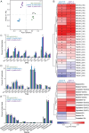
DAG and TAG profiles are altered in pme31‐5 seedlings. (A) PCA analysis of lipidomics data from 2‐day‐old seedlings shows limited variation among five biological replicates of each genotype. (B) Dendograms and heat maps showing log2 fold changes in DAG, TAG, and sterol levels in pme31 and miel1 mutants relative to Wt (mNG‐OLE1). (C–E) Bars show the mean levels of each DAG (C), TAG (D), or sterol (E) species for each genotype plotted as the percent of individual lipid species within the entire sample. p‐values were generated from Dunnett's multiple comparison tests of individual ANOVA of each lipid species among the five biological replicates (dots). Asterisks in panels (B)–(E) denote values significantly (p ≤ 0.001) different from Wt (mNG‐OLE1).
FIGURE 4
FIGURE 4
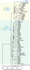
The PME31 catalytic domain is most similar to type II PMEs. Sequences of Arabidopsis PME proteins truncated to include only the catalytic domains were aligned with microbial PmeA from Dickeya dadantii and YbhC from E. coli to generate a phylogenetic tree. The catalytic domains of type I PMEs, which harbor a predicted cleavable signal peptide (SP) or transmembrane (TM) domain and an inhibitor domain on the N‐terminus, are distinct from the type II PMEs, which include a SP or TM domain on the N‐terminus but lack the inhibitor domain. PME31 lacks both N‐terminal domains. The percent identity to PME31 is shown in parentheses for selected proteins. An asterisk marks PME38, a probable pseudogene that groups with type I PMEs and has an inhibitor domain but lacks an N‐terminal SP or TM.
FIGURE 5
FIGURE 5
Predicted active‐site residues are conserved in PME31 and related enzymes. The alignment shows PME31 aligned with the catalytic domains of its closest type II (PME53) and type I (PME64) Arabidopsis homologs, PmeA from Dickeya dadantii , and YbhC from E. coli . Positions of newly identified mutations (pme31‐4 to pme31‐8) are highlighted in red and labeled above the alignment, and previously isolated alleles (pme31‐1 to pme31‐3; Rinaldi et al. 2016) are in maroon. Presumed active site residues, including catalytic residues labeled below the alignment, are highlighted in gold based on Dickeya dadantii PmeA (Fries et al. 2007). Five sequence motifs generally conserved in PMEs (Markovic and Janecek 2004) are noted in green below the alignment, with residues altered in PME31 depicted in blue. A short insertion (residues 84–87) in PME31 that is absent in other Arabidopsis PMEs is in blue in the PME31 sequence.
FIGURE 6
FIGURE 6
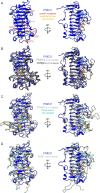
The predicted PME31 structure resembles PMEs and YbhC. (A) The PME31 structure was predicted by AlphaFold. Residues altered by pme31 mutations are highlighted in red (this work) and maroon (Rinaldi et al. 2016). The predicted active site residues from Figure 5 are highlighted in gold, with spheres designating predicted catalytic residues. The small insertion in PME31 that is absent in other Arabidopsis PMEs is highlighted in light blue. (B) Alignment of predicted structures of PME31 (blue) with PME53 (gray) and PME64 (charcoal). (C) Alignment of predicted structures of PME31 (blue) and D. dadantii PmeA (cream) (Fries et al. 2007). The identified PmeA active site residues are highlighted in teal, with spheres designating catalytic residues. (D) Alignment of the predicted structures of PME31(blue) with E. coli YbhC (mint). In B‐D, the root mean squared deviations (RMSD) across pruned atom pairs between PME31 and the comparison enzyme are shown in parentheses. The structures on the right in panels (A)–(D) are 90° counterclockwise rotations of the left structure around the vertical axis.
FIGURE 7
FIGURE 7
A PME31 reporter localizes to the cytosol and peroxisomes in developing Arabidopsis seedlings. Plants expressing a peroxisomal lumenal marker (tdTomato‐PTS1, magenta) and free mNeonGreen (mNeonGreen‐HA, green) (A, C) or fluorescently tagged PME31 (PME31‐HA‐mNeonGreen, green) expressed from PME31 5′ regulatory sequences (B, D) were imaged in radicles of stratified seeds (0 days) and roots of 1‐ and 2‐day‐old light‐grown seedlings (A, B) and in hypocotyls of 2‐ and 3‐day‐old light‐grown seedlings (C, D). Lipid droplets were stained with MDH (cyan); images are single 1‐μm slices. PME31‐mNeonGreen is cytosolic in younger tissue and becomes more peroxisome associated in older tissues. Arrows mark examples of peroxisomes with minimal mNeonGreen co‐localization; arrowheads mark examples of peroxisomes with substantial mNeonGreen co‐localization.

Similar articles

References

    1. Adham, A. R. , Zolman B. K., Millius A., and Bartel B.. 2005. “Mutations in Arabidopsis Acyl‐CoA Oxidase Genes Reveal Distinct and Overlapping Roles in β‐Oxidation.” Plant Journal 41: 859–874. - PubMed
    1. Anderson, C. T. 2016. “We Be Jammin': An Update on Pectin Biosynthesis, Trafficking and Dynamics.” Journal of Experimental Botany 67: 495–502. - PubMed
    1. Carrier, D. J. , van Roermund C. W. T., Schaedler T. A., et al. 2019. “Mutagenesis Separates ATPase and Thioesterase Activities of the Peroxisomal ABC Transporter, Comatose.” Scientific Reports 9: 10502. - PMC - PubMed
    1. Choudhary, V. , Ojha N., Golden A., and Prinz W. A.. 2015. “A Conserved Family of Proteins Facilitates Nascent Lipid Droplet Budding From the ER.” Journal of Cell Biology 211: 261–271. - PMC - PubMed
    1. Clough, S. J. , and Bent A. F.. 1998. “Floral Dip: A Simplified Method for Agrobacterium‐Mediated Transformation of Arabidopsis thaliana .” Plant Journal 16: 735–743. - PubMed

LinkOut - more resources