. 2024 May 23;4(8):2400097.
doi: 10.1002/smsc.202400097. eCollection 2024 Aug.

Light-Assisted 3D-Printed Hydrogels for Antibacterial Applications

Affiliations

Light-Assisted 3D-Printed Hydrogels for Antibacterial Applications

Liwen Zhang et al. Small Sci. .

Abstract

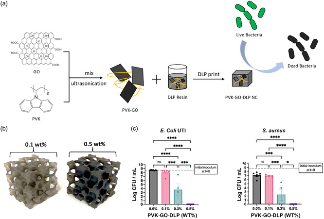
Light-assisted 3D printing technology, which uses a light source to solidify a photopolymerizable prepolymer solution, has shown great potential in the development of antibacterial hydrogels with high-resolution, specific features and functionalities. 3D-printed hydrogels with customized structures and antibacterial functions are widely used in tissue engineering, regenerative medicine, wound healing, and implants to advance the modeling and treatment of diseases. In the current review, an overview of light-assisted 3D printing technologies is first provided for the development of antibacterial hydrogels. Novel strategies involving the integration of inorganic nanomaterials, antibiotics, and functional polymers into 3D-printed hydrogels for the enhancement of antibacterial effects are then discussed. Finally, the perspective of advanced design using artificial intelligence and machine learning is proposed, providing a comprehensive yet succinct examination of 3D-printed hydrogels for antibacterial purposes.

Keywords: 3D printing; antibacterial and tissue engineering; hydrogel.


Conflict of interest statement

The authors declare no conflict of interest.

Figures

Figure 1
Figure 1
Summary of advantages and disadvantages of main light‐assisted 3D printing techniques.
Figure 2
Figure 2
a) Schematic illustration of the 3D printing process of methylcellulose and alginate hydrogel composites. b) Surface antibacterial activity of the hydrogel toward S. aureus and P. aeruginosa bacteria. Surviving S. aureus and P. aeruginosa colonies on agar plates after contact with 0.5 wt% gallium‐crosslinked hydrogels. The gallium‐crosslinked hydrogel kills almost all bacteria in contact with the hydrogel surface. c) Schematic illustration of the antibacterial activity of gallium‐crosslinked hydrogel. Accordingly, the antibacterial activity of the hydrogel stems from the presence of gallium cations on the hydrogel surface. Reproduced with permission.[ 87 ] Copyright 2021, Elsevier.
Figure 3
Figure 3
The schematic illustrates the fabrication process of double‐crosslinked materials, incorporating a UV‐crosslinker, HAMA, and click‐reaction crosslinked DTP‐modified HA. In vitro cytotoxicity testing revealed the potential of the drug‐loaded HA hydrogel for wound dressings. Reproduced with permission.[ 95 ] Copyright 2021, MDPI.
Figure 4
Figure 4
a) Schematic illustration of the fabrication of antibacterial 3D printing dressings. b) Biocidal results of GX2, GX2‐TiO2, and GX2‐TiO2‐PSPH‐Cl dressings with shake flask test against S. aureus and E. coli O157:H7. c) Cell viability of GX2 and GX2‐TiO2‐PSPH‐Cl dressings after 4, 12, 24, and 48 h incubation (*p ≤ 0.05, **p ≤ 0.01, and ***p ≤ 0.001). d) Representative photographs of wounds treated with gauze, GX2, and GX2‐TiO2‐PSPH‐Cl dressings at 0–16 days. Reproduced with permission.[ 104 ] Copyright 2021, Elsevier.
Figure 5
Figure 5
a) Schematic illustration of the formation of liposome hydrogel and Ag+. b) The formation of an adhesive, liposome‐laden, injectable, self‐healing, and antibacterial hydrogel via the Ag‐S coordination. c) The hydrogel was locally injected into an osteoporotic fracture and the bone marrow cavity for the release of drugs. Reproduced with permission.[ 119 ] Copyright 2019, Springer Nature.
Figure 6
Figure 6
a) The design strategy for the sustainable resin and antimicrobial hydrogel involves the following steps: i) Preparation of laccasepolymerized lignin in alkaline aqueous media from well‐defined lignin fractions. ii) Fabrication of lignin nanoparticles. iii) In situ reduction of Ag+ on the surface of alkali‐resistant lignin nanoparticles. iv) Scheme of the molecular structure of photo crosslinkable resin. v) Scheme of the molecular structure of photo crosslinkable hydrogel. b) Fabrication of 3D objects by projection lithography. c) In vitro release profiles of Ag+ from hydrogels. d) Viability assays of E. coli and S. aureus on hydrogels. Reproduced with permission.[ 34 ] Copyright 2022, Royal Society of Chemistry.
Figure 7
Figure 7
a) Schematics depict DLP printed PEGDA hydrogels containing ZnO NPs for antibacterial application; b) SEM images of DLP printed samples containing 1.5 wt% ZnO NPs; and c) E. coli and S. aureus viability percentage for different ZnO NP wt% in UV‐cured samples. Reproduced with permission.[ 138 ] Copyright 2023, MDPI.
Figure 8
Figure 8
a) DLP printing of polymer‐based GO nanocomposite as an efficient antibacterial coating for surgical implants; b) DLP printed gyroid structures with 0.1 and 0.5 wt% nanocomposites; Quantitative culture of c) E. coli UTI and (b) S. aureus at 24 h postinoculation onto DLP printed materials. Reproduced with permission.[ 33 ] Copyright 2023, Springer Nature.
Figure 9
Figure 9
a) Schematic illustration of the design and DLP 3D printing of GelMA microneedles containing amoxicillin; b) drug release profile from the amoxicillin‐loaded GelMA microneedles; and c) antibacterial activities of the amoxicillin‐loaded GelMA microneedles: 1) GelMA microneedles and 2) AMX‐loaded GelMA microneedles. Reproduced with permission.[ 157 ] Copyright 2023, Elsevier.

References

    1. Morens D. M., Taubenberger J. K., Fauci A. S., J. Infect. Dis. 2008, 198, 962. - PMC - PubMed
    1. Tan S. Y., Tatsumura Y., Singapore Med. J. 2015, 56, 366. - PMC - PubMed
    1. Frieri M., Kumar K., Boutin A., J. Infect. Public Health 2017, 10, 369. - PubMed
    1. Kalelkar P. P., Riddick M., García A. J., Nat. Rev. Mater. 2022, 7, 39. - PMC - PubMed
    1. Li S., Dong S., Xu W., Tu S., Yan L., Zhao C., Ding J., Chen X., Adv. Sci. 2018, 5, 1700527. - PMC - PubMed