

Mechanistic insights into the sleep-glymphopathy-cerebral small vessel disease loop: implications for epilepsy pathophysiology and therapy
- PMID: 40212714
- PMCID: PMC11983519
- DOI: 10.3389/fnins.2025.1546482
Mechanistic insights into the sleep-glymphopathy-cerebral small vessel disease loop: implications for epilepsy pathophysiology and therapy
Abstract
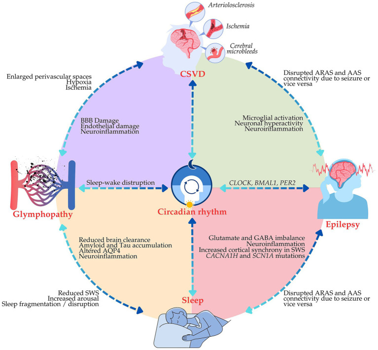
Epilepsy is the second most common neurological disorder and affects approximately 50 million people worldwide. Despite advances in antiepileptic therapy, about 30% of patients develop refractory epilepsy. Recent studies have shown sleep, glymphatic function, cerebral small vessel disease (CSVD), and epilepsy are interrelated by sharing a multidirectional relationship in influencing their severity and progression. Sleep plays a vital role in brain homeostasis and promotes glymphatic clearance responsible for the removal of metabolic wastes and neurotoxic substances from the brain. Disrupted sleep is a common feature in epilepsy and can lead to impairment in glymphatic efficiency or glymphopathy, promoting neuroinflammation and accrual of epileptogenic factors. CSVD, occurring in up to 60% of the aging population, further exacerbates neurovascular compromise and neurodegeneration by increasing seizure susceptibility and worsening epilepsy outcomes. This narrative review aims to discuss the molecular and pathophysiological inter-relationships between these factors, providing a new framework that places glymphopathy and CSVD as contributors to epileptogenesis in conditions of sleep disruption. We propose an integrative model wherein the glymphopathy and vascular insufficiency interact in a positive feedback loop of sleep disruption and increased seizure vulnerability mediated by epileptic activity. Acknowledging these interactions has significant impacts on both research and clinical practice. Targeting sleep modulation, glymphatic function, and cerebrovascular health presents a promising avenue for therapeutic intervention. Future research should focus on developing precision medicine approaches that integrate neuro-glial-vascular mechanisms to optimize epilepsy management. Clinically, addressing sleep disturbances and CSVD in epilepsy patients may improve treatment effectiveness, reduce seizure burden, and improve overall neurological outcomes. This framework highlights the need for interdisciplinary approaches to break the vicious cycle of epilepsy, sleep disturbance, and cerebrovascular pathology, paving the way for innovative treatment paradigms.
Keywords: cerebral small vessel disease; epilepsy; glymphatic system; sleep; therapy.
Copyright © 2025 Hein, Al-Zaghal, Muhammad Ghazali, Jaffer, Abdul Hamid, Mehat, Che Ramli and Che Mohd Nassir.
Conflict of interest statement
The authors declare that the research was conducted in the absence of any commercial or financial relationships that could be construed as a potential conflict of interest.
Figures
Similar articles
-
Cerebral small vessel disease: The impact of glymphopathy and sleep disorders.J Cereb Blood Flow Metab. 2025 May 5:271678X251333933. doi: 10.1177/0271678X251333933. Online ahead of print. J Cereb Blood Flow Metab. 2025. PMID: 40322968 Free PMC article. Review.
-
The Microbiota-Gut-Brain Axis: Key Mechanisms Driving Glymphopathy and Cerebral Small Vessel Disease.Life (Basel). 2024 Dec 24;15(1):3. doi: 10.3390/life15010003. Life (Basel). 2024. PMID: 39859943 Free PMC article. Review.
-
The Underlying Role of the Glymphatic System and Meningeal Lymphatic Vessels in Cerebral Small Vessel Disease.Biomolecules. 2022 May 25;12(6):748. doi: 10.3390/biom12060748. Biomolecules. 2022. PMID: 35740873 Free PMC article. Review.
-
Cerebral small vessel disease and glymphatic system dysfunction in multiple sclerosis: A narrative review.Mult Scler Relat Disord. 2024 Nov;91:105878. doi: 10.1016/j.msard.2024.105878. Epub 2024 Sep 5. Mult Scler Relat Disord. 2024. PMID: 39276600 Review.
-
Cerebral Small Vessel Disease: a Review of the Pathophysiological Mechanisms.Transl Stroke Res. 2024 Dec;15(6):1050-1069. doi: 10.1007/s12975-023-01195-9. Epub 2023 Oct 21. Transl Stroke Res. 2024. PMID: 37864643 Review.
References
Publication types
LinkOut - more resources
Full Text Sources