

Fluoroscopic guidance for bulla identification during ventral bulla osteotomy in eight French bulldogs
- PMID: 40212726
- PMCID: PMC11982915
- DOI: 10.1002/vro2.70008
Fluoroscopic guidance for bulla identification during ventral bulla osteotomy in eight French bulldogs
Abstract
Objective: To describe the technique and outcome of fluoroscopy to guide bulla identification in French bulldogs during ventral bulla osteotomy.
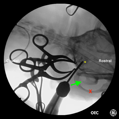
Materials and methods: Medical records of eight French bulldogs with otitis media that underwent a fluoroscopic-guided ventral bulla osteotomy between January 2020 and June 2023 were reviewed. Demographics, preoperative diagnostic findings, advanced imaging findings, surgical times, histopathology and culture results, as well as postoperative outcomes, were recorded.
Results: Following routine dissection of the bulla, fluoroscopic imaging was used to confirm the placement of a Steinmann pin before bulla penetration. The median surgical time was 152 minutes (range: 80-210 minutes). All dogs survived to discharge. Six out of eight dogs retained an ipsilateral head tilt postoperatively. Two dogs exhibited residual vestibular ataxia at 14 days postoperatively, which improved and resolved 7 months and 2 years postoperatively respectively. One dog developed recurring otitis media, and a total ear canal ablation and lateral bulla osteotomy were recommended.
Clinical significance: Intraoperative fluoroscopy can be used successfully to guide the identification of the bulla in ventral bulla osteotomies in brachycephalic breeds.
Keywords: canine; fluoroscopic‐guided; french bulldogs; otitis media; ventral bulla osteotomy.
© 2025 The Author(s). Veterinary Record Open published by John Wiley & Sons Ltd on behalf of British Veterinary Association.
Conflict of interest statement
The authors declare they have no conflicts of interest.
Figures


References
LinkOut - more resources
Full Text Sources
