. 2025 Mar 26:49:586-607.
doi: 10.1016/j.bioactmat.2025.03.017. eCollection 2025 Jul.

Synergistic effects of electrical and chemical cues with biodegradable scaffolds for large peripheral nerve defect regeneration

Affiliations

Synergistic effects of electrical and chemical cues with biodegradable scaffolds for large peripheral nerve defect regeneration

Rosalie Bordett et al. Bioact Mater. .

Abstract

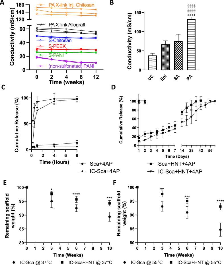
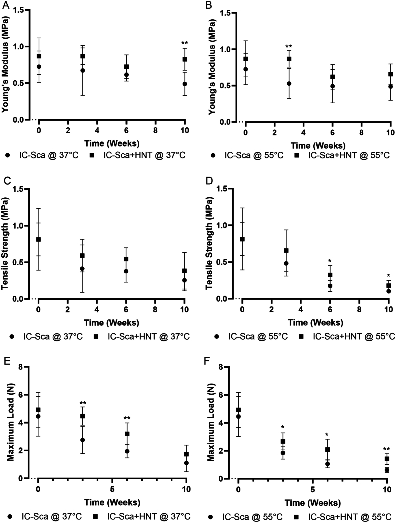
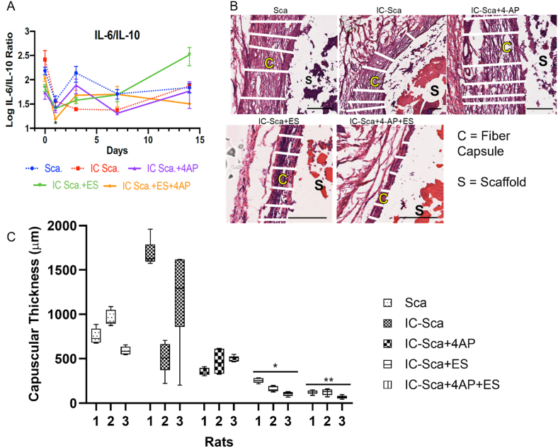
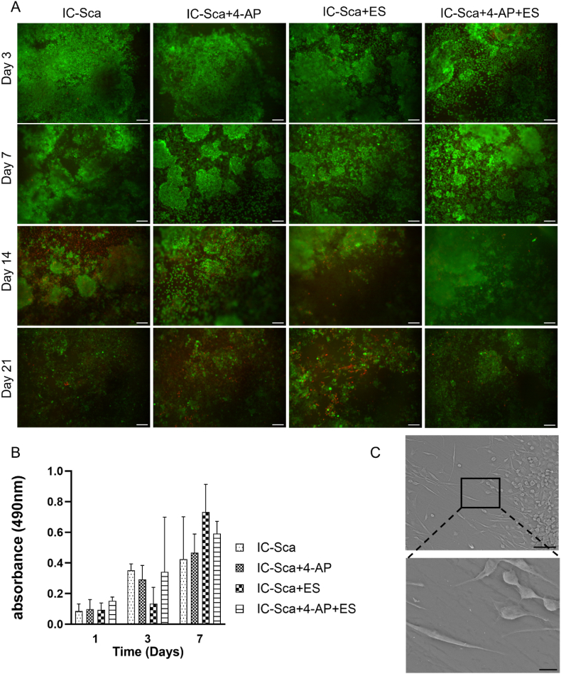
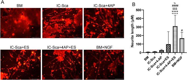
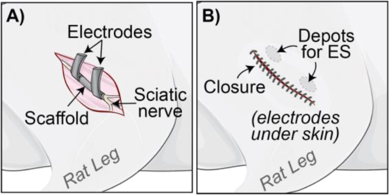
Large-gap peripheral nerve injuries (PNI) are often treated with autografts, allografts, or synthetic grafts to facilitate nerve regeneration, but these options are often limited in their availability or functionality. To address these issues, we developed ionically conductive (IC) nerve guidance conduits (NGCs) of sufficient biodegradability, mechanical strength, and bioactivity to support large-gap nerve regeneration. These chitosan-based NGCs release 4-aminopyridine (4-AP) from embedded halloysite nanotubes, and the NGC's IC properties enable transcutaneous electrical stimulation (ES) without invasive electrodes. In vitro, we found scaffolds with ES+4-AP synergistically enhanced Schwann cell adhesion, proliferation, and neurotrophin secretion, significantly improving axonal growth and neurite extension. In vivo, these scaffolds in large-gap PNI boosted neurotrophin levels, myelination, nerve function, and muscle weight while promoting angiogenesis and reducing fibrosis. Upregulated Trk receptors and PI3K/Akt and MAPK pathway highlight the regenerative potential. This study advances understanding of ES-mediated regeneration and supports innovative strategies for nerve and musculoskeletal repair.

Keywords: 4-Aminopyridine (4-AP); Ionically conductive nerve conduits; Neurotrophic factors; Peripheral nerve regeneration; Schwann cell proliferation; Sciatic nerve injury repair; Transcutaneous electrical stimulation.


Conflict of interest statement

Corresponding author Sangamesh G. Kumbar is an Associate Editor for Bioactive Materials and was not involved in the editorial review or the decision to publish this article. All authors declare that there are no competing interests.

Figures

Image 1
Graphical abstract
Schematic 1
Schematic 1
PNI types, and how ES and 4-AP delivery affects Schwann cell activity and nerve regeneration. (A) Crush and large-gap PNI models. (B) ES and 4-AP delivery timing, with (C) each treatment sustaining nerve depolarization and increased expression of intracellular Ca2+, neurotrophin BDNF, and TrkA, B, and C receptors. Secreted neurotrophins act on TrKA, B, and C to promote axon regeneration and remyelination. Both treatments enhance Schwann cell activities and motor function recovery in crush PNI.
Fig. 1
Fig. 1
Phytic acid (PA) cross-linked (X-link) chitosan, allografts, and sulfonated chitosan exhibit superior initial and sustained conductivity. PA X-link chitosan-HNT composites provide the highest ionic conductivity and sustained 4-AP release. (A) Ionic conductivity of sulfonated polyaniline (S-PANI), sulfonated poly(ether-ether-ketone) (S-PEEK), sulfonated chitosan (S-Chitosan), PA cross-linked chitosan, and nerve allografts measured in PBS (pH 7.4) over 12 weeks (N = 3 per group). (B) Effect of cross-linking agents on scaffold conductivity (UC = Uncross-linked, Epi-Epichlorohydrin, SA-Sulfuric Acid, PA-Phytic Acid). Significance levels: ∗∗∗∗p < 0.0001 vs. UC, ####p < 0.0001 vs. Epi, $$$$p < 0.0001 vs. SA. (C) Neat and uncross-linked scaffolds release all 4-AP within 8 h, while (D) chitosan/HNT and IC composite scaffolds provide sustained 4-AP release for over five weeks. (E) Scaffold degradation profiles in PBS (pH 7.4) at 37 °C and (F) under accelerated conditions at 55 °C for up to 10 weeks. The sample size was n = 6, and data presented as Mean ± SD (Significance: ∗p < 0.05, ∗∗p < 0.01, ∗∗∗p < 0.001, ∗∗∗∗p < 0.0001 vs. IC-Sca).
Fig. 2
Fig. 2
Composite scaffolds demonstrated superior tensile properties and a slower degradation rate than neat IC scaffolds at 37°C and 55°C. The tensile properties of scaffolds at 37 °C and 55 °C were assessed as a function of degradation time, including (A & B) Young's modulus, (C & D) UTS, and (E & F) maximum load at the break. However, the observed decrease in ultimate tensile strength (UTS) and load at break over time indicates a loss of mechanical integrity as degradation progresses. Statistical significance is indicated as ∗p < 0.05, ∗∗p < 0.01 compared to IC scaffolds (IC-Sca).
Fig. 3
Fig. 3
IC scaffolds with the combined 4-AP+ES treatment showed improved foreign body response compared to IC scaffolds alone in a 14-day subcutaneous rat implantation study. (A) Serum levels of IL-6 and IL-10 were measured via ELISA from blood samples collected over 14 days (mean ± SEM). (B) Representative H&E images highlight the fibrous capsule formation around the implant (marked by white lines, scale bar = 600 μm). (C) Fibrous capsule thickness was analyzed by group and individual rats, with data presented as box and whisker plots (median, interquartile range, and min/max). ∗p < 0.05 or ∗∗p < 0.01 compared to IC scaffold group, analyzed by two-way ANOVA and Tukey's multiple comparison test.
Fig. 4
Fig. 4
Composite IC scaffolds effectively supported Schwann cell viability and proliferation during chronic 4-AP and ES treatments. Schwann cells were seeded on the IC scaffolds and subjected to 4-AP (30 μg/mL) and/or ES (1 V, 20 Hz for 20 min). (A) Cell viability was assessed over 21 days, with live cells appearing green and dead cells red at 20× magnification (n = 3). Scale bar = 100 μm. (B) Cell metabolic activity was quantified using an MTS assay over 7 days (n = 5), and results are presented as Mean ± SD. (C) SEM images (n = 3) display Schwann cells' elongated morphology at 500x (top) and 2000x (bottom), with scale bars of 50 μm and 10 μm, respectively.
Fig. 5
Fig. 5
Schwann cells exhibited significantly higher expression of neurotrophic factors BDNF, NGF, and MPZ under the combined 4-AP+ES treatment than individual 4-AP or ES treatments. (A) Representative immunofluorescent images at day 14 show BDNF, NGF, and myelin protein zero (MPZ) expression on scaffolds (20× magnification), where blue fluorescence indicates cell nuclei stained with DAPI, and red fluorescence highlights the specific protein expression. Scale bar = 100 μm. (B) Quantification of immunofluorescent stains using total corrected cellular fluorescence (TCCF) (n = 15). Data are Mean ± SD. ∗p < 0.05, ∗∗p < 0.01 vs IC-Sca. ##p < 0.01 vs IC-Sca+4-AP. $p < 0.05 vs IC-Sca+ES.
Fig. 6
Fig. 6
Schwann cells exhibited significantly higher expression of neurogenic genes under the combined 4-AP+ES treatment than individual 4-AP or ES treatments. Cellular constructs harvested on day 14 were analyzed, with BM as the negative control and 100 ng/mL NGF as the positive control. Key neurogenic genes involved in nerve regeneration were quantified, including (A) Nerve growth factor (NGF), (B) Brain-derived neurotrophic factor (BDNF), (C) Glial cell line-derived neurotrophic factor (GDNF), (D) Neurotrophin-3 (NT-3), (E) Neurotrophin-4 (NT-4), and (F) S100 calcium-binding protein β (S100β). A sample size n = 5 was used for these assays, and the data is presented as Mean ± SD. ∗p < 0.05, ∗∗p < 0.01, ∗∗∗p < 0.001, and ∗∗∗∗p < 0.0001 vs basal media. #p < 0.05 vs IC-Sca+4-AP. $p < 0.05, $$p < 0.01 vs IC-Sca+ES.
Fig. 7
Fig. 7
Schwann cells expressed significantly higher levels of neurotrophic proteins under the combined 4-AP+ES treatment than individual 4-AP or ES treatments. Schwann cell-conditioned media, collected on day 14 from all treatments (BM as negative control and 100 ng/mL NGF as positive control), were subjected to proteomics analysis. Key proteins involved in nerve regeneration were quantified, including: (A) neural cell adhesion molecule 1 (NCAM1), (B) neuropilin 2 (NRP2), (C) carboxypeptidase E (CPE), (D) fibronectin (FN1), (E) insulin-like growth factor (IGF), and (F) transforming growth factor-β (TGF). A sample size n = 5 was used, and the data is presented as Mean ± SD. ∗p < 0.05, ∗∗p < 0.01, ∗∗∗p < 0.001, and ∗∗∗∗p < 0.0001 vs TCPS. #p < 0.05 vs IC-Sca+4-AP. $p < 0.05, $$p < 0.01 vs IC-Sca+ES. %p < 0.05, %%p < 0.01, %%%p < 0.001 vs NGF.
Fig. 8
Fig. 8
Schwann cell-conditioned media from the combined 4-AP+ES treatment significantly enhanced PC12 neurite outgrowth compared to individual 4-AP and ES treatments. Schwann cell culture media from all treatments, collected on day 7, were added to PC12 basal media (BM) to support neurite extension, with BM (negative control) and 100 ng/mL NGF (positive control). (A) Representative images of neurite outgrowth stained with InvitrogenTM Neurite Outgrowth Staining Kit at 10× magnification. (B) Quantification of average neurite length per cell (n = 13). ∗∗∗∗p < 0.0001 vs IC-Sca. #p < 0.05, ####p < 0.0001 vs IC-Sca+4-AP. ++++p < 0.0001 vs BM. $$$p < 0.001 vs IC-Sca+ES. %p < 0.05 vs BM + NGF.
Fig. 9
Fig. 9
Schwann cells express significantly higher amounts of BDNF, NGF, and S100β with combined 4-AP+ES treatment than individual 4-AP or ES treatments at days 7 and 14. (A) Representative western blots of S100, NGF, and BDNF proteins for 14-day samples. (TCPS; control n = 3 experiments of 6 pooled samples each). (B) Normalized western blot data is expressed as protein: actin ratio (ng/mL). Data: Mean ± SEM. One-way ANOVA with Tukey's multiple comparisons tests; %%%%p < 0.0001 vs. TCPS. ∗∗∗p < 0.001, and ∗∗∗∗p < 0.0001 vs IC-Sca. ###p < 0.001, and ####p < 0.0001 vs IC-Sca+4-AP. $$$$p < 0.0001 vs IC-Sca+ES.
Fig. 10
Fig. 10
Schwann cells express significantly higher amounts of NGF with combined 4-AP+ES treatment than individual 4-AP or ES treatments at days 7 and 14. Schwann cell-conditioned media collected from all treatments were quantified using NGF ELISA. NGF concentrations were measured on day (A) 7 and day (B) 14. Schwann cells treated with 100 ng/mL NGF were the positive control, while those treated with basal media were the negative control. A sample size of n = 3 was used for these assays, and the data is presented as Mean ± SD. ∗∗∗∗p < 0.0001 vs IC-Sca. ####p < 0.0001 vs IC-Sca+4-AP. $$$$p < 0.0001 vs IC-Sca+ES. %%%%p < 0.0001 vs NGF.
Fig. 11
Fig. 11
The combined 4-AP+ES treatment synergistically enhances CMAP compared to individual 4-AP or ES treatments for 2 cm and 4 cm long scaffolds. (A, C) Representative CMAP traces for 2 and 4-cm long scaffolds show a second waveform across all test groups. (B, D) Quantified CMAP amplitudes for the 2 cm and 4 cm scaffolds. (E) The Gastrocnemius muscle weight ratio increased with the combined 4-AP+ES treatment, reaching levels comparable to the autograft control for 2 cm and 4 cm scaffolds (2 cm data not shown). Mean ± SD; n = 5–6/group.; One-way ANOVA with Tukey's multiple comparisons test; ∗p < 0.05, ∗∗p < 0.001 vs IC-Sca. #p < 0.05 vs IC-Sca+4-AP.4.
Fig. 12
Fig. 12
Treatments 4-AP, ES, and 4-AP+ES—supported myelinated axon regeneration in 2 cm and 4 cm scaffolds at 12 weeks, with outcomes comparable to autografts. (A) TEM micrographs for 2 cm and (B) 4 cm long scaffolds, showing the mid-region cross sections of the scaffolds at 12 weeks post-surgery (Mag.-1000X; Scale 10 μm). Calculated G-ratio form TEM images for (C) 2 cm and (D) 4 cm long scaffolds. G-ratio is the inner axon diameter to outer fiber diameter ratio, where a lower ratio indicates thicker myelin. Data are expressed as mean ± SD. n = 35 measurements per group. ∗∗p < 0.01, ∗∗∗∗p < 0.0001 vs IC-Sca. ####p < 0.0001 vs IC-Sca+4-AP. $$p < 0.01 vs IC-Sca+ES.
Fig. 13
Fig. 13
The combined 4-AP+ES treatment synergistically enhances myelin sheath and blood vessel formation compared to individual 4-AP or ES treatments in the regenerated nerve for 2 cm and 4 cm long scaffolds. (A, B) (i)LFB Luxol Fast Blue staining for myelinated nerves (scale bars = 200 μm). (A, B) (ii) S100β immunostaining for Schwann cell activity (scale bars = 100 μm). (A, B) (iii) CD31 immunostaining for endothelial cell marker (scale bars = 100 μm). Image intensity quantification (C, F) S100β, (D, G) CD31, and (E, H) number of blood vessels. Blood vessels were counted N = 12 sections/Sample manually). One-way ANOVA with Dunnett's multiple comparisons test. ∗∗∗∗p < 0.0001; and (E) Quantification of CD31 immunostaining in terms of percentage of area stained. N = 10-12 sections. One-way ANOVA with Tukey's multiple comparisons test. ∗p < 0.05, ∗∗p < 0.01, ∗∗∗∗p < 0.0001 vs IC-Sca. ###p < 0.001, ####p < 0.0001 vs IC-Sca+4-AP. $$$p < 0.001, $$$$p < 0.0001 vs IC-Sca+ES. %%%p < 0.001, %%%%p < 0.0001 vs autograft.
Fig. 14
Fig. 14
Regenerated nerve samples from the 4 cm scaffolds showed significantly higher neurotrophic and endothelial marker gene expression under the combined 4-AP+ES treatment than individual 4-AP or ES treatments. (AH) At 12 weeks post-injury, 4-AP+ES treatment significantly upregulated neurotrophic factors (NGF, BDNF, GDNF & NT3) and their receptors (TRKA, TRKB & TRKC) and endothelial marker (CD31) compared to other groups. Data are Mean ± SD; n = 3–4. One-way ANOVA with Tukey's multiple comparisons test. ∗p < 0.05, ∗∗∗p < 0.001 vs IC-Sca. #p < 0.05, ###p < 0.001 vs IC-Sca+4-AP. $p < 0.05, $$p < 0.01, $$$p < 0.001 vs IC-Sca+ES. %p < 0.05, %%p < 0.01, %%%p < 0.001 vs autograft.
Schematic 2
Schematic 2
(A) Schematic of surgery with electrode wrapped around the implanted scaffold, and (B) showing closure with electrodes under the skin.

References

    1. Brattain K. Magellan Medical Technology Consultants, Inc.; 2013. ANALYSIS OF THE PERIPHERAL NERVE REPAIR MARKET IN THE UNITED STATES.
    1. Moore A.M., MacEwan M., Santosa K.B., Chenard K.E., Ray W.Z., Hunter D.A., Mackinnon S.E., Johnson P.J. Acellular nerve allografts in peripheral nerve regeneration: a comparative study. Muscle Nerve. 2011;44(2):221–234. - PMC - PubMed
    1. Chrząszcz P., Derbisz K., Suszyński K., Miodoński J., Trybulski R., Lewin-Kowalik J., Marcol W. Application of peripheral nerve conduits in clinical practice: a literature review. Neurol. Neurochir. Pol. 2018;52(4):427–435. - PubMed
    1. Manoukian O.S., Baker J.T., Rudraiah S., Arul M.R., Vella A.T., Domb A.J., Kumbar S.G. Functional polymeric nerve guidance conduits and drug delivery strategies for peripheral nerve repair and regeneration. J. Contr. Release. 2020;317:78–95. - PubMed
    1. Shapira Y., Sammons V., Forden J., Guo G.F., Kipp A., Girgulis J., Mishra T., de Villers Alant J.D., Midha R. Brief electrical stimulation promotes nerve regeneration following experimental in-continuity nerve injury. Neurosurgery. 2019;85(1):156–163. - PubMed

LinkOut - more resources