. 2025 Apr 1;29(5):255.
doi: 10.3892/ol.2025.15001. eCollection 2025 May.

Bone mesenchymal stem cell‑derived exosome‑encapsulated microRNA‑125b‑5p inhibits ovarian cancer progression via DDX5 downregulation

Affiliations

Bone mesenchymal stem cell‑derived exosome‑encapsulated microRNA‑125b‑5p inhibits ovarian cancer progression via DDX5 downregulation

Yuxia Wang et al. Oncol Lett. .

Abstract

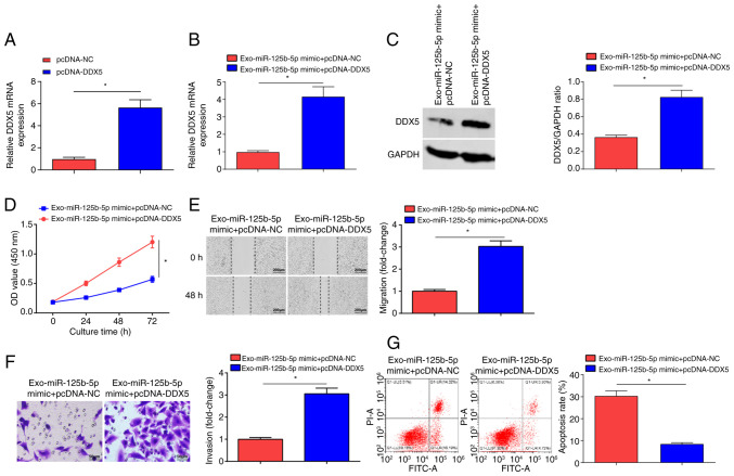
Exosomes can be used to mediate the delivery of nucleic acids such as microRNA-125b-5p (miR-125b-5p), a tumor-suppressor in certain types of cancer, into tumor cells. The present study investigated the use of bone mesenchymal stem cells-derived exosome (BMSCs-Exo) delivery of miR-125b-5p in ovarian cancer (OC). BMSCs were transfected with miR-125b-5p mimic, from which exosomes termed Exo-miR-125b-5p mimic were extracted. The expression levels of miR-125b-5p in OC tissue samples, BMSCs, exosomes and SKOV3 cells were quantified using reverse transcription-quantitative PCR. The influence of Exo-miR-125b-5p mimic on the biological functions of OC was evaluated through cell proliferation, invasion, migration and apoptosis assays. The targeting relationship between miR-125b-5p and DEAD-box helicase 5 (DDX5) was verified, and the expression levels of DDX5 in OC samples and SKOV3 cells were quantified using western blotting. miR-125b-5p was downregulated in tumor tissue samples from patients with OC. BMSCs-Exo reduced the malignant properties of SKOV3 cells in vitro, and these effects were be advanced by miR-125b-5p upregulation. miR-125b-5p targeted and inhibited DDX5 expression. DDX5 overexpression inhibited Exo-miR-125b-5p-induced suppression of OC development. Overall, this study highlights that BMSCs-Exo-encapsulated miR-125b-5p inhibited OC progression via DDX5 downregulation, providing insight into the molecular mechanisms underlying OC.

Keywords: DEAD-box helicase 5; bone mesenchymal stem cells; exosomes; microRNA-125b-5p; ovarian cancer; tumor.


Conflict of interest statement

The authors declare that they have no competing interests.

Figures

Figure 1.
Figure 1.
Identification of BMSCs and exosomes. (A) Stem cell markers detected using flow cytometry. (B) Oil Red O and Alizarin Red staining of BMSCs. Scale bar, 100 µm. (C) Representative transmission electron microscopy images of exosomes. Scale bar, 100 nm. (D) Nanoparticle tracking analysis detected the diameter and concentration of exosomes. (E) Western blot analysis of the exosomal markers, CD9 and CD81, with GAPDH as a loading controls. (F) The gene expression levels of miR-125b-5p in BMSCs transfected with miR-125b-5p mimic. (G) PKH26-labeled exosomes in SKOV3 cells. Scale bar, 25 µm. (H) miR-125b-5p gene expression levels in Exo-miR-125b-5p mimic and Exo-mimic were assessed. Data were represented by the mean ± standard deviation. *P<0.05. BMSCs, bone mesenchymal stem cells; miR-125b-5p, microRNA-125b-5p; NC, negative control; Exo-, exosome encapsulated.
Figure 2.
Figure 2.
BMSCs-Exo limits malignancy of OC cells. (A) miR-125b-5p gene expression levels evaluated in SKOV3 cells treated with BMSCs-Exo. (B) Proliferation of OC cells examined using the CCK-8 assay after BMSCs-Exo treatment. (C) Migration of OC cells assessed using a scratch test after BMSCs-Exo treatment (scale bar, 200 µm; magnification, ×100). (D) Invasion of OC cells evaluated using a Transwell assay after BMSCs-Exo treatment (scale bar, 50 µm; magnification, ×200). (E) Apoptosis of OC cells examined using flow cytometry after BMSCs-Exo treatment. Data were represented by the mean ± standard deviation. *P<0.05. BMSCs-Exo, bone mesenchymal stem cells-derived exosomes; OC, ovarian cancer; OD, optical density.
Figure 3.
Figure 3.
Exosome delivery of miR-125b-5p represses malignant progression of OC cells. (A) Gene expression levels of miR-125b-5p in normal ovarian tissues and OC tissues detected. (B) miR-125b-5p gene expression levels in SKOV3 cells treated with Exo-miR-125b-5p mimic and Exo-mimic NC. (C) Proliferation of OC cells examined using the CCK-8 assay after Exo-miR-125b-5p mimic treatment. (D) Migration of OC cells evaluated using a scratch test after Exo-miR-125b-5p mimic treatment (scale bar, 200 µm; magnification, ×100). (E) Invasion of OC cells assessed using a Transwell assay after Exo-miR-125b-5p mimic treatment (scale bar, 50 µm; magnification, ×200). (F) Apoptosis of OC cells examined using flow cytometry after Exo-miR-125b-5p mimic treatment. Data were represented by the mean ± standard deviation. *P<0.05. OC, ovarian cancer; OD, optical density; miR-125b-5p, microRNA-125b-5p; NC, negative control; Exo-, exosome encapsulated.
Figure 4.
Figure 4.
Targeting relationship between miR-125b-5p and DDX5. (A) DDX5 mRNA and protein expression in normal ovarian and tumor tissues. (B) Targeted sites between miR-125b-5p and DDX5 using the ENCORI software. (C) Correlation between miR-125b-5p and DDX5 mRNA expression levels. The binding of miR-125b-5p and DDX5 was analyzed using (D) dual luciferase reporter and (E) RNA immunoprecipitation assays. (F) DDX5 mRNA and protein expression levels were evaluated in mimic NC and miR-125b-5p mimic group. (G) DDX5 mRNA expression levels were evaluated. Data were represented by the mean ± standard deviation. *P<0.05. OC, ovarian cancer; OD, optical density; miR-125b-5p, microRNA-125b-5p; NC, negative control; Exo-, exosome encapsulated; WT, wild-type; Mut, mutant; DDX, DEAD-box helicase 5.
Figure 5.
Figure 5.
DDX5 knockdown inhibited the malignancy of OC cells. DDX5 (A) mRNA and (B) protein expression levels upon transfection of si-DDX5. (C) Proliferation of OC cells evaluated by CCK-8 assay upon transfection of si-DDX5. (D) Migration of OC cells assessed using a scratch test upon transfection of si-DDX5 (Scale bar, 200 µm; magnification, ×100). (E) Invasion of OC cells tested using a Transwell assay upon transfection of si-DDX5 (scale bar, 50 µm; magnification, ×200). (F) Apoptosis of OC cells evaluated using flow cytometry upon transfection of si-DDX5. Data were represented by the mean ± standard deviation. *P<0.05. OC, ovarian cancer; OD, optical density; NC, negative control; DDX, DEAD-box helicase 5; si, small interfering.
Figure 6.
Figure 6.
DDX5 overexpression inhibits miR-125b-5p-induced suppression of OC development. (A) DDX5 mRNA expression in the pcDNA-NC group and pcDNA-DDX5 group. DDX5 (B) mRNA and (C) protein expression levels in the Exo-miR-125b-5p mimic + pcDNA-NC and Exo-miR-125b-5p mimic + pcDNA-DDX5 groups. (D) Proliferation of OC cells examined using the CCK-8 assay in the Exo-miR-125b-5p mimic + pcDNA-NC and Exo-miR-125b-5p mimic + pcDNA-DDX5 groups. (E) Migration of OC cells evaluated using a scratch test in the Exo-miR-125b-5p mimic + pcDNA-NC and Exo-miR-125b-5p mimic + pcDNA-DDX5 groups (scale bar, 200 µm; magnification, ×100). (F) Invasion of OC cells assessed using a Transwell assay in the Exo-miR-125b-5p mimic + pcDNA-NC and Exo-miR-125b-5p mimic + pcDNA-DDX5 groups (scale bar, 50 µm; magnification, ×200). (G) Apoptosis of OC cells examined using flow cytometry in the Exo-miR-125b-5p mimic + pcDNA-NC and Exo-miR-125b-5p mimic + pcDNA-DDX5 groups. Data were represented by the mean ± standard deviation. *P<0.05. OC, ovarian cancer; OD, optical density; miR-125b-5p, microRNA-125b-5p; NC, negative control; Exo-, exosome encapsulated; WT, wild-type; Mut, mutant; DDX, DEAD-box helicase 5; transfected using the pcDNA3.1 plasmid.

References

    1. Sung H, Ferlay J, Siegel RL, Laversanne M, Soerjomataram I, Jemal A, Bray F. Global cancer statistics 2020: GLOBOCAN estimates of incidence and mortality worldwide for 36 cancers in 185 countries. CA Cancer J Clin. 2021;71:209–249. doi: 10.3322/caac.21660. - DOI - PubMed
    1. Stewart C, Ralyea C, Lockwood S. Ovarian cancer: An integrated review. Semin Oncol Nurs. 2019;35:151–156. doi: 10.1016/j.soncn.2019.02.001. - DOI - PubMed
    1. Rooth C. Ovarian cancer: Risk factors, treatment and management. Br J Nurs. 2013;22:S23–S30. doi: 10.12968/bjon.2013.22.Sup17.S23. - DOI - PubMed
    1. Narod S. Can advanced-stage ovarian cancer be cured? Nat Rev Clin Oncol. 2016;13:255–261. doi: 10.1038/nrclinonc.2015.224. - DOI - PubMed
    1. Crapnell K, Blaesius R, Hastings A, Lennon DP, Caplan AI, Bruder SP. Growth, differentiation capacity, and function of mesenchymal stem cells expanded in serum-free medium developed via combinatorial screening. Exp Cell Res. 2013;319:1409–1418. doi: 10.1016/j.yexcr.2013.04.004. - DOI - PubMed

LinkOut - more resources