. 2023 Apr 24;3(5):2300024.
doi: 10.1002/smsc.202300024. eCollection 2023 May.

Vertex-Shared Linear Superatomic Molecules: Stepping Stones to Novel Materials Composed of Noble Metal Clusters

Affiliations

Vertex-Shared Linear Superatomic Molecules: Stepping Stones to Novel Materials Composed of Noble Metal Clusters

Yoshiki Niihori et al. Small Sci. .

Abstract

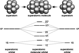
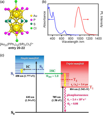
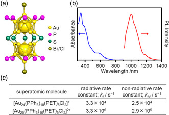
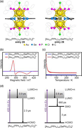
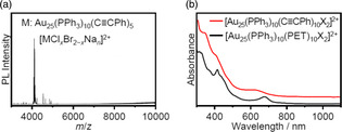
Extremely small metal clusters composed of noble metal atoms (M) have orbitals similar to those of atoms and therefore can be thought of as artificial atoms or superatoms. If these superatoms can be assembled into molecular analogs, it might be possible to create materials with new characteristics and properties that are different from those of existing substances. Therefore, the concept of superatomic molecules has attracted significant attention. The present review focuses on vertex-shared linear M12n+1 superatomic molecules formed via the sharing of a single metal atom between M13 superatoms having icosahedral cores and summarizes the knowledge obtained to date in this regard. This summary discusses the most suitable ligand combinations for the synthesis of M12n+1 superatomic molecules along with the valence electron numbers, stability, optical absorption characteristics, and luminescence properties of the M12n+1 superatomic molecules fabricated to date. This information is expected to assist in the production of many M12n+1 superatomic molecules with novel structures and physicochemical properties in the future.

Keywords: connections; geometrical structures; ligand-protected metal clusters; superatomic molecules.


Conflict of interest statement

The authors declare no conflict of interest.

Figures

Figure 1
Figure 1
Diagram showing the formation of a superatomic molecule and the associated orbitals. [3b]
Figure 2
Figure 2
a–c) Diagrams of vertex‐sharing (a), edge‐sharing (b), and face‐sharing (c) superatomic molecules.
Figure 3
Figure 3
a) The anatomy of an M25 superatomic molecule comprising an M25 core and three types of ligands. b) The five different metal sites in an M25 core. c) Characteristic geometrical parameters of an M25 superatomic molecule. In these figures, carbon and hydrogen atoms in the phosphine ligands are omitted for clarity.
Figure 4
Figure 4
Classification of ligands used for the formation of superatomic molecules based on a Venn diagram. L1, L2, and L3 represent characteristic ligand sites (see also Figure 3). a) Ethanethiolate, b) 2‐phenylethanethiolate, c) benzenethiolate, d) benzeneselenolate, e) triphenylphosphine, f) tri(p‐tolyl)phosphine, g) methyldiphenylphosphine, h) bis(diphenylphosphino)methane, i) 1,1′‐bis(diphenylphosphino)ferrocene, j) 1,3‐di‐i‐propylbenzimidazol‐2‐ylidene, k) 1,3‐di(2,4,6‐trimethylbenzyl)benzimidazol‐2‐ylidene, l) bromide, m) chloride, n) 1‐adamantanethiolate, o) O,O‐dipropyldithiophosphate, p) 4‐tert‐buthylbenzenethiolate, and q) dipyridylaminide ligands.
Figure 5
Figure 5
Framework structures of bimetallic M25 superatomic molecules protected by PR3 and X ligands. a) [Ag12Au13(P(p‐tol)3)10Cl7]+ (entry 1), [10a] b) [Ag12Au13(PPh3)10Br8]+ (entry 2), [10b] c) [Ag12Au13(PPh3)10Br8]+ (entry 3), [10c] d) [Ag12Au13(P(p‐tol)3)10Br8]+ (entry 4), [10d] e) [Ag12Au13(PPh3)10Cl8]+ (entry 5), [10e,f] f) [Ag12Au13(PPh3)10Cl8]+ (entry 6), [10f] g) [Ag12Au13(P(p‐tol)3)10Cl8]+ (entry 7), [10g] h) [Ag12+x Au13−x (PPh3)10Cl8]2+ (entry 8), [10h] i) [Ag13Au12(PPh2Me)10Br9]0 (entry 9), [10i] and j) [Ag17Au8(PPh3)10Cl10]0 (entry 10). [10j] In each structure, the left and right images indicate a side and top view, respectively. In the top views, only the MC1 and ML1 site metal atoms and L1 atoms are shown for the clarity. These structures are classified by the number of halogen ligands and bonding motifs.
Figure 6
Figure 6
Framework structures of Ag23M2 cores protected by PPh3 and X ligands. a) [Ag23Pd2(PPh3)10Br7]0 (entry 11), [10k] b) [Ag23Pd2(PPh3)10Cl7]0 (entry 12), [10l] c) [Ag23Pt2(PPh3)10Br7]0 (entry 13), [10k] and d) [Ag23Pt2(PPh3)10Cl7]0 (entry 14). [10m] In each structure, the left and right figures indicate the side and top view, respectively.
Figure 7
Figure 7
a) The geometrical structure of [Au23Pd2(PPh3)10Br7]0 (entry 15). [10n] b) Electrospray ionization‐mass spectra of [Au23Pt2(PPh3)10Br7]0 and [Au23Pd2(PPh3)10Cl7]0. b) Reproduced with permission. [10n] Copyright 2017, American Chemical Society.
Figure 8
Figure 8
Framework structures of trimetallic M25 superatomic molecules. a) [Ag12Au12Ni(PPh3)10Cl7]+ (entry 16), [10o] b) [Ag12Au12Pt(PPh3)10Cl7]+ (entry 17), [10p] c) [Ag12Au11Pt2(PPh3)10Cl7]0 (entry 18), [10q] and d) [Ag13Au10Pt2(PPh3)10Cl7]0 (entry 20). [10r]
Figure 9
Figure 9
a) Framework structure of [Au25(PPh3)10(SR)5Cl2]2+ (SR = SEt (entry 20), [10s] PET (entry 21), [10t] or SPh (entry 22) [10u] ). b) UV–visible absorption/PL spectra of [Au25(PPh3)10(PET)5Cl2]2+. b) Reproduced with permission.[ 23 ] Copyright 2021, Wiley‐VCH. c) Excited state deactivation pathway of [Au25(PPh3)10(SR)5Cl2]2+. Reproduced with permission.[ 24 ] Copyright 2022, Royal Society of Chemistry.
Figure 10
Figure 10
a) Framework structure of [Au24(PPh3)10(PET)5X2]+ (X = Br or Cl)[ 26 ] and b) UV–vis absorption/PL spectra of [Au24(PPh3)10(PET)5Cl2]+. b) Reproduced with permission.[ 23 ] Copyright 2021, Wiley‐VCH. c) The effect of a central Au atom on the excitation relaxation rate.[ 23 ]
Figure 11
Figure 11
A) Framework structures of: a) [Au25(PPh3)10(PET)5Cl2]2+ (entry 21), [10t] b), [Ag x Au25−x (PPh3)10(PET)5X2]2+ (x ≤ 12), c) [Ag x Au25−x (PPh3)10(PET)5X2]2+ (x ≤ 13; entry 23), [10v] and d) [Au25−x Cu x (PPh3)10(PET)5Cl2]2+ (entry 24). [10w] B) UV–vis absorption and C) PL spectra of [Ag x Au25−x (PPh3)10(SR)5X2]2+. Data in (B,C) are replotted from ref. [10v].
Figure 12
Figure 12
Framework structures of monometal doped [Au24M(PPh3)10(PET)5Cl2] z . a) [AgAu24(PPh3)10(PET)5Cl2]2+ (entry 25), [10x] b) [Au24Cu(PPh3)10(PET)5Cl2]2+ (entry 26), [10x] and c) [Au24Pd(PPh3)10(PET)5Cl2]+ (entry 27). [10y] d) Comparison of UV–vis absorption spectra of [Au24M(PPh3)10(PET)5Cl2] z incorporating various metals. Data in (d) are replotted from refs. [10x] (yellow, gray, and brown lines) and ref. [10y] (blue line).
Figure 13
Figure 13
a) Geometrical structures, b) EPR spectra at 5 K, and c) magnetic susceptibilities versus temperature plots for [Au25(PPh3)10(SePh)5Cl2]2+/+ (entries 28 and 29). [10z] b,c) Reproduced with permission. [10z] Copyright 2016, American Chemical Society. d) Excited state relaxation dynamics for these same molecules. Reproduced with permission.[ 30 ] Copyright 2021, The Royal Society of Chemistry.
Figure 14
Figure 14
a) The electrospray ionization mass spectrum of [Au25(PPh3)10(PA)5X2]2+ and b) the UV–vis absorption spectra of [Au25(PPh3)10(PET)5X2]2+ and [Au25(PPh3)10(PA)5X2]2+. a,b) Reproduced with permission. [14r] Copyright 2014, American Chemical Society.
Figure 15
Figure 15
Framework structures of NHC‐protected Au25 superatomic molecules. a) [Au25( i Pr2‐bimy)10Br7]2+ (entry 30), [10aa] b) [Au25(MesCH2bimy)10Br7]2+ (entry 31), and c) [Au25(MesCH2bimy)10Br8]+ (entry 32). [10ab] Side views of the main structures and top views around the MC1, ML1, and L1 atoms are depicted in the upper and lower part of each figure, respectively.
Figure 16
Figure 16
Geometrical structures and the anatomies of: a) [Ag31−x Au x (dppm)6(S‐Adm)6Cl7]2+ (entry 33), [10h] b) [Au29Cd2(dppf)2(TBBT)17]0 (entry 34), [10ac] and c) [Ag33Pt2(dpt)17]0 (entry 35). [10ad]
Figure 17
Figure 17
A) Framework structures of: a) the [Au13(dppe)5Cl2]3+ superatom, [5a] b) the [Au25(PPh3)10(PET)5Cl2]2+ superatomic molecule (entry 21), [10t] and c) the [Au37(PPh3)10(PET)10X2]+ superatomic molecule (entry 37). [10t] B) UV–vis–NIR absorption and C) PL spectra of these compounds. Note that each PL spectra was recorded on a different spectrometer and so these spectra cannot be directly compared. Data in (B) and (C) are replotted from refs. [10ae] (red lines), [23] (green lines), and [27] (blue lines).
Figure 18
Figure 18
A) The geometrical structures of: a) [Ag20Pt(dpt)12]0, b) [Ag33Pt2(dpt)17]0 (entry 35), and c) [Ag44Pt3(dpt)22] (entry 36). [10ad] B) The UV–vis–NIR absorption spectra of [Ag20Pt(dpt)12]0, [Ag33Pt2(dpt)17]0 (entry 35), and [Ag44Pt3(dpt)22] (entry 36). B) Data in (B) are replotted from ref. [10af].
Figure 19
Figure 19
A) Geometrical structures of: a) the [Ag21(dpa)12]+ superatom[ 36 ] and b) the [Ag61(dpa)27]4+ superatomic molecule (entry 38). [10af] B) The binding motifs of the dpa ligands. Reproduced with permission. [10af] Copyright 2021, American Chemical Society. C) The UV–vis–NIR absorption spectra of [Ag21(dpa)12]+ and [Ag61(dpa)27]4+. Data in (C) are replotted from ref. [10af].

Similar articles

References

    1. Cohen M. L., Knight W. D., Phys. Today 1990, 43, 42.
    1. de Heer W. A., Rev. Mod. Phys. 1993, 65, 611.
    1. a) Nishigaki J., Koyasu K., Tsukuda T., Chem. Rec. 2014, 14, 897; - PubMed
    2. b) Mingos D. M. P., Dalton Trans. 2015, 44, 6680. - PubMed
    1. Briant C. E., Theobald B. R. C., White J. W., Bell L. K., Mingos D. M. P., Welch A. J., J. Chem. Soc., Chem. Commun. 1981, 201.
    1. a) Shichibu Y., Konishi K., Small 2010, 6, 1216; - PubMed
    2. b) Zhang J., Zhou Y., Zheng K., Abroshan H., Kauffman D. R., Sun J., Li G., Nano Res. 2018, 11, 5787.

LinkOut - more resources