. 2025 Mar 27:16:1531344.
doi: 10.3389/fimmu.2025.1531344. eCollection 2025.

Improved CAR internalization and recycling through transmembrane domain optimization reduces CAR-T cytokine release and exhaustion

Affiliations

Improved CAR internalization and recycling through transmembrane domain optimization reduces CAR-T cytokine release and exhaustion

Shufeng Xie et al. Front Immunol. .

Abstract

Background: Anti-CD19 chimeric antigen receptor T (CAR-T) cell therapy has proven effective for treating relapsed or refractory acute B cell leukemia. However, challenges such as cytokine release syndrome, T cell dysfunction, and exhaustion persist. Enhancing CAR-T cell efficacy through changing CAR internalization and recycling is a promising approach. The transmembrane domain is the easiest motif to optimize for modulating CAR internalization and recycling without introducing additional domains, and its impact on CAR internalization and recycling has not yet been thoroughly explored. In this study, we aim to enhance CAR-T cell function by focusing on the solely transmembrane domain design.

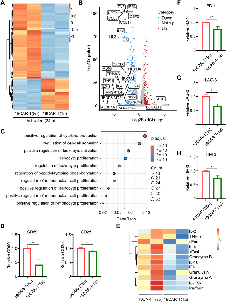
Methods: Utilizing plasmid construction and lentivirus generation, we get two different transmembrane CAR-T cells [19CAR-T(1a) and 19CAR-T(8α)]. Through co-culture with tumor cells, we evaluate CAR dynamic change, activation levels, exhaustion markers, mitochondrial function, and differentiation in both CAR-T cells. Furthermore, immunofluorescence microscopy analysis is performed to reveal the localization of internalized CAR molecules. RNA sequencing is used to detect the transcriptome of activated CAR-T cells. Finally, a mouse study is utilized to verify the anti-tumor efficacy of 19CAR-T(1a) cells in vivo.

Results: Our findings demonstrate that 19CAR-T(1a) has lower surface CAR expression, faster internalization, and a higher recycling rate compared to 19CAR-T(8α). Internalized 19CAR(1a) co-localizes more with early and recycling endosomes, and less with lysosomes than 19CAR(8α). These features result in lower activation levels, less cytokine release, and reduced exhaustion markers in 19CAR-T(1a). Furthermore, CAR-T cells with CD1a transmembrane domain also exhibit a superior anti-tumor ability and reduced exhaustion in vivo.

Conclusion: Overall, we demonstrate that the transmembrane domain plays a critical role in CAR-T cell function. An optimized transmembrane domain can alleviate cytokine release syndrome and reduce CAR-T cell exhaustion, providing a direction for CAR design to enhance CAR-T cell function.

Keywords: car-t; cytokine release; exhaustion; internalization; recycling; transmembrane domain.


Conflict of interest statement

The authors declare that the research was conducted in the absence of any commercial or financial relationships that could be construed as a potential conflict of interest.

Figures

Figure 1
Figure 1
CD1a transmembrane CAR-T cells exhibit lower surface CAR levels. (A) The schematic of CAR structure. (B) The positive rate of CAR-T cells is indicated by GFP and anti-FLAG antibody. (C) The relative total CAR on CAR-T cells is indicated by GFP expression. Normalized to 19CAR-T(8α) GFP MFI. (D) The surface CAR expression on CAR-T cells is indicated by anti-FLAG antibody. (E) The relative surface CAR expression on CAR-T cells is indicated by anti-FLAG antibody. Normalized to 19CAR-T(8α) surface CAR. (F) The total CAR expression on CAR-T cells was indicated by staining intracellular FLAG expression with anti-FLAG antibody. (G) The relative total CAR expression on CAR-T cells is indicated by staining intracellular FLAG expression with anti-FLAG antibody. Normalized to 19CAR-T(8α) total CAR. (H, I) CAR-T cells were co-cultured with SEM cells at 1:1, 1:2 or 1:4 for 4 h (H) or 24 h (I). The apoptosis of target cells was detected by using the Annexin V kit. Two-tailed Student t-test, *** for P < 0.001, **** for P < 0.0001, the ns indicate no significant difference. Error bars reflect ± SD of three independent experiments.
Figure 2
Figure 2
CD1a transmembrane CAR shows rapid internalization and recycling rate. (A) CAR-T cells were co-cultured with SEM cells at 1:1 for 1, 2, or 4 hThe surface CAR on CAR-T cells was detected by anti-FLAG antibody. Normalized to each CAR-T cell non-cocultured surface CAR, respectively. (B) Antibody-based assay for CAR internalization. (C) Brefeldin A-based assay for CAR internalization. CAR-T cells were treated with 10 μM Brefeldin A at 37°C for 0, 1, 2, or 4 h, then the surface CAR was detected by anti-FLAG antibody. Normalized to each CAR-T cell Brefeldin A non-treated surface CAR, respectively. (D) Antibody-based assay for CAR recycling. (E) CAR-T cells were co-cultured with SEM cells at 1:1 for 0, 1, 2, or 4 h The transferred surface CAR on SEM cells was detected by anti-FLAG antibody. (F) CAR-T cells were co-cultured with SEM cells at 1:1 for 0, 1, 2, or 4 h The transferred CD19 on CAR-T cells was detected by anti-CD19 antibody. (G, H) CAR-T cells (G) or CAR-T cells co-cultured with SEM at a 1:1 ratio (H) were treated with 50 µg/mL cycloheximide at 37°C for 0, 1, 2, or 4 h The total CAR was staining intracellular FLAG expression by anti-FLAG antibody after using BD Cytofix/Cytoperm Buffer System. The decrease in the percentage of total CAR on CAR-T cells was quantified by flow cytometry as an indication of degradation rate. Two-tailed Student t-test, * for P < 0.05, ** for P < 0.01, *** for P < 0.001, **** for P < 0.0001, the ns indicate no significant difference. Error bars reflect ± SD of three independent experiments.
Figure 3
Figure 3
Internalized CD1a transmembrane CAR co-localizes more with early and recycling endosomes. (A–C) CAR-T cells were co-cultured with SEM cells at 1:1 for 4 h, and the cells were stained with mouse anti-FLAG antibody in combination with one of the following rabbit antibodies: anti-human Rab5 (A), anti-human Rab11 (B), or anti-human LAMP1 (C). Then Goat anti-Mouse IgG (H+L) Secondary Antibody, DyLight™ 650 was used to stain mouse anti-FLAG antibody, and Goat anti-Rabbit IgG (H+L) Highly Cross-Adsorbed Secondary Antibody, Alexa Fluor™ 568 was used to stain rabbit anti- human Rab5, Rab11, or LAMP1. Positive CAR-T cells were identified by staining with the anti-FLAG antibody. The co-localization of CAR with Rab5, Rab11, or LAMP1 was indicated by the Pearson correlation coefficient, calculated by ImageJ. Ten cells with CAR and Rab5, Rab11, or LAMP1 positive, which were used to calculate the Pearson correlation coefficient were selected from three independent experiments. Two-tailed Student t-test, ** for P < 0.01. Error bars reflect ± SD.
Figure 4
Figure 4
CD1a transmembrane CAR-T cells exhibit low activation levels and reduced exhaustion markers. (A) CAR-T cells were sorted after co-culture with SEM cells at 1:1 for 24 h, and then RNA-seq was performed. The heatmap of DEGs between 19CAR-T(1a) and 19CAR-T(8α) selecting based on |log2FoldChange| > 1 and Q-value < 0.05 were shown. (B) The volcano plot of DEGs between 19CAR-T(1a) and 19CAR-T(8α). The cutoff was set based on |log2FoldChange| > 1 and Q-value < 0.05. Some significant up or down regulation genes were marked. (C) Pathway analysis by Gene Ontology (GO) shows the top 10 pathways of the Biological Process of downregulated DEGs. (D) CAR-T cells were co-cultured with SEM at 1:1 for 24 h Then CD69 and CD25 were detected as an indication of CAR-T activation. Normalized to 19CAR-T(8α) expression of CD69 or CD25. (E) CAR-T cells were co-cultured with SEM cells at a 1:1 ratio for 24 h, and the supernatant was collected for detection of cytokines by LEGENDplex™ Human CD8/NK Panel. (F, G) CAR-T cells were co-cultured with SEM cells at 1:1 for 3 days, and exhaustion markers on CAR-T cells were detected by anti-human CD279 (PD-1) antibody (F), anti-human CD223 (LAG-3) antibody (G), and anti-human CD366 (TIM-3) antibody (H). Normalized to 19CAR-T(8α) expression of exhaustion markers MFI. Two-tailed Student t-test, * for P < 0.05, ** for P < 0.01. Error bars reflect ± SD of three independent experiments. The RNA-seq analysis and CAR-T cytokines release detection were performed with two independent experiments.
Figure 5
Figure 5
CD1a transmembrane CAR-T cells differentiate more into memory T cells. (A) CAR-T or T cells co-cultured with SEM at a 1:1 ratio for 24 h, and 200 nM TMRE was used to detect CAR-T cell mitochondrial activity. Normalized to T cells MFI of TMRE. (B) CAR-T or T cells were co-cultured with SEM at a 1:1 ratio for 24 h, and 200 nM Mitotracker was used to detect CAR-T cell mitochondrial mass. Normalized to T cells MFI of Mitotracker. (C) CAR-T cells TMRE/Mitotracker MFI ratio, respectively. (D) CAR-T or T cells were co-cultured with SEM at a 1:1 ratio for 24 h, and 5 μM MitoSOX was used to detect CAR-T cell mitochondrial ROS. Normalized to T cells MFI of MitoSOX. (E) CAR-T cells MitoSOX/Mitotracker MFI ratio, respectively. (F, G) CAR-T cells were co-cultured with SEM cells at 1:1 for 3 days, and CD62L or CD45RA on CAR-T cells were detected by anti-human CD62L or anti-human CD45RA antibodies. The percentage of Tcm (CD62L+CD45RA-) (F) and Teff (CD62L-CD45RA+) (G) were shown. Two-tailed Student t-test, * for P < 0.05, ** for P < 0.01, *** for P < 0.001, **** for P < 0.0001, the ns indicate no significant difference. Error bars reflect ± SD of three independent experiments.
Figure 6
Figure 6
CD1a transmembrane CAR-T cells exhibit superior anti-tumor ability in vivo. (A) Schematic of mouse survival assay. (B) Representative bioluminescence images of tumor growth over time. (C) Total radiances post tumor cell engraftment of different treatment groups. Two-tailed Student t-test, * for P < 0.05, *** for P < 0.001, Error bars reflect ± SD of four mice. (D) The survival curve of mice post-transplantation was shown. Mouse survival was monitored regularly and statistically analyzed using a long-rank test, * for P < 0.05, ** for P < 0.01. (E) Schematic of CAR-T detection in vivo. (F) The PD-1 and LAG-3 on CAR-T cells from bone marrow were shown. Normalized to MFI of exhaustion markers expression on 19CAR-T(8α). Two-tailed Student t-test, *** for P < 0.001, **** for P < 0.0001, Error bars reflect ± SD of three mice.
Figure 7
Figure 7
The Schematic for the mechanism of dynamic change of CD1a transmembrane CAR. 19CAR-T(1a) has lower surface CAR expression than 19CAR-T(8α). Internalized 19CAR(1a) co-localizes with early and recycling endosomes more than 19CAR(8α) and co-localizes with lysosomes less than 19CAR(8α). This phenotype results in lower activation levels, less cytokine release, and reduced exhaustion markers in 19CAR-T(1a) cells.

Similar articles

Cited by

References

    1. Othman T, Logan AC, Muffly L, Leonard J, Park J, Shah B, et al. . The role of CAR T-cell therapy in relapsed/refractory adult B-ALL. J Natl Compr Canc Netw. (2024) 22(8):e247065. doi: 10.6004/jnccn.2024.7065 - DOI - PubMed
    1. Safarzadeh Kozani P, Safarzadeh Kozani P, Ahmadi Najafabadi M, Yousefi F, Mirarefin SMJ, Rahbarizadeh F. Recent advances in solid tumor CAR-T cell therapy: driving tumor cells from hero to zero? Front Immunol. (2022) 13:795164. doi: 10.3389/fimmu.2022.795164 - DOI - PMC - PubMed
    1. Xiao X, Huang S, Chen S, Wang Y, Sun Q, Xu X, et al. . Mechanisms of cytokine release syndrome and neurotoxicity of CAR T-cell therapy and associated prevention and management strategies. J Exp Clin Cancer Res. (2021) 40:367. doi: 10.1186/s13046-021-02148-6 - DOI - PMC - PubMed
    1. Freyer CW, Porter DL. Cytokine release syndrome and neurotoxicity following CAR T-cell therapy for hematologic Malignancies. J Allergy Clin Immunol. (2020) 146:940–8. doi: 10.1016/j.jaci.2020.07.025 - DOI - PubMed
    1. Posey AD, Jr., Young RM, June CH. Future perspectives on engineered T cells for cancer. Trends Cancer. (2024) 10:687–95. doi: 10.1016/j.trecan.2024.05.007 - DOI - PubMed

LinkOut - more resources