

Free glutaraldehyde gelatin microsphere loaded mesenchymal stem cells alleviate osteoarthritis by promoting Ext1 expression
- PMID: 40213660
- PMCID: PMC11980673
- DOI: 10.7150/thno.109468
Free glutaraldehyde gelatin microsphere loaded mesenchymal stem cells alleviate osteoarthritis by promoting Ext1 expression
Abstract
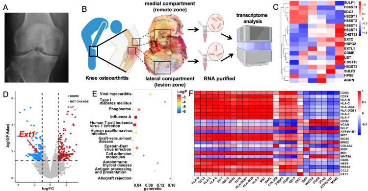
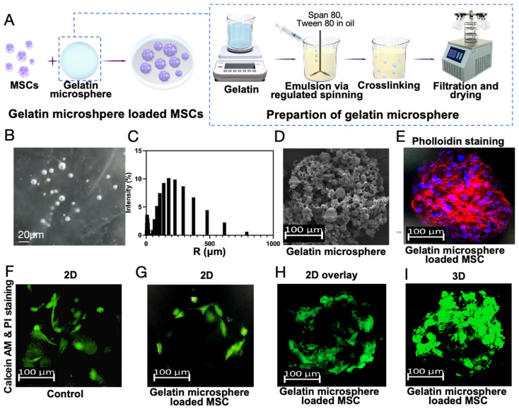
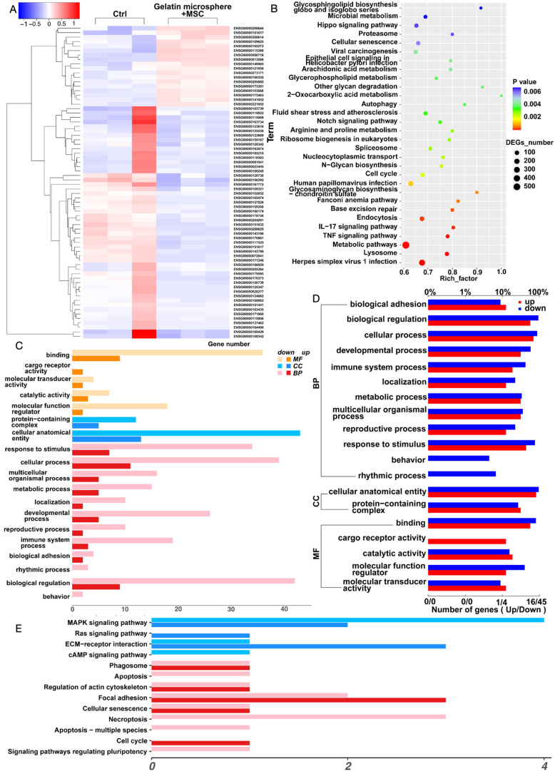
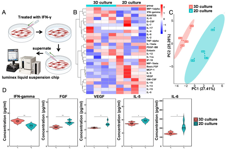
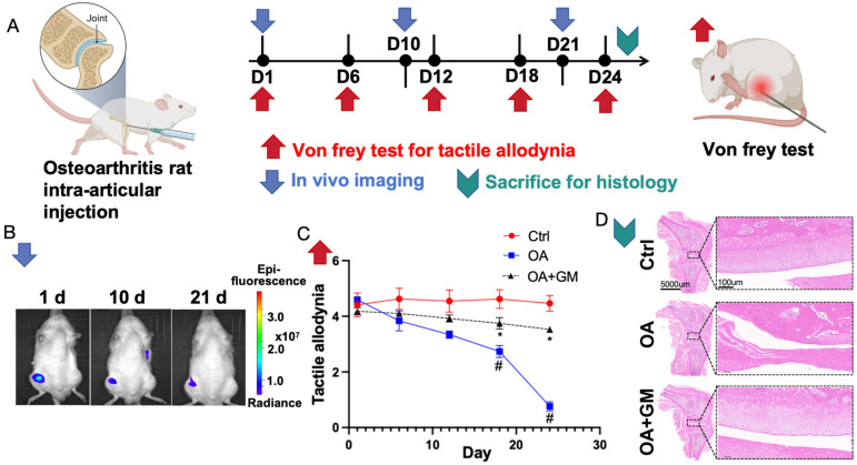
Rationale: Osteoarthritis (OA) is a chronic joint disorder with limited treatment efficacy, necessitating innovative therapeutic strategies. This study explores one-pot-synthesized gelatin microspheres devoid of glutaraldehyde as a novel biomaterial for OA management. Focusing on the Ext1 gene, critical for cartilage development and downregulated in OA, we investigated its restoration and immune regulation using gelatin microspheres cultured with mesenchymal stem cells (MSCs). Methods: OA patients undergoing knee replacement surgery have their lateral compartment (remote zone) and medial compartment (lesion zone) cartilage collected for transcriptomic testing. The differential gene Ext1 is identified, and the expression of immune regulatory genes is examined. MSCs were cultured with gelatin microspheres to evaluate their compatibility and ability to promote cell attachment. The effects of the gelatin microspheres on Ext1 gene overexpression, immune regulation, and OA symptom mitigation were investigated through in vitro and in vivo experiments. Results: OA patients exhibit decreased expression of the Ext1 gene in the medial compartment (lesion zone) cartilage area, accompanied by abnormal expression of immune regulatory genes. The study demonstrated that the gelatin microspheres exhibited excellent compatibility with MSCs and facilitated their attachment. Culturing MSCs with the microspheres led to enhanced overexpression of the Ext1 gene, which is crucial for cartilage growth and development. Additionally, the microspheres regulated immune responses, contributing to a reduction in OA symptoms. Conclusion: This study introduces an innovative therapeutic strategy for osteoarthritis using gelatin microspheres cultured with MSCs. By promoting Ext1 gene overexpression and regulating immune responses, these microspheres effectively mitigate OA symptoms. The findings highlight the potential of this biomaterial as a promising treatment option for OA.
Keywords: Ext1; Gelatin microsphere; Immune response; Mesenchymal stem cells; Osteoarthritis.
© The author(s).
Conflict of interest statement
Competing Interests: The authors have declared that no competing interest exists.
Figures

Similar articles
-
Injectable porous microspheres for articular cartilage regeneration through in situ stem cell recruitment and macrophage polarization.Acta Biomater. 2024 Sep 1;185:429-440. doi: 10.1016/j.actbio.2024.07.007. Epub 2024 Jul 10. Acta Biomater. 2024. PMID: 38997077
-
Transplantation of Gelatin Microspheres Loaded with Wharton's Jelly Derived Mesenchymal Stem Cells Facilitates Cartilage Repair in Mice.Tissue Eng Regen Med. 2024 Jan;21(1):171-183. doi: 10.1007/s13770-023-00574-5. Epub 2023 Sep 9. Tissue Eng Regen Med. 2024. PMID: 37688747 Free PMC article.
-
Regulation of fibroblast phenotype in osteoarthritis using CDKN1A-loaded copper sulfide nanoparticles delivered by mesenchymal stem cells.Am J Physiol Cell Physiol. 2025 Feb 1;328(2):C679-C698. doi: 10.1152/ajpcell.00573.2024. Epub 2025 Jan 16. Am J Physiol Cell Physiol. 2025. PMID: 39819042
-
Harnessing knee joint resident mesenchymal stem cells in cartilage tissue engineering.Acta Biomater. 2023 Sep 15;168:372-387. doi: 10.1016/j.actbio.2023.07.024. Epub 2023 Jul 21. Acta Biomater. 2023. PMID: 37481194 Review.
-
Mesenchymal stem cell-based treatment for cartilage defects in osteoarthritis.Mol Biol Rep. 2012 May;39(5):5683-9. doi: 10.1007/s11033-011-1376-z. Epub 2011 Dec 20. Mol Biol Rep. 2012. PMID: 22183306 Review.
References
-
- Yuan Y, Xu F, Cao Y, Xu L, Yu C, Yang F. et al. Iron Accumulation Leads to Bone Loss by Inducing Mesenchymal Stem Cell Apoptosis Through the Activation of Caspase3. Biol Trace Elem Res. 2019;187:434–441. - PubMed
-
- Xu X, Xu L, Xia J, Wen C, Liang Y, Zhang Y. Harnessing knee joint resident mesenchymal stem cells in cartilage tissue engineering. Acta Biomater. 2023;168:372–387. - PubMed
-
- McIntyre JA, Jones IA, Han B, Vangsness CT Jr. Intra-articular Mesenchymal Stem Cell Therapy for the Human Joint: A Systematic Review. Am J Sports Med. 2018;46:3550–3563. - PubMed
MeSH terms
Substances
LinkOut - more resources
Full Text Sources
Medical
Miscellaneous
