. 2025 Apr 1;152(7):dev204533.
doi: 10.1242/dev.204533. Epub 2025 Apr 11.

Targets of the transcription factor Six1 identify previously unreported candidate deafness genes

Affiliations

Targets of the transcription factor Six1 identify previously unreported candidate deafness genes

Ramya Ranganathan et al. Development. .

Abstract

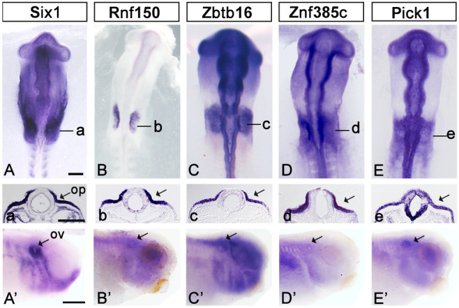
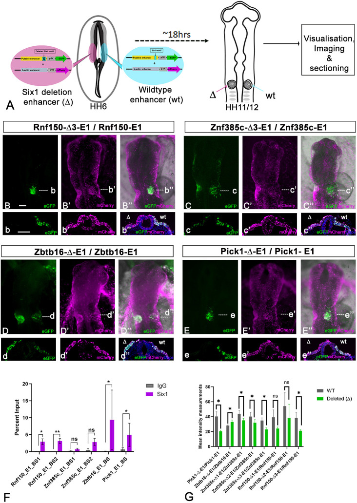
Branchio-otic (BOS) and branchio-oto-renal (BOR) syndromes are autosomal dominant disorders featuring multiple birth defects including ear, renal and branchial malformations. Mutations in the homeodomain transcription factor SIX1 and its co-factor EYA1 have been identified in about 50% of individuals with BOS or BOR, while causative mutations are unknown in the other half. We hypothesise that SIX1 target genes represent new BOS and BOR candidates. Using published transcriptomic and epigenomic data from chick ear progenitors, we first identify putative Six1 targets. Next, we provide evidence that Six1 directly regulates some of these candidates: Six1 binds to their enhancers, and functional experiments in Xenopus and chick confirm that Six1 controls their expression. Finally, we show that most putative chick Six1 targets are also expressed in the human developing ear and are associated with known deafness loci. Together, our results not only characterise the molecular mechanisms that mediate Six1 function in the developing ear, but also provide new candidates for human congenital deafness.

Keywords: Branchio-oto-renal Syndrome; Deafness; Ear development; Otic placode.


Conflict of interest statement

Competing interests The authors declare no competing or financial interests.

Figures

Fig. 1.
Fig. 1.
Expression of putative Six1 targets. (A-E′) In situ hybridisation for Six1 (A,A′), Rnf150 (B,B′), Zbtb16 (C,C′), Znf385c (D,D′) and Pick1 (E,E′) shows their expression in the chick otic placode (op) at HH10-11 (A-E) and in wild-type Xenopus laevis otic vesicles (ov) (A′-E′, arrows) at stages 30-32. Black lines in A-E indicate the level of sections shown in a-e. Arrows in a-e indicate the otic placode. Scale bars: 100 µm in A-E; 400 µm in A′-E′.
Fig. 2.
Fig. 2.
Six1 occupies and regulates enhancer activity in the chick otic placode. (A) Experimental strategy for bilateral electroporation of HH6/7 chick embryos with wild-type reporter constructs (right, blue) and eGFP reporters with the Six1 motif deleted (left, pink); each construct was co-electroporated with ubiquitously active mCherry. Embryos were assessed for enhancer activity at HH11-12. (B-B″) Rnf150-E1 activity in the otic placode with corresponding sections shown in b-b″; deletion of both Six1-binding sites (BSs) (Rnf150-Δ3-E1) leads to loss of enhancer activity (n=6). (C-C″) Znf385c-E1 activity in the otic placode with corresponding sections shown in c-c″; deletion of both Six1 motifs (Znf385c-Δ3-E1) leads to decreased enhancer activity (n=6). (D-D″) Zbtb16-E1 activity is moderate in the otic placode; corresponding sections shown in d-d″; Six1 motif deletion (Zbtb16-Δ-E1) leads to increased enhancer activity (n=5). (E-E″) Pick1-E1 activity in the otic placode with corresponding sections shown in e-e″; Six1 motif deletion (Pick1-Δ-E1) leads to reduced enhancer activity (n=4). Dashed white lines in B-E″ indicate the levels of sections shown in b-e″. Scale bars: 100 µm. (F) ChIP using Six1 and IgG control antibodies was performed on dissected chick otic placodes followed by qPCR for the enhancers indicated on the x-axis. *P≤0.05, **P≤0.01; ns, not significant; P≤0.09 for Znf385cBS1 and P≤0.08 for Znf385cBS2 (paired Student's t-test). Data are mean±s.e.m. (G) Quantification of GFP intensity as a proxy for enhancer activity in the otic placode before and after Six1 motif deletion. *P≤0.05 (paired Student's t-test). Data are mean±s.e.m.
Fig. 3.
Fig. 3.
Six1 is required for the expression of putative target genes. (A-D′) Rnf150 (A), Zbtb16 (B), Znf385c (C) and Pick1 (D) are expressed in the otic vesicle on the control side of Xenopus laevis larvae (A-D, arrows), but their expression is greatly reduced on the Six1 MO-injected side of the same embryo (A′-D′, arrows). (E-H) Injection of mRNA encoding EnR-Six1 into Xenopus laevis embryos leads to loss of the expression of all four genes; pink indicates lineage tracer demarking the injected side of the larva. (I-L) Six1 loss-of-function in Xenopus tropicalis F0 mutants created by CRISPR-Cas9 (Coppenrath et al., 2021) leads to loss of Rnf150 (n=15) (I), Zbtb16 (n=6) (J), Znf385c (n=12) (K) and Pick1 (n=16) (L) expression in the otic vesicle (arrows). Scale bars: in A, 400 µm for A,C-H,A′,Cʹ,D′; in B, 400 µm in B,Bʹ; in I, 200 µm for I-L.
Fig. 4.
Fig. 4.
Six1 regulates putative target gene expression in chick. (A) Experimental design: EnR-Six1 and eGFP vectors were electroporated on one side of HH6/7 embryos (pink) and control eGFP vectors (blue) on the other. Embryos were grown until they reached HH11-12, and processed for HCR in situ hybridisation and imaging. (B-E) Whole-mount HCR in situ hybridisation for Rnf150 (B), Zbtb16 (C), Znf385c (D) and Pick1 (E) in magenta; nuclei are stained using DAPI (blue). (F-I) Embryos in B-E with eGFP channel to visualise EnR-Six1 (left side of the embryo) and control GFP (right side of the embryo). The images shown in C,G and D,H represent the same embryo that was processed for double HCR in situ hybridisation. (J-Q) Higher magnifications of the otic placode territory of embryos shown in B-I. (R-Y) The outlined areas in B-E are imaged at a higher magnification and single confocal slices of these regions are shown. HCR signal is in magenta; GFP is in green. (Z-C′) Quantification of fluorescent gene expression signals on the control and experimental side. Dots indicate individual data points; data are mean±s.e.m.; *P<0.05, **P<0.01, ***P<0.001 (paired Student's t-test). Scale bars: 100 µm (bar in B applies to B-I; bar in J applies to J-Q; bar in R applies to R-Y).
Fig. 5.
Fig. 5.
Gene expression in human otic vesicles and candidate deafness genes. (A) Volcano plot showing genes enriched in the otic vesicle (OV) of CS14 human embryos compared to the dorsal hindbrain (DHB). (B) Venn diagram showing putative Six1 targets located in human deafness loci. (C) In situ hybridisation for human ZNF385C on CS14 sections through the otic vesicle (OV; arrow). (c) Higher magnification of the OV shown in C. (D,E) Section through the OV of a human CS14 embryo stained using ZBTB16 antibodies (green); nuclei are visualised with DAPI (blue). (d,e) Higher magnification of the OVs shown in D and E. Scale bars: 500 µm in C,D; 100 µm in c,d.

Similar articles

Cited by

References

    1. Alessi, D. R., Zhang, J., Khanna, A., Hochdörfer, T., Shang, Y. and Kahle, K. T. (2014). The WNK-SPAK/OSR1 pathway: master regulator of cation-chloride cotransporters. Sci. Signal. 7, re3. 10.1126/scisignal.2005365 - DOI - PubMed
    1. Barald, K. F. and Kelley, M. W. (2004). From placode to polarization: new tunes in inner ear development. Development 131, 4119-4130. 10.1242/dev.01339 - DOI - PubMed
    1. Basch, M. L., Brown, R. M., II, Jen, H. I. and Groves, A. K. (2016). Where hearing starts: the development of the mammalian cochlea. J. Anat. 228, 233-254. 10.1111/joa.12314 - DOI - PMC - PubMed
    1. Bok, J., Chang, W. and Wu, D. K. (2007). Patterning and morphogenesis of the vertebrate inner ear. Int. J. Dev. Biol. 51, 521-533. 10.1387/ijdb.072381jb - DOI - PubMed
    1. Breuskin, I., Bodson, M., Thelen, N., Thiry, M., Borgs, L., Nguyen, L., Lefebvre, P. P. and Malgrange, B. (2009). Sox10 promotes the survival of cochlear progenitors during the establishment of the organ of Corti. Dev. Biol. 335, 327-339. 10.1016/j.ydbio.2009.09.007 - DOI - PubMed

MeSH terms