

Reversible High-Affinity Binding of Coagulation Factor Xa to Zeolites Induces Accelerated Blood Coagulation
- PMID: 40213827
- PMCID: PMC12165020
- DOI: 10.1002/advs.202417099
Reversible High-Affinity Binding of Coagulation Factor Xa to Zeolites Induces Accelerated Blood Coagulation
Abstract
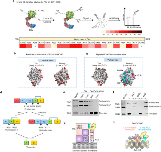
Zeolite is recognized as an essential hemostatic material for controlling massive bleeding. Elucidating the procoagulant mechanism of zeolite is critically important, as it will facilitate the rational design of more effective zeolite-based hemostatic materials. In this study, it is discovered an extremely strong, calcium-dependent interaction between coagulation factor Xa (FXa) and zeolite-termed target-specific biorecognition-that mimics the FXa/factor Va (FXa/FVa) interface formed during the natural coagulation cascade. This interaction alters the prothrombin activation pathway to a more efficient mechanism, significantly amplifying FXa activity. Notably, the complex structure and FXa activity can be reversibly modulated through Na+/Ca2+ ion exchange of zeolites, offering a novel strategy for dynamically tuning enzymatic activity. Furthermore, this protein-zeolite based biorecognition system, mediated by reversible interactions, represents a promising biomimetic platform for regulating protein bioactivity in cell-free applications, extending its utility beyond hemostatic material development.
Keywords: biorecognition; coagulation; factor Xa; protease; zeolite.
© 2025 The Author(s). Advanced Science published by Wiley‐VCH GmbH.
Conflict of interest statement
The authors declare no conflict of interest.
Figures


Similar articles
-
Innovative Three-Step Microwave-Promoted Synthesis of N-Propargyltetrahydroquinoline and 1,2,3-Triazole Derivatives as a Potential Factor Xa (FXa) Inhibitors: Drug Design, Synthesis, and Biological Evaluation.Molecules. 2020 Jan 23;25(3):491. doi: 10.3390/molecules25030491. Molecules. 2020. PMID: 31979319 Free PMC article.
-
Understanding the anchoring interaction of coagulation factor Va light chain on zeolites: A molecular dynamics study.J Colloid Interface Sci. 2022 Feb 15;608(Pt 1):435-445. doi: 10.1016/j.jcis.2021.09.129. Epub 2021 Sep 24. J Colloid Interface Sci. 2022. PMID: 34626987
-
Factor Va-factor Xa interactions: molecular sites involved in enzyme:cofactor assembly.Scand J Clin Lab Invest Suppl. 2002;237:5-11. doi: 10.1080/003655102762377439. Scand J Clin Lab Invest Suppl. 2002. PMID: 12570161 Review.
-
Synergistic Procoagulant Mechanism and Application of Kaolin-Zeolite Composite Hemostat for Effective Hemorrhage Control.ACS Appl Mater Interfaces. 2024 Sep 18;16(37):49186-49196. doi: 10.1021/acsami.4c12623. Epub 2024 Sep 10. ACS Appl Mater Interfaces. 2024. PMID: 39252609
-
Blood coagulation factor Xa as an emerging drug target.Expert Opin Ther Targets. 2011 Mar;15(3):341-9. doi: 10.1517/14728222.2011.553608. Epub 2011 Jan 21. Expert Opin Ther Targets. 2011. PMID: 21250873 Review.
References
-
- Lozano R., Naghavi M., Foreman K., Lim S., Shibuya K., Aboyans V., Abraham J., Adair T., Aggarwal R., Ahn S. Y., AlMazroa M. A., Alvarado M., Anderson H. R., Anderson L. M., Andrews K. G., Atkinson C., Baddour L. M., Barker‐Collo S., Bartels D. H., Bell M. L., Benjamin E. J., Bennett D., Bhalla K., Bikbov B., Abdulhak A. B., Birbeck G., Blyth F., Bolliger I., Boufous S., Bucello C., et al., Lancet 2012, 380, 2095.
-
- Zhang S., Li J., Chen S., Zhang X., Ma J., He J., Carbohydr. Polym. 2020, 230, 115585. - PubMed
-
- Masoudi M., Wiseman J., Wiseman S. M., Expert. Rev. Med. Devices. 2023, 20, 741. - PubMed
-
- Malette W. G., Quigley H. J., Gaines R. D., Johnson N. D., Rainer W. G., Ann. Thorac. Surg. 1983, 36, 55. - PubMed
-
- Azargoon H., Williams B. J., Solomon E. S., Kessler H. P., He J., Spears R., J. Endod. 2011, 37, 807. - PubMed
MeSH terms
Substances
Grants and funding
LinkOut - more resources
Full Text Sources
Miscellaneous