. 2025 Jan 2;19(1):wraf066.
doi: 10.1093/ismejo/wraf066.

Episomal virus maintenance enables bacterial population recovery from infection and promotes virus-bacterial coexistence

Affiliations

Episomal virus maintenance enables bacterial population recovery from infection and promotes virus-bacterial coexistence

Rodrigo Sanchez-Martinez et al. ISME J. .

Abstract

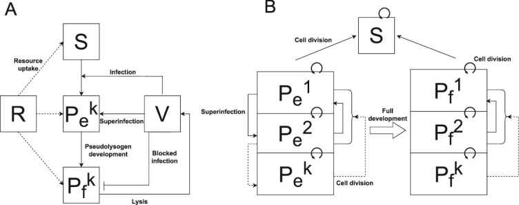
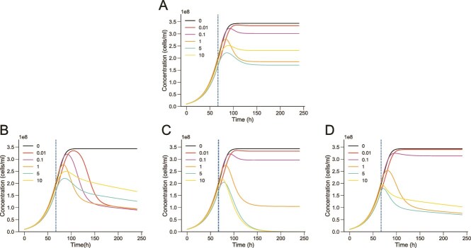
Viruses are ubiquitous in aquatic environments with total densities of virus-like particles often exceeding 107/ml in surface marine oligotrophic waters. Hypersaline environments harbor elevated prokaryotic population densities of 108/ml that coexist with viruses at even higher densities, approaching 1010/ml. The presence of high densities of microbial populations and viruses challenge traditional explanations of top-down control exerted by viruses. At close to saturation salinities, prokaryotic populations are dominated by Archaea and the bacterial genus Salinibacter. In this work we examine the episomal maintenance of a virus within a Salinibacter ruber host. We found that infected cultures of Sal. ruber M1 developed a population-level resistance and underwent systematic and reproducible recovery post infection that was counter-intuitively dependent on the multiplicity of infection, where higher viral pressures led to better host outcomes. Furthermore, we developed a nonlinear population dynamics model that successfully reproduced the qualitative features of the recovery. Together, experiments and models suggest that episomal virus maintenance and lysis inhibition enable host-virus co-existence at high viral densities. Our results emphasize the ecological importance of exploring a spectrum of viral infection strategies beyond the conventional binary of lysis or lysogeny.

Keywords: Salinibacter; acquired phage resistance; modeling; pseudolysogeny; virus–host interactions.


Conflict of interest statement

None declared.

Figures

Figure 1
Figure 1
Infection experiments show a reproducible recovery after viral lysis. Infection experiments of Sal. ruber strain M1 with the EM1 virus were performed using two different MOIs. Black lines represent uninfected control and red (A, MOI = 0.01) and purple (B, MOI = 0.1) lines represent virus-infected cultures. Each of the individual replicates used are shown as dotted lines. The dots show the mean and the vertical bars the standard error. The experiments were performed with 3 replicates for MOI 0.01 and 10 replicates for MOI 0.1 for both the control and infected cultures. Viruses were mixed with the host at time = 0 h.
Figure 2
Figure 2
The EM1 virus establishes a pseudolysogenic state in Sal. ruber M1. (A) Three randomly selected cultures of the 20 colonies of CF, PCR positive for the EM1 virus, that we named 1R, 2R, and 3R, were stained with SYBR gold and observed under epifluorescence microscope to test the presence of extracellular viruses. No extracellular viruses were seen in any of them. Thirty fields were examined per sample. The photos shown here are a representation of each sample. Scale bar: 10 μm. (B) A PFGE was performed (left) comparing initial cells (C0) with final colony 1R (lanes 3 and 4 of the gel, respectively). The DNA of the virus was also loaded as a control (lane 5). The PFGE showed a band in 1R with the size of EM1 (35 kb) which was not found in C0. A Southern blot (right) with a labeled probe targeting the genome of EM1 confirmed that this band belonged to the virus. Both the PFGE gel and the southern blot images were cropped for visualization purposes, as intermediate lanes contained samples from experiments conducted in the lab unrelated to this work.
Figure 3
Figure 3
Viral acquisition protects Sal. ruber M1 from further infection and has MOI dependent properties. (A) Growth curves (performed in triplicate) of Sal. ruber M1 wild-type (black dotted line) and pseudolysogen 1R (blue line) were performed and their growth dynamics were found to have no statistical differences (Modeling supplementary information: Sal. ruber M1 wild-type and 1R (pseudolysogen) growth comparison). (B) Susceptibility to the virus was assessed using a spot test. Sal. ruber M1 wild-type (left) and pseudolysogen 1R (right) were exposed to 3 μl of the EM1 virus tittered at 1010 PFUs/ml (left part of each plate) and diluted 102, 104, and 106 (left to right) times. (C) An adsorption assay of the EM1 virus to Sal. ruber M1 wild-type (black dotted line) and pseudolysogen 1R (blue line) was carried out with a MOI of 0.01, quantifying the quantity of extracellular viruses through PFU/ml for 120 min. The experiment was also carried out with the virus alone as a decay control (gray line). The experiment was performed by triplicate. Error bars represent the standard error. (D) Infection curves of Sal. ruber M1 and the EM1 virus were performed at different MOI. The M1 cultures were infected at 66 h, during the exponential growth phase. The black dotted line represents the non-infected control, and the different colors indicate the MOI, as is indicated in the legend. The experiment results exhibit three key features (see text).
Figure 4
Figure 4
Schematic of nonlinear dynamics population model. (A) Schematic representation of the ODE model, illustrating the resource level and denotes the free virus concentration. The host population is split into three classes: sensitive, early pseudolysogen, and fully developed pseudolysogen, where k is the number of viral genome copies within the host cell. (B) Dynamics of pseudolysogen growth and superinfection assuming primary infection has already occurred (primary infection: S → Pe1). Early pseudolysogens get superinfected causing → transitions, whereas fully developed pseudolysogens are resistant to superinfection at the surface level (denoted by “blocked infection” arrow). Pseudolysogen cell division for both early and fully developed pseudolysogens randomly splits the viral genome of the parent, causing → transitions where k’ is a randomly chosen positive integer that is ≤ k (P0 is virus-free and is therefore equivalent to S). Early stage pseudolysogens eventually transition into fully developed pseudolysogens that still cell divide but are resistant to superinfection.
Figure 5
Figure 5
Simulated population dynamics recapitulate experimental features through implementation of lysis inhibition and pseudolysogenic cell division. (A) Simulated host population dynamics (using the ODE model proposed (Modeling supplementary information: Main model) to successfully recapitulate the experimental features highlighted in Fig. 3D. Subfigures B, C, and D graphically show how decoupling features from the model prevents simulations from capturing experimental features. (B) Simulated host population dynamics with lysis rates independent of resource levels. (C) Simulated host population dynamics with lysis rates independent of cellular MOI. (D) Simulated host population dynamics without pseudolysogen cell division.

References

    1. Shiklomanov IA. World Water Resources: A New Appraisal and Assessment for the 21st Century. Paris: UNESCO-IHP, 1998.
    1. Mutlu MB, Martínez-García M, Santos F. et al. Prokaryotic diversity in Tuz Lake, a hypersaline environment in inland Turkey. FEMS Microbiol Ecol 2008;65:474–83. 10.1111/j.1574-6941.2008.00510.x - DOI - PubMed
    1. Ramos-Barbero MD, Martínez JM, Almansa C. et al. Prokaryotic and viral community structure in the singular chaotropic salt Lake Salar de Uyuni. Environ Microbiol 2019;21:2029–42. 10.1111/1462-2920.14549 - DOI - PubMed
    1. Boujelben I, Yarza P, Almansa C. et al. Virioplankton community structure in Tunisian solar salterns. Appl Environ Microbiol 2012;78:7429–37. - PMC - PubMed
    1. Ventosa A. Unusual Micro-Organisms from Unusual Habitats: Hypersaline Environments. Cambridge: Cambridge University Press, 2006.. 10.1017/CBO9780511754913.015 223 54. - DOI

MeSH terms

LinkOut - more resources