. 2025 May 27;10(5):e0096124.
doi: 10.1128/msphere.00961-24. Epub 2025 Apr 11.

Toward a deeper understanding of dengue: novel method for quantification and isolation of envelope protein epitope-specific antibodies

Affiliations

Toward a deeper understanding of dengue: novel method for quantification and isolation of envelope protein epitope-specific antibodies

Sokchea Lay et al. mSphere. .

Abstract

The dengue viruses (DENV) envelope (E) protein is the main target of the antibody (Ab) response. Abs target different epitopes on the E-protein, including sE-dimer, E domain III (EDIII), and fusion loop (FL). Anti-EDIII Abs are mainly serotype-specific, whereas anti-FL Abs can induce antibody-dependent enhancement (ADE) in vitro. Abs targeting sE-dimer epitopes can cross-neutralize different DENV serotypes. However, the involvement of each Ab subset in disease pathogenicity and/or protection remains unclear. We aimed to optimize the quantification and purification of DENV E-protein epitope-specific Abs from human samples. C-terminal biotinylated DENV2 E recombinant proteins (EDIII, soluble E [sE], and sE-dimer) were coupled to color-coded magnetic microspheres for a multiplex immunoassay (MIA), testing different antigen concentrations. Assay performance was evaluated using well-characterized anti-DENV monoclonal antibodies (mAbs) and total IgG from DENV seronegative and seropositive human plasma. Specific FL epitopes were blocked with mouse mAb clone 4G2 to quantify anti-FL- and sE-dimer-specific Abs, measuring antigen-antibody reactions as median fluorescence intensity (MFI). For isolation of E-protein epitope-specific antibodies, sE-proteins were conjugated to streptavidin resin beads. Total IgG from human plasma was incubated with immobilized EDIII to elute anti-EDIII Abs. The flow-through was incubated with sE-dimer resin beads to elute sE-dimer specific Ab enriched fraction, and the flow-through was applied to immobilized sE to elute anti-FL Abs. In conclusion, we have developed a serological assay to detect E-protein epitope-specific Abs in DENV-infected humans. Additionally, we successfully isolated anti-EDIII, anti-FL, and an enriched fraction of sE-dimer specific Abs from human samples.IMPORTANCEThe development of effective dengue virus (DENV) vaccines has been hampered by limited insights into the immunological mechanisms of protection. Our study addresses this gap by introducing a refined multiplex microsphere-based immunoassay (MIA) to quantify and isolate antibodies (Abs) targeting specific E-protein epitopes, such as E domain III (EDIII), the fusion loop (FL), and the sE-dimer specific Abs. This method provides detailed epitope-specific Ab profiling with high sensitivity and requires minimal sample volumes. The ability to isolate specific Ab subsets from human plasma also enables detailed investigations into their roles in protection or pathogenesis, paving the way for more effective dengue interventions.

Keywords: antibody response to dengue; antibody-dependent enhancement; dengue virus; flavivirus anti-envelope antibodies.


Conflict of interest statement

The authors declare no conflict of interest.

Figures

Fig 1
Fig 1
DENV E-protein domains and antibody response. (A) Structural organization of DENV E protein (PDB:7A37) in soluble E (sE)-dimeric and sE-monomeric form. Each monomer consists of three ecto-domains: EDI (red), EDII (yellow), EDIII (blue), and fusion loop (FL) (green) (3, 4). (B) Antibodies targeting different epitopes on DENV E-protein: anti-EDIII Abs (blue) bind to both sE-monomer and sE-dimer due to the availability of the EDIII epitope on both forms. Anti-FL Abs (green) bind only to sE-monomer as the FL is not exposed in the sE-dimer. sE-dimer specific Abs (orange) bind to quaternary epitopes and hence only bind to sE-dimer protein.
Fig 2
Fig 2
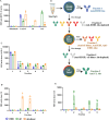
Validation of the coupling of DENV E recombinant proteins to the microsphere beads. (A) Scheme of MIA to detect anti-DENV E antibodies in human plasma. (B) Anti-DENV E mAb pool (containing mAb clone C8, 1A1D-2, 1A5) was serially diluted and tested for binding to EDIII (blue line), sE (green line), and sE-dimer (orange line) coupled beads. (C) Site-specific-biotinylated EDIII proteins were loaded onto streptavidin-coupled beads to generate a linker. EDIII-coupled beads with (dashed line) and without (solid line) linker were compared to detect anti-EDIII Abs in the anti-DENV E mAbs pool. Mean ± standard error of the mean (s.e.m). are shown of two independent experiments. To determine the threshold (D–F), three DENV IgG seropositive (red lines) and three DENV IgG seronegative (black lines) human plasma samples were used to determine the positivity threshold of anti-EDIII, anti-sE, and total anti-sE-dimer Abs. All data were expressed as MFI per 50 beads.
Fig 3
Fig 3
Detection of E-protein epitope-specific mAbs by MIA. (A) Rituximab, an anti-CD20 human mAb, was used as a negative control, and well-characterized anti-DENV E (B) mAb clone C8, (C) mAb clone C10, (D) mAb clone 1A1D-2, (E) FL-specific mAb clone 1A5, and (F) FL-mAb clone 4G2 were used to confirm binding specificity. (G) To determine anti-4G2-like Abs, mouse mAb clone 4G2 was used at a concentration of 10 µg/mL to block the FL epitope in sE. Mean ± standard error of the mean (s.e.m). are shown of two independent experiments.
Fig 4
Fig 4
Detection of E-protein epitope-specific antibodies in human plasma by MIA. Detection of E-protein epitope-specific Abs using MIA in plasma of dengue fever patients (n = 25) obtained 4-6 days after onset of symptoms. All patients had confirmed secondary infection with DENV2. (A) The binding of Abs against EDIII (blue dots), sE (green dots), total sE-dimer (orange dots), and sE pre-blocked with mouse mAb clone 4G2 (green circles) were reported as MFI. (B) 4G2-like Abs (green triangles) are measured by subtracting the MFI signal of antibodies bound to sE pre-incubated with mouse mAb clone 4G2 from the total signal of antibodies bound to sE. sE-dimer specific Abs (orange triangles) were quantified by subtracting the MFI signal of antibodies bound to sE pre-incubated with mouse mAb clone 4G2 from the antibodies bound to sE-dimer protein. The data is shown as ∆MFI IgG. Each dot/circle/triangle represents an individual, and whiskers show maximum, minimum, and mean values. The dotted red line represents the threshold for positivity, The Wilcoxon test was used to compare two groups, while the Friedman test was used to compare multiple groups (*P < 0.05; **P < 0.01, ***P < 0.001; ****P < 0.0001).
Fig 5
Fig 5
Confirmation of purification of E-protein epitope-specific Abs using mAbs. (A) The binding properties of mAbs (Rituximab, 1A1D-2, C8, and 1A5) before isolation were assessed against DENV E-proteins. (B) Isolation strategy. C-terminal biotinylated EDIII, sE, and sE-dimer proteins from DENV2 were coupled to streptavidin-resin beads for isolating E-protein epitope-specific Abs. Ab fractions were selected based on their specificity, employing both negative and positive selection techniques. (C–E) The purity of each fraction obtained from the isolation of anti-EDIII clone 1A1D-2 (C), anti-EDE specific mAb clone C8 (D), and anti-FL mAb clone 1A5 (E) was evaluated by MIA. Blue indicates the MFI of binding to the EDIII-coupled beads, green indicates the MFI of binding to the sE-coupled beads, and orange indicates MFI of binding to the sE-dimer-coupled beads. Mean ± standard error of the mean (s.e.m) are shown of two independent experiments.
Fig 6
Fig 6
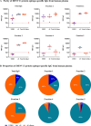
Purification of E-protein epitope-specific Abs from human plasma. Total IgG isolated from five DENV2 seropositive individuals was subjected to the Ab isolation strategy. (A) The IgG binding properties in each sample pre- and post-isolation were tested against all DENV E-proteins. Each dot represents an individual sample and is reported as MFI. Whiskers show max, min and mean value. The dotted red line represents the threshold for positivity. (B) Proportion of each E-protein epitope-specific Ab subset was calculated using the following formula: 100 × (MFI EDIII, sE, or total sE-dimer)/(MFI EDIII + sE + total sE-dimer).

References

    1. Paz-Bailey G, Adams LE, Deen J, Anderson KB, Katzelnick LC. 2024. Dengue. Lancet 403:667–682. doi:10.1016/S0140-6736(23)02576-X - DOI - PubMed
    1. Murugesan A, Manoharan M. 2020. Dengue virus, p 281–359. In Emerging and reemerging viral pathogens. 10.1016/B978-0-12-819400-3.00016-8. - DOI
    1. Rey FA, Stiasny K, Heinz FX. 2017. Flavivirus structural heterogeneity: implications for cell entry. Curr Opin Virol 24:132–139. doi:10.1016/j.coviro.2017.06.009 - DOI - PMC - PubMed
    1. Sevvana M, Kuhn RJ. 2020. Mapping the diverse structural landscape of the flavivirus antibody repertoire. Curr Opin Virol 45:51–64. doi:10.1016/j.coviro.2020.07.006 - DOI - PMC - PubMed
    1. Rey FA, Stiasny K, Vaney M-C, Dellarole M, Heinz FX. 2018. The bright and the dark side of human antibody responses to flaviviruses: lessons for vaccine design. EMBO Rep 19:206–224. doi:10.15252/embr.201745302 - DOI - PMC - PubMed

LinkOut - more resources