Review
. 2025 Oct 1;105(4):1947-1988.
doi: 10.1152/physrev.00010.2024. Epub 2025 Apr 11.

UTERINE FIBROIDS

Affiliations
Review

UTERINE FIBROIDS

Serdar E Bulun et al. Physiol Rev. .

Abstract

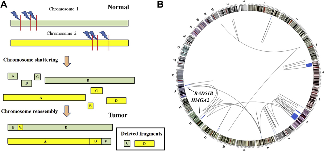
Uterine fibroids (leiomyomas), the most common tumors in women and those assigned female at birth, originate from myometrial smooth muscle cells and cause heavy menstrual bleeding, anemia, pelvic discomfort, pregnancy loss, and obstruction of labor in approximately a quarter of reproductive-age women. During each ovulatory cycle, the myometrium responds to the ovarian steroids, estradiol, and progesterone, via increased tissue stem cell proliferation to prepare for impending pregnancy, during which a somatic mutation may arise to initiate a tumor. Both the mutated smooth muscle cells and the adjacent tumor-associated fibroblasts lay down excessive quantities of extracellular matrix, providing a unique feature that led to naming these tumors "fibroids." The most common somatic mutations in fibroids affect the Mediator complex subunit 12 (MED12; 77%) and high-mobility group AT-hook 2/1 (HMGA2/1; 10%) genes. Heterozygous mutations in MED12, a chromatin-associated protein, disrupt the attached CDK8 kinase module in the Mediator complex. MED12 mutations are associated with increased genomic instability, altered chromatin landscape and enhancer engagement, and increased responsiveness to progesterone. Progesterone and a small stem cell population in a fibroid are essential for tumor survival and growth. Progesterone, via its receptors in differentiated fibroid cell populations, activates the production of Wingless-type MMTV integration site family (WNT) ligands, cytokines, and other growth substances to act on adjacent stem cells in a paracrine fashion to support tumor growth. Suppression of estrogen or progesterone production and progesterone antagonists have been used for temporary shrinkage of these tumors and symptom relief. Here we provide an overview of these mechanisms and future approaches for prevention and medical management of uterine fibroids.

Keywords: HMGA2; MED12; progesterone; stem cell; uterine fibroid.


Figures

Figure 1.
Figure 1.. Pathogenesis of a uterine fibroid.
Mutation-free myometrial tissue and leiomyoma (fibroid) tissue harbor distinct populations of cells with the capacity for regeneration and proliferation. These are called tissue progenitor or stem cells. A. Under physiological circumstances, estrogen and progesterone stimulate the proliferation of a stem-cell population in the myometrium. This repetitious and physiological phenomenon is necessary for the striking growth of the myometrium during pregnancy. Compared with the stem cells, mature myometrial cells express extraordinarily higher levels of the nuclear receptors, estrogen receptor α (ERα) and progesterone receptor (PGR). Estrogen- and progesterone- related cell proliferation is predominantly mediated by the ERα and PGR that reside in mature myometrial cells. Paracrine factors released by mature cells may act on stem cells to induce their regeneration and proliferation. B. A mutation that affects MED12 or HMGA2 fundamentally alters a myometrial stem cell and converts it into a fibroid stem cell. This tumor stem cell regenerates and proliferates in an unrestrained manner and eventually differentiates into a mature fibroid smooth-muscle cell. ERα and PGR are present largely in differentiated fibroid cells and convey estrogen or progestogen action to stem cells through paracrine signals. C. Extracellular-matrix that is laid down via various cell types in the heterogeneous fibroid tissue significantly contributes to tumor expansion. Therefore, the originally mutated fibroid stem cell has the capacity to construct a clinically recognizable fibroid tumor, which develops and grows within the myometrium. (Reproduced with permission from Bulun SE. Uterine Fibroids. N Engl J Med 2013;369:1344–55. Copyright © 2013 Massachusetts Medical Society.) (9)
Figure 2.
Figure 2.. Distribution of tumor driver mutations in a cohort of 2,263 fibroids from 726 in the Finland Myoma Study.
OM, other member (of the genes that encode the SRCAP complex proteins). (Reproduced with permission from Berta DG, et al. Deficient H2A.Z deposition is associated with genesis of uterine leiomyoma. Nature. 2021;596:398–403. Copyright © 2021 Springer Nature Limited.) (18)
Figure 3.
Figure 3.. Usual-type leiomyoma with MED12 mutation.
A. A hysterectomy specimen with multiple intrauterine leiomyomas. B. Usual-type leiomyoma with leiomyoma smooth muscle cells with uniform-appearing nuclei without atypia and abundant extracellular matrix bundles. C. Usual-type leiomyoma with a MED12 mutation consists of thick bundles of tumor-smooth muscle cells and tumor-associated fibroblasts.
Figure 4.
Figure 4.. Leiomyoma with HMGA2 overexpression.
A. A bivalved hysterectomy specimen containing two large lobulated-appearing leiomyomas with HMGA2 overexpression. B. These tumors are highly cellular with a trabecular organization of smooth muscle cells separated by modest amounts of extracellular matrix. In contrast to the MED12-mutant fibroid in Fig 3, a conspicuous fibroblast cell population is not visible. C. Leiomyoma smooth muscle cells are strongly immunoreactive for HMGA2 (brown stain).
Figure 5.
Figure 5.. FH-deficient leiomyoma.
A. These tumors can present as the usual type leiomyoma smooth muscle cells showing uniform-appearing nuclei without atypia and moderate extracellular matrix. B. FH-deficient leiomyoma may also contain neoplastic smooth muscle cells with nuclear atypia. C. Immunoreactive FH (brown stain) is lacking in the majority of the smooth muscle cells. D. Increased quantities of immunoreactive 2-Succinocysteine (2SC, brown stain) that accumulates in FH-deficient leiomyomas. 2SC has been used a marker for FH-deficient fibroids. Owing to elevated levels of fumarate that alter the cysteine residues in a number of proteins, increased succination causes overproduction of 2SC.
Figure 6.
Figure 6.. A. Leiomyoma with bizarre nuclei.
This variant of uterine leiomyoma displays remarkable nuclear atypia. There is also abundant extracellular matrix. B. Leiomyosarcoma. This malignant tumor also contains pleomorphic nuclei with atypia and hypercellular with minimal extracellular matrix. What separates a leiomyosarcoma from a leiomyoma with bizarre nuclei are the high number of mitotic figures in the former (arrows).
Figure 7.
Figure 7.. Paracrine interactions between LM tissue cell populations.
Sorting of leiomyoma cells based on the cell surface antigen CD34 and CD49b generated 3 cell populations: +/+ cells make up 5% of the tumor cell population and act as the progenitor/stem cells with self-renewal and high proliferation capabilities; +/+ cells express very low levels of estrogen receptor-a (ESR1) and progesterone receptor (PGR). +/− cells represent an intermediately differentiated population (7%) and express high levels of ESR1 and PGR. Finally −/− cell population (88%) possibly represents the terminally differentiated cells with minimal proliferative capacity and high steroid receptor expression. The intermediate cells express the highest levels of the paracrine factors WNT4, RANKL, IGF1 and IGF-2, whereas the corresponding receptors of these ligands, FZL6, RANK and IR-A are highly expressed in the stem cell population. Thus, the intermediately differentiated cell population is likely to be physically proximal to the stem cells and essential for providing estradiol (E2) plus progesterone (P4)-induced paracrine support for them.
Figure 8.
Figure 8.. Role of leiomyoma stem cells.
Human CD34CD49b cells isolated from a fibroid comprise 88% of the tumor cells; they are well differentiated with very low proliferative activity. When grafted under the mouse kidney capsule, CD34CD49b cells produce small tumors with low cellularity and high collagen content. On the other hand, human fibroid CD34+CD49b+ cells comprise 5% of a fibroid tumor and demonstrate self-renewal capacity and high proliferative activity. Grafts of these leiomyoma stem cells produce much larger tumors. (Modified with permission from Yin P, et al., Human uterine leiomyoma stem/progenitor cells expressing CD34 and CD49b initiate tumors in vivo. 2015; J Clin Endocrinol Metab. 100:E601–E606. Copyright © 2015 by the Endocrine Society.) (45)
Figure 9.
Figure 9.. Human equivalent of a heterozygous MED12 mutation in mouse uterus.
A. A subset of uterine cells conditionally expresses the mutant (mt) Med12 (c.131G>A) from an autosomal locus (Amt/+), while X chromosome–derived wild-type Med12 will either be conditionally excised (XcKO) or silenced by X chromosome inactivation indicated by red color. B. mRNA (cDNA) from mouse uterine tumor demonstrating predominant presence of the mutant c.131G>A variant (green chromatogram peak, black arrow) in the absence of the WT Med12 allele, consistent with human leiomyomas carrying MED12 mutations. C. 18-week-old multiparous Med12fl/+ Med12Rmt/+Amhr2-Cre uterus exhibiting a prominent fibroid-like a tumor outlined by dotted lines. D. Histological sections showed a leiomyoma-like tumor nodule originated from the uterine smooth muscle layer. E. A higher-magnification image of the black-box from D demonstrates the neoplastic smooth muscle cells and with areas of ECM deposition. F. 24-week-old Med12fl/+ Med12Rmt/+ Amhr2-Cre multiparous female uterus showing multiple tumors (white arrows). G. Nodular tumors from F are outlined by black dotted lines, and the black box, shown at higher magnification in H, contains sheets of neoplastic smooth muscle cells separated by ECM deposits. Note that only multiparous mice developed these tumors, suggesting the essential roles of the ovarian hormones estradiol and progesterone for LM formation. LM, leiomyoma; ES, endometrial stroma; MY, myometrium. (Reproduced with permission from Mittal P, et al. Med12 gain-of-function mutation causes leiomyomas and genomic instability. J Clin Invest. 2015;125(8):3280–4. Copyright © 2015, American Society for Clinical Investigation) (125)
Figure 10.
Figure 10.. Complex chromosomal rearrangements in uterine fibroids.
A. Chromothripsis is a biological event, whereby one or more chromosomes are smashed to pieces and randomly reconstructed. This disruption frequently results in the loss of DNA fragments and the formation of highly complex chromosomes. B. The picture depicts rearrangements involving many chromosomes in a single uterine fibroid tissue. Lines between and within chromosomes indicate exchanges of DNA fragments between various chromosomes, whereas the blue boxes indicate deleted DNA material. Chromothripsis in this fibroid tissue affected chromosomes 6, 8, 12, and 14, and led to a rearrangement between HMGA2 and RAD51B. (Reproduced with permission from Mehine M, et al. Characterization of uterine leiomyomas by whole-genome sequencing. N Engl J Med 369:43–53. Copyright © 2013, Massachusetts Medical Society.) (47)
Figure 11.
Figure 11.. Defective epigenetic programming of DNA in uterine fibroids and possible therapeutic targets.
Histone modification (1), aberrant promoter methylation (2) and altered enhancer architecture (3) are three categories of epigenetic defects that are implicated in the pathogenesis of uterine LMs. Epi-drugs (HDACi indicates histone deacetylase inhibitor; HATi, histone acetyltransferase inhibitor; BETi, bromodomain and extra-terminal motif protein inhibitor; HDMi, histone demethylase inhibitor; HMTi, histone methyltransferase inhibitor; TETi, ten-eleven translocation protein inhibitor; DNMTi, DNA methyltransferase inhibitor), which target the enzymes involved in modulating epigenomic marks (HDAC indicates histone deacetylase; HAT, histone acetyltransferase; BRD, bromodomain protein; HDM, histone demethylase; HMT, histone methyltransferase; TET, ten-eleven translocation protein; DNMT, DNA methyltransferase), may play a critical role in managing tumor development. (Reproduced with permission from Mlodawska OW, et al. Epigenomic and enhancer dysregulation in uterine leiomyomas. Human Reproduction Update. 28:4:518–47. Copyright © The Authors.) (168)
Figure 12.
Figure 12.. Production and action of estrogen and progesterone on in uterine LM.
The ovary directly secretes estrogen and progesterone that arrive at fibroid tissue by circulation. Moreover, the ovary, adrenal, skin and adipose tissue also produce the estrogen precursor androstenedione that is converted by the aromatase enzyme in fibroid tissue to estrone that is further converted to the biologically active estrogen estradiol. Estradiol stimulates the production of progesterone receptor (PR) by means of estrogen receptor-a (ERα). PGR is essential for the response of fibroid tissue to progesterone secreted by the ovaries. Progesterone and PGR are essential to tumor growth, increasing cell proliferation and survival and enhancing extracellular-matrix formation. Immunohistochemical staining in fibroid tissue (insets, brown) indicates nuclear presence of ERα or PGR in smooth-muscle cells. This mechanism pertains because an aromatase inhibitor or an antiprogestin decrease the size of uterine fibroids and provides symptom relief. (Reproduced with permission from Bulun SE. Uterine Fibroids. N Engl J Med 2013;369:1344–55. Copyright © 2013 Massachusetts Medical Society.) (9)
Figure 13.
Figure 13.. Human LM tissues grafted under mouse kidney capsule.
Treatment of ovariectomized mice containing xenografts of human LM tissue with estradiol and progesterone for 8 weeks. The most robust tumor growth was observed only in the presence of estradiol and progesterone only. Further experiments revealed that the central role of estradiol is to induce the expression of progesterone receptors, enabling fibroid tissue to maximally respond to progesterone.
Figure 14.
Figure 14.. DNA methylation inhibitor 5’-Aza sensitizes LM tissue to antiprogestin treatment.
A. Top panel: workflow of primary human LM cell xenograft experiments in mouse kidneys. Bottom left: representative pictures of regenerated human LM tumors under each treatment. Bottom right: tumor volume quantification (asterixes indicate statistically significant results). B. Left panel, representative images of immunohistochemical staining of Cyclin D1 (proliferation marker); right panel, percentage of Cyclin D1-positive cells among total cells counted at 403 magnification (asterixes indicate statistically significant results). C. Bar graphs show mRNA levels of the stem cell markers (NANOG, KLF4) and progesterone receptor (PGR) in xenograft tumors treated with vehicle or 5’-Aza (asterixes indicate statistically significant results). D. Model of the proposed role of PGR signaling and DNA methylation interaction in the complex regulation of LM stem cell maintenance and LM tissue growth. LSC, LM stem cells; LIC, LM intermediate cells; LDC, LM differentiated cells. RU486, progesterone antagonist. (Reproduced with permission from Liu S, et al. Progesterone receptor-DNA methylation crosstalk regulates depletion of uterine leiomyoma stem cells: a potential therapeutic target. Stem Cell Reports 2021;16:2099–2106. Copyright © 2021 The Authors.) (220)
Figure 15.
Figure 15.. Regulation of the RANKL gene in myometrial cells (MM) and leiomyoma cells (LM).
In MM, higher DNA methylation at a progesterone response element (PRE) at the RANKL gene distal enhancer region (RDRE) blocks progesterone receptor (PR) binding, resulting in low RANKL expression levels. In LM, PRE at RDRE is hypomethylated, leading to higher PR-binding activity. G44D-mutated MED12 further stabilizes PGR binding at the RDRE. (Reproduced with permission from Liu S, et al. Progesterone receptor integrates the effects of mutated MED12 and altered DNA methylation to stimulate RANKL expression and stem cell proliferation in uterine leiomyoma Oncogene [2019] 38:2722–2735. Copyright © 2018 The Authors.) (207)
Figure 16.
Figure 16.. Targeting RANK in LM.
Xenografted human LM tumor tissues from mice treated with the RANK inhibitor RANK-Fc showed decreased tumor volume compared with mice treated with vehicle. E, estradiol; P, progesterone. (Modified with permission from Ikhena DE, et al. RANKL/RANK pathway and its inhibitor RANK-Fc in uterine leiomyoma growth. 2018; J Clin Endocrinol Metab 103: 1842–49. Copyright © 2018 by the Endocrine Society.) (52)
Figure 17.
Figure 17.. Mediator kinase, a regulatory hub for signal-dependent gene regulation, is disrupted by uterine fibroid driver mutations in MED12.
A. Model for Mediator function. Mediator facilitates enhancer-promoter interactions and links signal-activated and enhancer-bound transcription factors with RNA polymerase II (indicated by light blue color). The dissociable 4-subunit Cdk8 kinase module (CKM) can regulate transcription factor and polymerase stability and activity, as well as histone chemistry and chromatin organization (indicated by purple arrows). Additional functions attributed to the CKM (Other) include DNA replication and repair. B. Overall structure of the four-subunit S. Cerevisiae CKM with individual subunits indicated. The lavender box indicates the region highlighted in panel C. C. Surface representation of the Med12 N-terminus (gold) bound to CycC-Cdk8 (cyan-purple). The corresponding region (residues 27–51) on human MED12 carrying recurrent uterine fibroid driver mutations is shaded in red. The T-loop and RHYT segment of Cdk8 are rendered in pink and green surfaces, respectively. Residues in yeast Med12 (G53, K52, L46) orthologous to three human residues most frequently mutated in uterine fibroids (G44, Q43, L36) are indicated by red circles.
Figure 18.
Figure 18.. Model for Mediator kinase dysfunction in the pathogenesis of MED12-mutant uterine fibroids.
MED12 mutation-induced Mediator kinase disruption triggers pathogenic changes along the continuum of leiomyoma development from tumor initiation to progression. In myometrial stem cells (MM SC), Mediator kinase disruption reprograms the enhancer landscape and core transcription factor circuitry driving fibrotic transformation into a uterine fibroid-initiating stem cell (UF SC) with altered myogenic and enhanced tumorigenic potential. In transformed (proliferating) cells, Mediator kinase disruption triggers replication stress and/or DNA repair defects leading to chromosomal instability that drives tumor progression.
Figure 19.
Figure 19.. TDO2-Kynurenine-AHR pathway in LM smooth muscle cells with MED12 mutations.
Mut-MED12 leads to extraordinarily increased mRNA and protein levels of the enzyme TDO2 in LM cells. TDO2 catalyzes increased conversion of tryptophan to kynurenine, which serves as an endogenous ligand for the nuclear receptor AHR. Upon binding of kynurenine to AHR, its partner ARNT joins the complex that translocates to the nucleus and regulates gene expression favoring tumor growth. The expression of the prototype targets of AHR, CYP1A1 and CYP1B1, are also induced in these LM cells.
Figure 20.
Figure 20.. Schematic of MEHHP effect in LM cells.
The phthalate MEHHP exposure significantly increases the risk for having a symptomatic fibroid. MEHHP stimulates tryptophan metabolism and increases the production of ligands for AHR, which contributes to LM cell survival. MEHHP enhances this pathway at a number of checkpoints. 1- MEHHP induces the membrane amino acid transporter proteins SLC7A5/7A8, which increase tryptophan uptake. 2- MEHHP also stimulates the expression of the enzyme TDO2. Thus, MEHHP increases kynurenine production that activates AHR. This leads to increased LM cell viability and decreased apoptosis. These actions of MEHHP were demonstrated using siRNA knockdown and pharmacologic inhibition. XRE, Xenobiotic response element. (Reproduced with permission from Iizuka T, et al. Mono-[2-ethyl-5-hydroxyhexyl] phthalate promotes uterine leiomyoma cell survival through tryptophan-kynurenine-AHR pathway activation. PNAS [2022] 119: e2208886119. Copyright © 2022 The Authors.) (283)

References

    1. Day Baird D, Dunson DB, Hill MC, Cousins D, and Schectman JM. High cumulative incidence of uterine leiomyoma in black and white women: ultrasound evidence. Am J Obstet Gynecol 188: 100–107, 2003. - PubMed
    1. Eltoukhi HM, Modi MN, Weston M, Armstrong AY, and Stewart EA. The health disparities of uterine fibroid tumors for African American women: a public health issue. Am J Obstet Gynecol 210: 194–199, 2014. - PMC - PubMed
    1. Farquhar CM, and Steiner CA. Hysterectomy rates in the United States 1990–1997. Obstet Gynecol 99: 229–234, 2002. - PubMed
    1. Cardozo ER, Clark AD, Banks NK, Henne MB, Stegmann BJ, and Segars JH. The estimated annual cost of uterine leiomyomata in the United States. Am J Obstet Gynecol 206: 211 e211–219, 2012. - PMC - PubMed
    1. Stewart EA, Laughlin-Tommaso SK, Catherino WH, Lalitkumar S, Gupta D, and Vollenhoven B. Uterine fibroids. Nat Rev Dis Primers 2: 16043, 2016. - PubMed

LinkOut - more resources