. 2025 Mar 21;14(7):470.
doi: 10.3390/cells14070470.

Identification of Nuclear Localization Sequence (NLS) Sites in R2R3-MYB Transcription Factor Involved in Anther Development

Affiliations

Identification of Nuclear Localization Sequence (NLS) Sites in R2R3-MYB Transcription Factor Involved in Anther Development

Si-Da Zhou et al. Cells. .

Abstract

The R2R3-MYB family of transcription factors (TFs) plays a crucial role in cell specification and secondary metabolism regulation during plant development. In Arabidopsis, MS188, a typical R2R3-MYB protein, is essential for tapetal development and pollen wall formation. However, the nuclear localization sequence (NLS) responsible for directing MS188 into the nucleus has not been fully elucidated. In this study, the subcellular localization of the NLS-containing proteins was determined by GFP tagging in tobacco leaves, and three NLS regions within MS188 were identified: two located at the N-terminus of R2-MYB and one at the C-terminus of R3-MYB. We further narrowed the NLSs located at amino acids (AAs) 12-15, 18-22, and 96-107 via point mutation analysis. Combined with the cytoplasmic protein FBA6, these NLSs fusion proteins could localize in the nucleus. Importantly, the proteins with mutations in AAs 18-22 exhibited completely cytoplasmic signals, whereas other mutated sites partially abolished the nuclear signals. These findings suggest that the NLS at AAs 18-22 is sufficient for nuclear localization. To confirm the NLS functions in vivo, we constructed the vectors including the MS188 gene without the NLS sites, which failed to complement the male sterile phenotype of ms188. We also searched the highly conserved NLSs in other R2R3-MYB TFs and showed they are required for nuclear localization. Collectively, these findings revealed the specific NLS regions within R2R3-MYB transcription factors and highlighted their critical role for subcellular localization in plant developmental regulation.

Keywords: R2R3-MYB transcription factor; anther development; nuclear localization sequence.


Conflict of interest statement

The authors declare that they have no conflicts of interest.

Figures
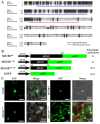
Figure 1
Figure 1
MS1881–168-GFP is located in the nucleus. (A) Amino acid sequence alignment of two R2R3-MYB genes: MS188 and TDF1. The R2R3 domain is marked by the blue bar, and the necessary 44 AAs of MS188 are marked by the red bar. Dark shading, identical residues; light shading, similar residues. * represents the identical animo acids between two sequences. (B,C) The complete MS188cds and two fragments, AAs 1–168 and AAs 169–320, were fused with GFP and infiltrated into tobacco leaves. MS188cds and MS1881–168-GFP were detected only in the nucleus. MS188169–320-GFP was distributed throughout the cytoplasm and the nucleus. N, nucleus; C, cytoplasm. Bars = 20 μm. The number in the bottom right corner of the Merge panel represents the statistical number of observations.
Figure 2
Figure 2
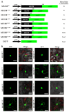
MS188 has NLSs at both the N- and C-termini of the R2R3-MYB domain. (A) To determine the NLS of MS188, MS1881–168 was truncated into additional short segments: MS1881–168, MS1881–124, MS1881–103, MS1886–168, MS18811–168, MS18816–168, and MS18821–168 fusions with GFP were infiltrated into tobacco leaves. N, nucleus; C, cytoplasm. (B) The fluorescence of MS1881–168-GFP, MS1881–124-GFP, MS1886–168-GFP, and MS18811–168-GFP was detected only in the nucleus. MS1881–103-GFP, MS18816–168-GFP, and MS18821–168-GFP were distributed throughout the cytoplasm and the nucleus. Bars = 20 μm. The number in the Merge panel represents the statistical number of observations.
Figure 3
Figure 3
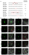
MS18812–15, MS18818–22, and MS18896–107 contribute to MS188 nuclear localization. (A) Point mutation of MS188 was performed to refine the NLS of MS188. The selected amino acids are shown in red font and were mutated to alanine (A). M1: 12VKRG15; M2: W17; M3: 18TEEED22; M4: 24KI25; M5: 96LPGRTDNDVKNH107; M6: 109NTKLKKKL116. All the mutated proteins were fused with GFP and infiltrated into tobacco leaves. N, nucleus; C, cytoplasm. (B) The fluorescence of MS188M2-GFP, MS188M4-GFP, and MS188M6-GFP was detected only in the nucleus. MS188M1-GFP, MS188M3-GFP, and MS188M5-GFP were distributed throughout the cytoplasm and the nucleus. Bars = 20 μm. (C) The fluorescence of MS188M1–13K14R-GFP and MS188M5–99R105R-GFP was distributed throughout the cytoplasm and the nucleus. Bars = 20 μm. The number in the Merge panel represents the statistical number of observations.
Figure 4
Figure 4
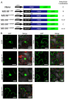
MS18812–15, MS18818–22, and MS18896–107 are the NLSs of MS188. (A) The cytoplasmic protein FBA6 was used to confirm the function of the MS188 NLSs: MS18812–15, MS18818–22, and MS18896–107. Linker peptides (PTPTPTPTP) were added between MS188 and FBA6. The chimeric proteins were fused with GFP and infiltrated into tobacco leaves. N, nucleus; C, cytoplasm. (B) The fluorescence of FBA6-GFP was detected only in the cytoplasm, but the MS1881–168 N-terminal fusion was transferred into the nucleus, whereas for the MS188169–320 fusion, the fluorescence was still distributed in the cytoplasm. Fusions with mutated MS1881–168 at the N-terminus, MS1881–168M1F-GFP and MS1881–168M5F-GFP, were distributed throughout the cytoplasm and the nucleus. MS1881–168M3F-GFP was detected only in the cytoplasm. Bars = 20 μm. The number in the Merge panel represents the statistical number of observations.
Figure 5
Figure 5
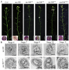
The function of MS188 was impaired when its NLS sites were mutated. (A) Phenotypes of the wild-type (Col), ms188 mutant, and transgenic plants. A wild-type Col plant presented normal fertility. An ms188 mutant presented very small siliques containing no seeds. All of the MS188-M1, MS188-M3, MS188-M5, and MS188-M135 transgenic lines presented small siliques without seeds. Bars = 2 cm. Insets in A show the pollen grains in the anthers stained with Alexander’s stain. Bars = 50 μm. (B) Semithin sections of ms188 and MS188-M135 anthers at different stages. dMSp, degraded microspores; MC, meiocytes; T, tapetum; Td, tetrad.
Figure 6
Figure 6
The NLS is conserved in R2R3-MYB family proteins. (A) Amino acid sequence alignment of the R2R3-MYB domain of three R2R3-MYB genes: MS188, TDF1, and MYB2. The refined NLS motif is marked by the red bar. Dark shading, identical residues; light shading, similar residues. * represents the identical animo acids between two sequences. (B) Point mutations of all the NLSs (MS18812–15, MS18818–22, and MS18896–107) in MS188, TDF1, and MYB2 were constructed and fused with GFP and then infiltrated into tobacco leaves. N, nucleus; C, cytoplasm. (C) The fluorescence of MS188-GFP, TDF1-GFP, and MYB2-GFP was detected only in the nucleus. Fluorescence of the point mutations MS188M-GFP, TDF1M-GFP, and MYB2M-GFP was distributed throughout the cytoplasm and the nucleus. Bars = 20 μm. The number in the Merge panel represents the statistical number of observations.

References

    1. Latchman D.S. Transcription factors: An overview. Int. J. Biochem. Cell Biol. 1997;29:1305–1312. - PubMed
    1. Amoutzias G.D., Robertson D.L., Van de Peer Y., Oliver S.G. Choose your partners: Dimerization in eukaryotic transcription factors. Trends Biochem. Sci. 2008;33:220–229. - PubMed
    1. Klempnauer K.-H., Gonda T.J., Michael Bishop J. Nucleotide sequence of the retroviral leukemia gene v-myb and its cellular progenitor c-myb: The architecture of a transduced oncogene. Cell. 1982;31:453–463. - PubMed
    1. Rosinski J.A., Atchley W.R. Molecular Evolution of the Myb Family of Transcription Factors: Evidence for Polyphyletic Origin. J. Mol. Evol. 1998;46:74–83. - PubMed
    1. Ogata K., Kanei-Ishii C., Sasaki M., Hatanaka H., Nagadoi A., Enari M., Nakamura H., Nishimura Y., Ishii S., Sarai A. The cavity in the hydrophobic core of Myb DNA-binding domain is reserved for DNA recognition and trans-activation. Nat. Struct. Biol. 1996;3:178–187. - PubMed

Publication types

MeSH terms

Substances

LinkOut - more resources