. 2025 Mar 22;14(7):481.
doi: 10.3390/cells14070481.

Opposite Roles of IL-32α Versus IL-32β/γ Isoforms in Promoting Monocyte-Derived Osteoblast/Osteoclast Differentiation and Vascular Calcification in People with HIV

Affiliations

Opposite Roles of IL-32α Versus IL-32β/γ Isoforms in Promoting Monocyte-Derived Osteoblast/Osteoclast Differentiation and Vascular Calcification in People with HIV

Hardik Ramani et al. Cells. .

Abstract

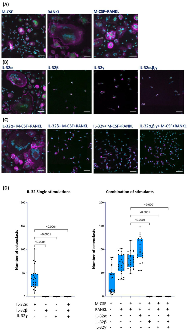
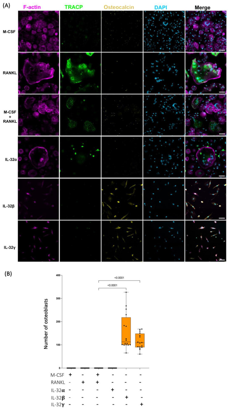
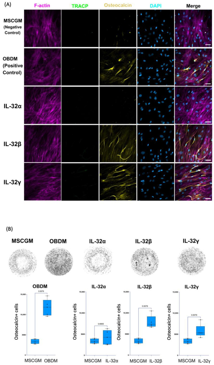
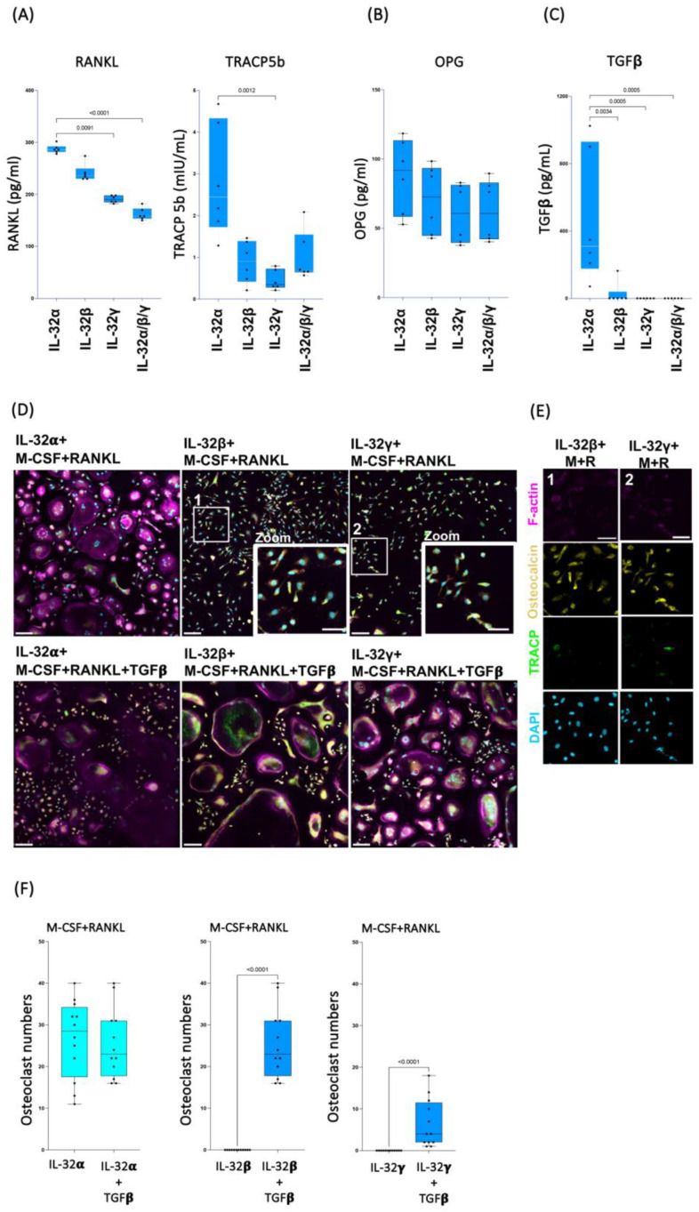
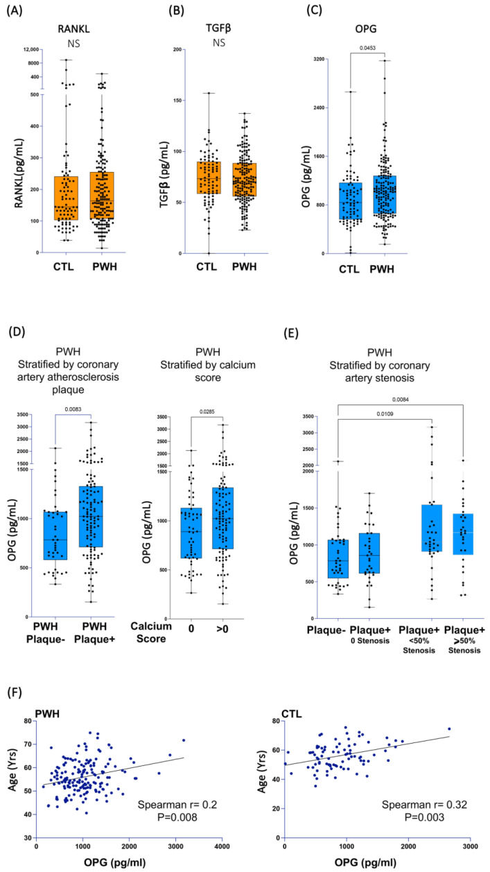
People with HIV (PWH) have an increased risk of developing cardiovascular disease (CVD). Our recent data demonstrated that the multi-isoform proinflammatory cytokine IL-32 is upregulated in PWH and is associated with arterial stiffness and subclinical atherosclerosis. However, the mechanisms by which IL-32 contributes to the pathogenesis of these diseases remain unclear. Here, we show that while the less expressed IL-32α isoform induces the differentiation of human classical monocytes into the calcium-resorbing osteoclast cells, the dominantly expressed isoforms IL-32β and IL-32γ suppress this function through the inhibition of TGF-β and induce the differentiation of monocytes into the calcium-depositing osteocalcin+ osteoblasts. These results aligned with the increase in plasma levels of osteoprotegerin, a biomarker of vascular calcification, and its association with the presence of coronary artery subclinical atherosclerosis and calcium score in PWH. These findings support a novel role for the proinflammatory cytokine IL-32 in the pathophysiology of CVD by increasing vascular calcification in PWH.

Keywords: HIV; IL-32; TGF-β; arterial calcification; atherosclerosis; cardiovascular diseases; inflammation; osteoblasts; osteoclasts; osteoprotegerin.


Conflict of interest statement

J.R.K. reports stock ownership in Abbott, AbbVie, Bristol Myers Squibb, Johnson & Johnson, Eli Lilly, Medtronic, Merck, and Pfizer.

Figures

Figure 1
Figure 1
Impact of IL-32 isoforms on monocyte differentiation into osteoclasts. Representative multiplexed spinning-disk confocal (20×/0.8) images of monocytes isolated from HIV-negative cells stimulated with either (A) M-CSF, RANKL, or M-CSF+ RANKL; or (B) IL-32α, IL-32β, IL-32γ, or with combination of three isoforms; or (C) combinations of individual IL-32 isoforms with M-CSF + RANKL. Osteoclasts are defined as cells expressing TRACP5 (green), formation of F-actin rings (magenta), and presence of three or more nuclei (DAPI, blue) as shown in (A) (all panels) and (B,C) (left panels). (D) Analysis of osteoclast numbers from monocytes isolated from n = 6 donors and induced with individual IL-32 isoforms alone (left panel) or combined with RANKL and M-CSF (right panel). Data represent analyses by counting 4 image clusters (3 × 3 tiles per cluster) representing 36 image tiles per condition per donor using Zeiss Zen 3.2 software. Nonparametric Kruskal–Wallis test and Dunn’s subtests were performed for statistical analysis. Comparisons were carried out with respect to positive control conditions M-CSF + RANKL. Scale bars represent 50 µm.
Figure 2
Figure 2
Distinct effects of IL-32 isoform IL-32α, IL-32β, and IL-32γ on differentiation of primary human monocytes into either osteoclasts or osteoblast-like cells. (A) Representative multiplex spinning-disk confocal (20×/0.8) images illustrating influence of individual IL-32 isoforms as well as osteoclastogenic molecules RANKL and/or M-CSF on monocyte differentiation. Osteoblast-like cells are defined as single-nucleated cells with expression of osteocalcin (yellow). Single color images are used to show F-actin, TRACP5, osteocalcin, and DAPI (from left to right) followed by merged color images (right panels). (B) Quantification of osteocalcin+ osteoblast-like cell numbers under different stimulation conditions and counted by automated counting using FIJI ImageJ software (Fiji, ImageJ, 64-bit). Data analysis was performed on 4 image clusters (3 × 3 tiles per cluster) representing 36 image tiles per condition per donor (cells from n = 4 HIV-negative donors). Nonparametric Kruskal–Wallis test and Dunn’s subtests were performed for statistical analysis. Scale bars represent 50 µm.
Figure 3
Figure 3
Impact of individual IL-32 isoforms IL-32α, IL-32β, and IL-32γ on differentiation of human mesenchymal stem cells (hMSCs) into osteoblasts. (A) Representative multiplex spinning-disk confocal (20×/0.8) images demonstrating impact of osteoblast differentiation medium/OBDM used as positive control (upper panels), IL-32 isoforms (middle panels), or negative control mesenchymal stem cell growth medium/MSCGM (lower panels) on hMSC differentiation. Single color images are used to show F-actin, TRACP5, osteocalcin, and DAPI (from left to right) followed by merged color images on far-right. Differentiated hMSC cells were identified by expression of osteocalcin. (B) Upper panels: representative full-well images of each condition at 10×/0.3 depicting osteocalcin expression in different conditions (dark areas indicate osteocalcin expression in cells). Lower panels: Comparing numbers of osteocalcin+ mononucleated osteoblasts from hMSCs stimulated with positive control conditions (osteoblast differentiation medium/OBDM) or with individual IL-32 isoforms compared to same negative control conditions using cell growth MSCG medium (experiments were performed simultaneously with single negative control experiment). Quantification was performed using FIJI ImageJ software, analyzing entire well for each condition using customized osteocalcin and DAPI expression measurement macro for ImageJ software (n = 5 experimental replicates). Statistical analysis was conducted using nonparametric Mann–Whitney test. Scale bars represent 50 µm.
Figure 4
Figure 4
TGF-β promotes osteoclast formation in presence of IL-32β and IL-32γ. (AC) Effect of IL-32 isoforms IL-32α, IL-32β, and IL-32γ on expression of soluble RANKL, TRACP5b, OPG, and TGF-β. Soluble proteins were measured by ELISA in supernatant of primary monocytes stimulated with IL-32 isoforms for 21 days (n = 6). Statistical analyses were performed using nonparametric Kruskal–Wallis test and Dunn’s subtest. (D,E) Representative multiplex spinning-disk confocal (20×/0.8), 3 × 3 tile images showing differentiation of monocytes into osteoclasts or osteoblasts in response to IL-32 isoforms (IL-32α, IL-32β, or IL-32γ) combined with osteoclastogenic molecules M-CSF(M)/RANKL(R) (D, upper panels, and E), and with TGF-β (D, lower panels). The 2 zoomed-in regions (D, 1 and 2) in panels show typical single-nucleated osteocalcin+ osteoblast cells induced by IL-32β and IL-32γ in absence of TGF-β. In (E), single-color images are used to show F-actin, osteocalcin, TRACP5, and DAPI (from top to bottom) of 2 zoomed-in regions in (D). (F) Comparison of osteoclast numbers induced by individual IL-32 isoforms IL-32α, IL-32β, or IL-32γ in combination with RANKL and M-CSF, with or without TGF-β (30 ng/mL). Osteoclasts were counted using Zeiss Zen 3.2 software from 4 image clusters (3 × 3 tiles per cluster), representing 36 image tiles per condition per donor, from three independent donors. Statistical analysis was performed using nonparametric Mann–Whitney U test. Scale bars in tile images represent 100 µm in (D) and 50 µm in (D,E).
Figure 5
Figure 5
Plasma biomarkers associated with vascular calcification and subclinical atherosclerosis in PWH. Plasma collected from PWH (n = 168) and a control group (n = 84) were used to measure soluble RANKL (A), TGFβ (B), and OPG (C). (D) Left panel: OPG plasma levels in plasma from PWH stratified by presence (n = 114) or absence (n = 43) of measurable subclinical atherosclerotic plaque in coronary artery. Right panel: OPG plasma levels in plasma from PWH stratified by calcium score (data available for n = 166/n = 107 with positive calcium score and n = 59 with score = 0). (E) OPG plasma levels in plasma from PWH participants for whom coronary artery maximum stenosis was calculated (data available for n = 139/n = 42 with no coronary artery plaque, n = 33 with measurable plaque but zero % stenosis, n = 35 with measurable plaque and stenosis levels < 50%, and n = 29 with measurable plaque and stenosis levels ≥ 50%). (F) Correlation between plasma OPG levels and age of PWH (n = 168, left panel) and control group participants (n = 84, right panel). Data were analyzed using non-parametric Mann–Whitney test in (AD), Kruskal–Wallis and Dunn’s subtest in (E) and Spearman’s correlations in (F).

Similar articles

References

    1. Aberg J.A. Aging, inflammation, and HIV infection. Top. Antivir. Med. 2012;20:101–105. - PMC - PubMed
    1. Hunt P.W. HIV and aging: Emerging research issues. Curr. Opin. HIV AIDS. 2014;9:302–308. doi: 10.1097/COH.0000000000000072. - DOI - PMC - PubMed
    1. Ruzicka D.J., Imai K., Takahashi K., Naito T. Comorbidities and the use of comedications in people living with HIV on antiretroviral therapy in Japan: A cross-sectional study using a hospital claims database. BMJ Open. 2018;8:e019985. doi: 10.1136/bmjopen-2017-019985. - DOI - PMC - PubMed
    1. Zicari S., Sessa L., Cotugno N., Ruggiero A., Morrocchi E., Concato C., Rocca S., Zangari P., Manno E.C., Palma P. Immune Activation, Inflammation, and Non-AIDS Co-Morbidities in HIV-Infected Patients under Long-Term ART. Viruses. 2019;11:200. doi: 10.3390/v11030200. - DOI - PMC - PubMed
    1. Guzman-Fulgencio M., Medrano J., Rallon N., Echeverria-Urabayen A., Miguel Benito J., Restrepo C., Garcia-Alvarez M., Vispo E., San Roman J., Sanchez-Piedra C., et al. Soluble markers of inflammation are associated with Framingham scores in HIV-infected patients on suppressive antiretroviral therapy. J. Infect. 2011;63:382–390. doi: 10.1016/j.jinf.2011.08.006. - DOI - PubMed

Publication types

MeSH terms

LinkOut - more resources