. 2025 Mar 27;14(7):501.
doi: 10.3390/cells14070501.

The Impact of TRPM8 on Prostate Cancer Transcriptomic Dynamics

Affiliations

The Impact of TRPM8 on Prostate Cancer Transcriptomic Dynamics

Swapna Asuthkar et al. Cells. .

Abstract

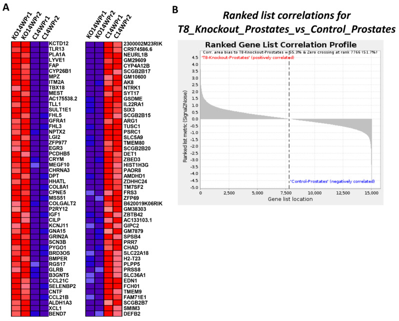
Prostate cancer (PC) remains a significant health challenge, with androgen receptor (AR) signaling playing a pivotal role in its progression. This study investigates the expression and functional implications of the transient receptor potential melastatin 8 (TRPM8) channel in PC, focusing on its interaction with AR and its impact on oncogenic pathways. We analyzed mRNA expression levels of TRPM8 and AR in PC tissues, revealing that TRPM8 is upregulated in benign and early-stage tumors but significantly downregulated in metastatic samples. This decline correlates with increased AR expression, suggesting a compensatory mechanism that enhances AR-driven tumorigenesis. RNA sequencing and pathway enrichment analyses demonstrated that TRPM8 knockout (KO) prostates exhibited significant alterations in gene expression, particularly in pathways related to extracellular matrix (ECM) remodeling, cell proliferation, and survival signaling. Notably, genes associated with metastasis, such as MMP2 and FAP, were upregulated in TRPM8 KO samples, indicating a potential role for TRPM8 in inhibiting tumor invasion. Furthermore, Gene Set Enrichment Analysis (GSEA) revealed positive enrichment of androgen response, angiogenesis, and epithelial-mesenchymal transition (EMT) pathways in TRPM8 KO prostates, reinforcing the notion that TRPM8 loss creates a pro-tumorigenic environment. Our findings suggest that TRPM8 functions as a molecular brake on PC progression, and its loss may contribute to the development of aggressive disease phenotypes. This study underscores the importance of TRPM8 as a potential therapeutic target and biomarker in PC, warranting further investigation into its role in cancer biology and treatment response.

Keywords: TRPM8; androgen; androgen receptor (AR); mRNA expression; prostate cancer.


Conflict of interest statement

Jai Velpula volunteers at Pringle Robotics, learning the skill sets for analysis. The remaining authors declare that the research was conducted in the absence of any commercial or financial relationships that could be construed as a potential conflict of interest.

Figures

Figure 1
Figure 1
mRNA expression of TRPM8 and AR in PC patients from the UIC Cancer Center. (A) TRPM8 mRNA expression: bar graphs display fragments per kilobase of transcript per million mapped reads (FPKM) for TRPM8 in benign, tumor, and metastatic prostate tissues. The right panel provides FPKM values for individual patient samples. (B) AR mRNA expression: bar graphs show FPKM values for AR across benign, tumor, and metastatic tissues, with individual sample data on the right (Gleason score = GS, GS6 = low grade, GS7 = intermediate, and GS8–9 = high grade) (ANOVA with post hoc Tukey multiple comparisons of different tissues (*, p < 0.05, ***, p < 0.0001; n = 3–51)).
Figure 2
Figure 2
Differential gene expression and pathway enrichment analysis of TRPM8 KO and WT mouse prostates. (A) Hierarchical clustering of differentially expressed genes between TRPM8 KO (n = 2) and wild-type (WT) (n = 2) mouse prostates. Red indicates upregulated genes and green indicates downregulated genes. (B) Visualization of differentially expressed genes with log2 fold change on the x-axis and −log10 p-value on the y-axis. Red and green dots represent significantly upregulated and downregulated genes, respectively, with a fold change cutoff of 1.5 and p-value ≤ 0.05. (C) Displays the top ten enriched pathways of differentially expressed genes (DEGs) in TRPM8 KO compared to WT mouse prostates. The enrichment score is presented as −log10 (p-value), highlighting significant pathways, such as focal adhesion and PI3K-Akt signaling. (D) Displays the top ten downregulated pathways in TRPM8 KO compared to WT mouse prostates. The enrichment score is presented as −log10 (p-value), highlighting significant pathways, such as small cell lung cancer and N-glycan biosynthesis.
Figure 3
Figure 3
Gene ontology enrichment analysis of differentially expressed genes in TRPM8 KO mouse prostates. (A) The top panel displays the enriched GO terms for biological processes (BPs), cellular components (CCs), and molecular functions (MFs) for upregulated genes in TRPM8 KO compared to WT mouse prostates. Significant processes include the regulation of cell adhesion and ECM organization. (B) The bottom panel shows the enriched GO terms for BPs, CCs, and MFs for downregulated genes. Key processes involve metabolic pathways and structural components.
Figure 4
Figure 4
Hallmark pathways and gene ontology enrichment analysis in TRPM8 KO prostates. Displays the enrichment plots for hallmark pathways significantly altered in TRPM8 KO compared to WT mouse prostates. Key pathways include androgen response, angiogenesis, epithelial–mesenchymal transition, and oxidative phosphorylation.
Figure 5
Figure 5
Gene Set Enrichment Analysis (GSEA) of specific TRPM8 KO mouse prostate signaling pathways. Displays enrichment plots for specific signaling pathways significantly altered in TRPM8 KO compared to WT mouse prostates. Key pathways include AKT, Cyclin D1, EGFR, KRAS, MEK, MTOR, P53, PTEN, and VEGF.
Figure 6
Figure 6
Retinoblastoma (RB) pathway enrichment analysis in TRPM8 KO mouse prostates. Displays the enrichment plot for the RB pathway, showing negative enrichment in TRPM8 KO compared to WT mouse prostates.
Figure 7
Figure 7
Ranked list correlations and heatmap analysis in TRPM8 KO vs. WT mouse prostates. (A) Displays the top 50 features for each phenotype in TRPM8 KO and WT mouse prostates. Red and blue colors indicate high and low expression levels, respectively. (B) Shows the ranked list correlations for TRPM8 KO prostates versus WT prostates. The plot illustrates the correlation bias toward TRPM8 KO prostates, with a positive correlation area of 55.3% and zero crossing at rank 7766 (51.7%).
Figure 8
Figure 8
A 3D scatter plot of coding potential distribution in TRPM8 KO mouse prostates. The plot displays the distribution of coding and non-coding transcripts in TRPM8 KO mouse prostates. Red dots represent coding transcripts, while blue dots indicate non-coding transcripts. The axes show ORF size (bp, log10), Fickett score, and Hexamer score, which are used to assess coding potential.
Figure 9
Figure 9
Gene expression in LAPC9 CSPC vs. CRPC and LNCaP CSPC vs. CRPC. The transcripts per kilobase million were obtained from the data extracted from the GSE88752 dataset and used to graph and compare the expression of the genes of interest: AR and TRPM8. (A) Expression of AR in LAPC9 CSPC is significantly higher than in CRPC (Student’s t-test: ***, p < 0.0001; n = 5). (B) Expression of TRPM8 in LAPC9 CSPC is significantly higher than in CRPC (Student’s t-test: *, p < 0.05; n = 5). (C) Expression of AR in LNCaP CSPC is significantly lower than in both primary and secondary CRPC (ANOVA with post hoc Tukey multiple comparisons of different cell types (ns, p > 0.05, *, p < 0.05, ***, p < 0.0001; n = 4). (D) Expression of TRPM8 LNCaP CSPC is significantly lower than in both primary and secondary CRPC (ANOVA with post hoc Tukey multiple comparisons of different cell types (*, p < 0.05, ***, p < 0.0001; n = 4).

References

    1. Tan M.H.E., Li J., Xu H.E., Melcher K., Yong E. Androgen receptor: Structure, role in prostate cancer and drug discovery. Acta Pharmacol. Sin. 2015;36:3–23. - PMC - PubMed
    1. Jacob A., Raj R., Allison D.B., Myint Z.W. Androgen receptor signaling in prostate cancer and therapeutic strategies. Cancers. 2021;13:5417. doi: 10.3390/cancers13215417. - DOI - PMC - PubMed
    1. Yanagisawa T., Rajwa P., Quhal F., Kawada T., Bekku K., Laukhtina E., von Deimling M., Chlosta M., Karakiewicz P.I., Kimura T., et al. Neoadjuvant androgen receptor signaling inhibitors before radical prostatectomy for non-metastatic advanced prostate cancer: A systematic review. J. Pers. Med. 2023;13:641. doi: 10.3390/jpm13040641. - DOI - PMC - PubMed
    1. Pagliarulo V., Bracarda S., Eisenberger M.A., Mottet N., Schroder F.H., Sternberg C.N., Studer U.E. Contemporary role of androgen deprivation therapy for prostate cancer. Eur. Urol. 2012;61:11–25. - PMC - PubMed
    1. Harris W.P., Mostaghel E.A., Nelson P.S., Montgomery B. Androgen deprivation therapy: Progress in understanding mechanisms of resistance and optimizing androgen depletion. Nat. Clin. Pract. Urol. 2009;6:76–85. - PMC - PubMed

Publication types

MeSH terms