Review
. 2025 Mar 29;14(7):511.
doi: 10.3390/cells14070511.

Oxidative Stress and Redox Imbalance: Common Mechanisms in Cancer Stem Cells and Neurodegenerative Diseases

Affiliations
Review

Oxidative Stress and Redox Imbalance: Common Mechanisms in Cancer Stem Cells and Neurodegenerative Diseases

Nikhil Raj Selvaraj et al. Cells. .

Abstract

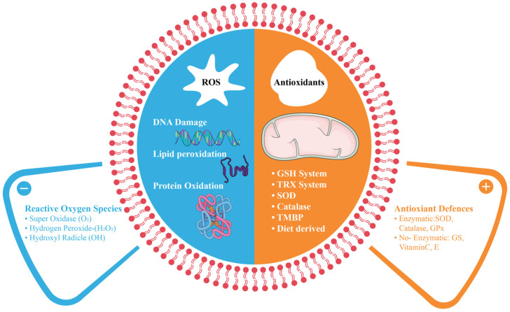
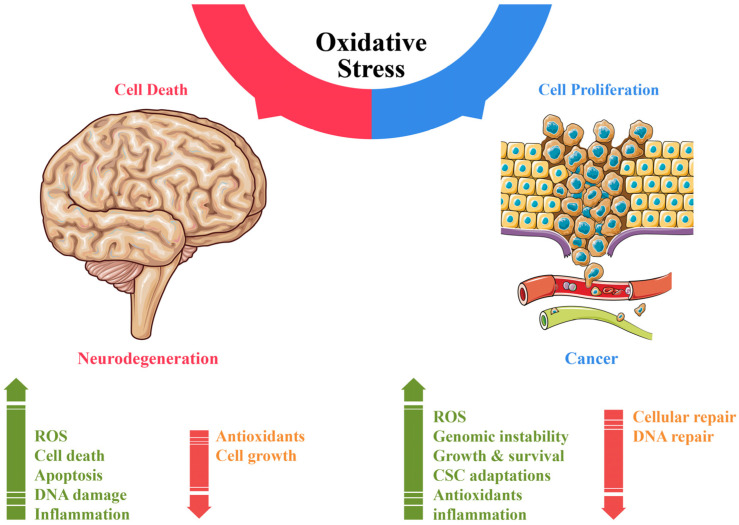
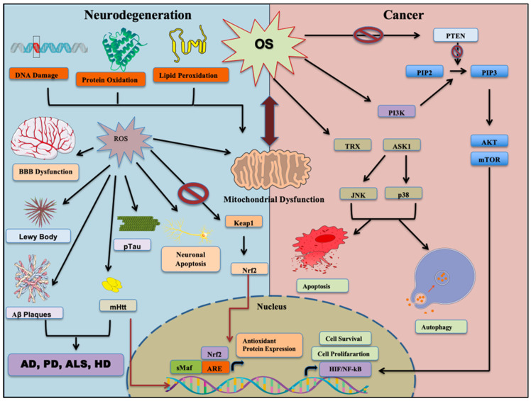
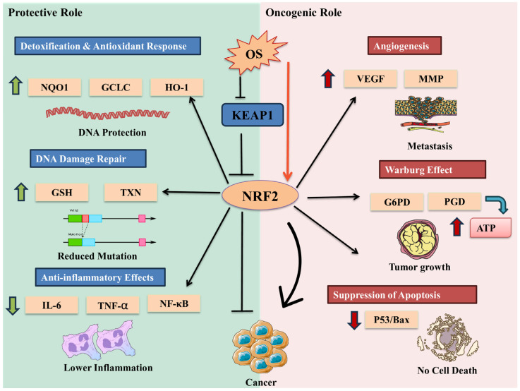
Oxidative stress (OS) is an established hallmark of cancer and neurodegenerative disorders (NDDs), which contributes to genomic instability and neuronal loss. This review explores the contrasting role of OS in cancer stem cells (CSCs) and NDDs. Elevated levels of reactive oxygen species (ROS) contribute to genomic instability and promote tumor initiation and progression in CSCs, while in NDDs such as Alzheimer's and Parkinson's disease, OS accelerates neuronal death and impairs cellular repair mechanisms. Both scenarios involve disruption of the delicate balance between pro-oxidant and antioxidant systems, which leads to chronic oxidative stress. Notably, CSCs and neurons display alterations in redox-sensitive signaling pathways, including Nrf2 and NF-κB, which influence cell survival, proliferation, and differentiation. Mitochondrial dynamics further illustrate these differences: enhanced function in CSCs supports adaptability and survival, whereas impairments in neurons heighten vulnerability. Understanding these common mechanisms of OS-induced redox imbalance may provide insights for developing interventions, addressing aging hallmarks, and potentially mitigating or preventing both cancer and NDDs.

Keywords: antioxidant; autophagy; cancer stem cells; ferroptosis; mitochondrial dysfunction; neurodegenerative diseases; oxidative phosphorylation; oxidative stress; reactive oxygen species; redox imbalance.


Conflict of interest statement

The authors declare no conflicts of interest.

Figures

Figure 1
Figure 1
The composition of the antioxidant system and oxidative stress. The core antioxidant systems include glutathione and thioredoxin system (GSH/TRX), superoxide dismutase (SOD), catalase (CAT), transition metal ion binding proteins (TMBP), and diet-derived antioxidants. These work together to prevent ROS-mediated oxidative damage by preventing protein carbonylation, lipid peroxidation, and DNA damage.
Figure 2
Figure 2
An overview of oxidative stress-mediated overlapping mechanisms of neurodegeneration and cancer.
Figure 3
Figure 3
Schematic representing the effect of oxidative stress in neurodegenerative diseases and cancer. An imbalance between ROS and antioxidants results in oxidative stress, which damages essential cellular components such as lipids, proteins, and DNA. Mitochondrial dysfunction, along with microglial activation, triggers the release of inflammatory cytokines and chemokines, ultimately leading to cell apoptosis and tissue degeneration. During oxidative stress, Keap1 is inhibited, and Nrf2 translocates into nucleus, upregulating ARE genes (antioxidant response elements) to counteract OS. Reactive oxygen species (ROS) play a crucial role in regulating cancer-related signaling pathways. An increase in ROS inhibits PTEN, leading to sustained activation of the PI3K/Akt pathway, which promotes cell survival and proliferation. In parallel, oxidative stress triggers the release of ASK-1 from the Trx-ASK1 complex, activating downstream p38 and JNK signaling, which can induce apoptosis and autophagy. Additionally, ROS-mediated inactivation of PP2A supports persistent NF-κB signaling, driving processes such as cell migration, invasion, and uncontrolled growth. These interconnected pathways highlight ROS as a key regulator of both cancer progression and neurodegeneration. Nrf2: Nuclear factor erythroid 2-related factor 2; JNK: c-Jun N-terminal kinase; Keap1: Kelch-like ECH-associated protein 1; mTOR: mammalian target of rapamycin; PI3K: phosphoinositide 3-kinase; PIP: phosphatidylinositol phosphate; PTEN: phosphatase and tensin homolog; ASK1: Apoptosis Signal-Regulating Kinase 1; TRX: thioredoxin; HIF: hypoxia-inducible facto; NF-κB: nuclear factor-kappa B.
Figure 4
Figure 4
Contradictory roles of Nrf2 in cancer: The contradictory role of Nrf2 in cancer is evident through its diverse functions. On one hand, Nrf2 exhibits protective properties by activating DNA repair mechanisms, regulating antioxidant enzymes, and suppressing pro-inflammatory pathways. These actions potentially reduce cancer-causing mutations, protect cells from oxidative stress, and mitigate chronic inflammation associated with cancer development. On the other hand, Nrf2 demonstrates oncogenic functions by promoting angiogenesis, enhancing metabolic reprogramming in cancer cells, suppressing apoptosis, and potentially promoting tumor growth and chemoresistance when constitutively activated. This dual nature underscores the complexity of Nrf2’s role in cancer biology.

Similar articles

Cited by

References

    1. Sies H., Berndt C., Jones D.P. Oxidative stress. Annu. Rev. Biochem. 2017;86:715–748. doi: 10.1146/annurev-biochem-061516-045037. - DOI - PubMed
    1. Aramini B., Masciale V., Grisendi G., Bertolini F., Maur M., Guaitoli G., Chrystel I., Morandi U., Stella F., Dominici M., et al. Dissecting Tumor Growth: The Role of Cancer Stem Cells in Drug Resistance and Recurrence. Cancers. 2022;14:976. doi: 10.3390/cancers14040976. - DOI - PMC - PubMed
    1. Vera-Ramirez L., Sanchez-Rovira P., Ramirez-Tortosa M.C., Ramirez-Tortosa C.L., Granados-Principal S., Lorente J.A., Quiles J.L. Free radicals in breast carcinogenesis, breast cancer progression and cancer stem cells. Biological bases to develop oxidative-based therapies. Crit. Rev. Oncol./Hematol. 2011;80:347–368. doi: 10.1016/j.critrevonc.2011.01.004. - DOI - PubMed
    1. García-Blanco A., Baquero M., Vento M., Gil E., Bataller L., Cháfer-Pericás C. Potential oxidative stress biomarkers of mild cognitive impairment due to Alzheimer disease. J. Neurol. Sci. 2017;373:295–302. doi: 10.1016/j.jns.2017.01.020. - DOI - PubMed
    1. Zabel M., Nackenoff A., Kirsch W.M., Harrison F.E., Perry G., Schrag M. Markers of oxidative damage to lipids, nucleic acids and proteins and antioxidant enzymes activities in Alzheimer’s disease brain: A meta-analysis in human pathological specimens. Free. Radic. Biol. Med. 2018;115:351–360. doi: 10.1016/j.freeradbiomed.2017.12.016. - DOI - PMC - PubMed

Publication types

Substances

LinkOut - more resources