. 2025 Mar 30;14(7):514.
doi: 10.3390/cells14070514.

A Broad-Spectrum Chemokine Inhibitor Drives M2 Macrophage Polarization Through Modulation of the Myometrial Secretome

Affiliations

A Broad-Spectrum Chemokine Inhibitor Drives M2 Macrophage Polarization Through Modulation of the Myometrial Secretome

Adam Boros-Rausch et al. Cells. .

Abstract

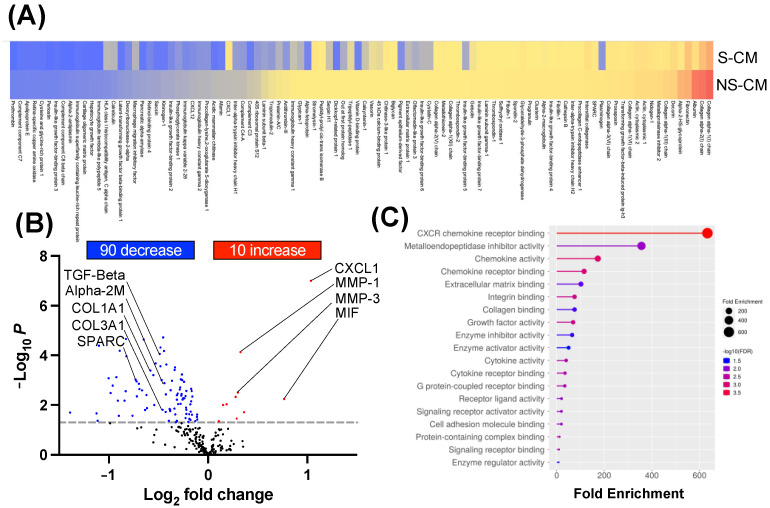
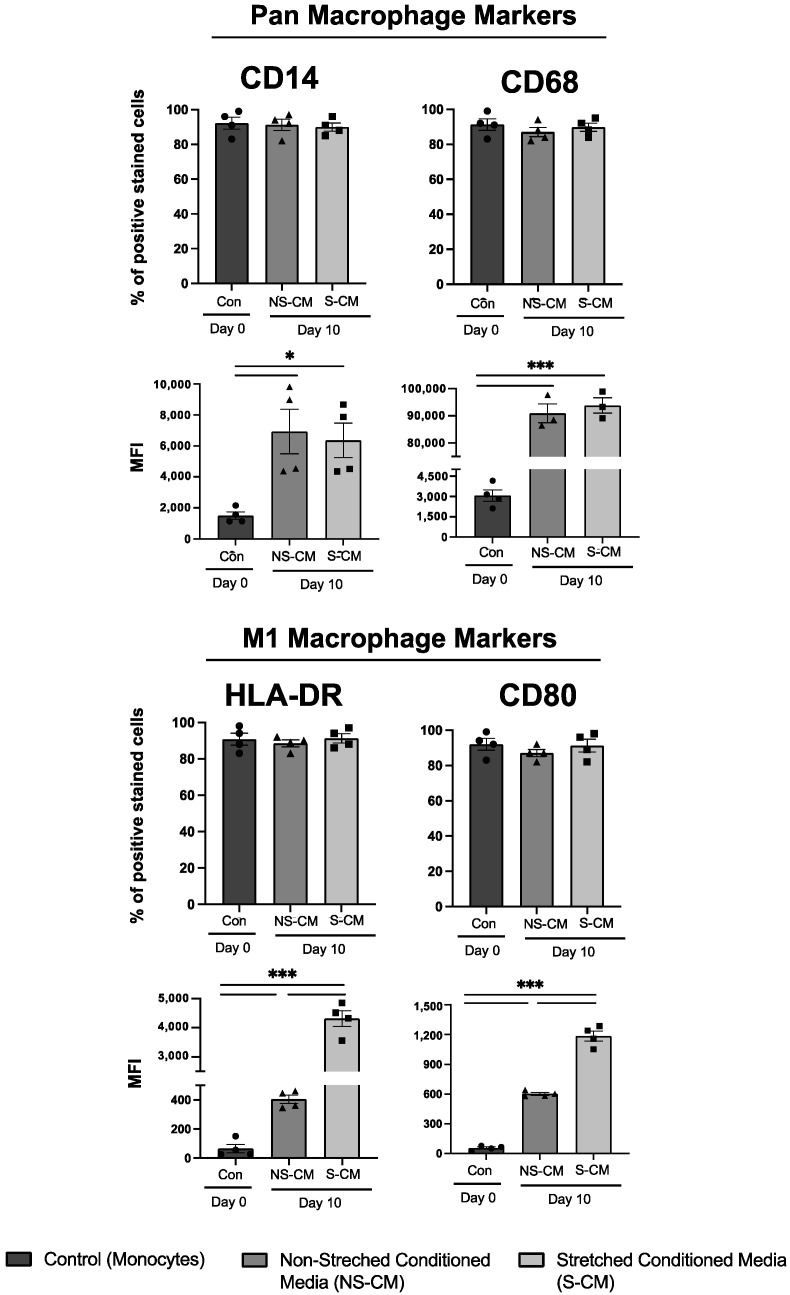
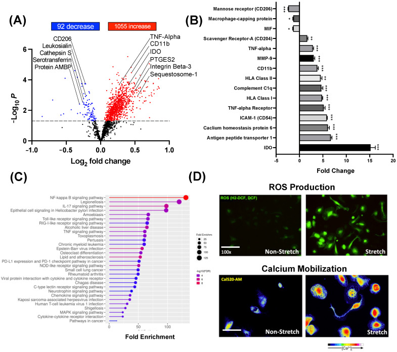
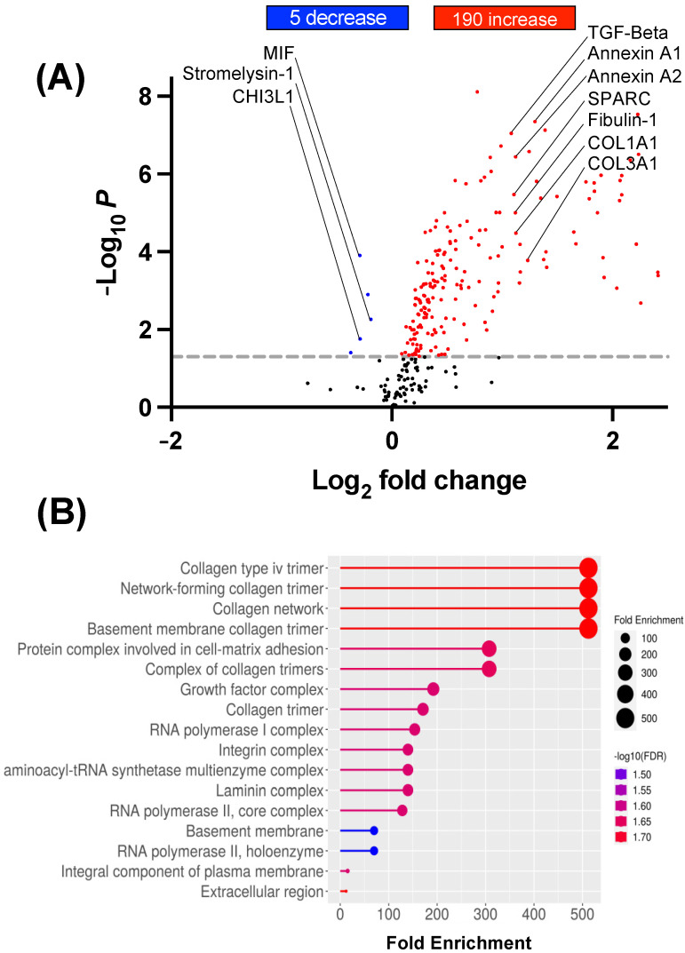
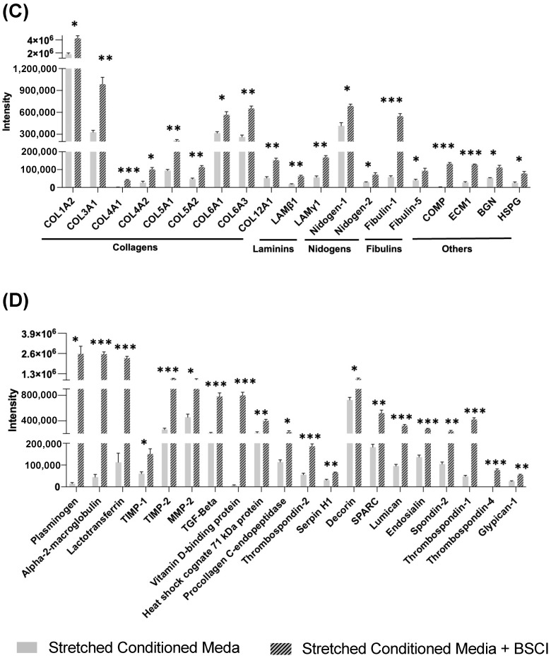
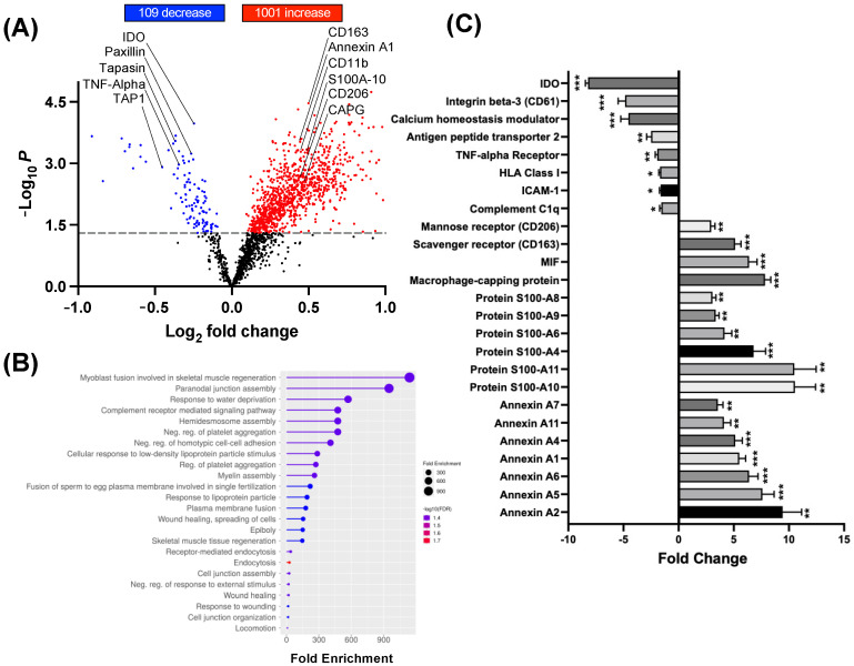
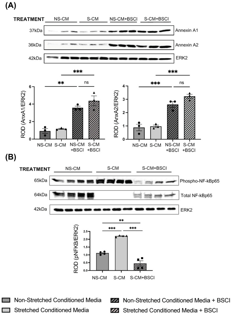
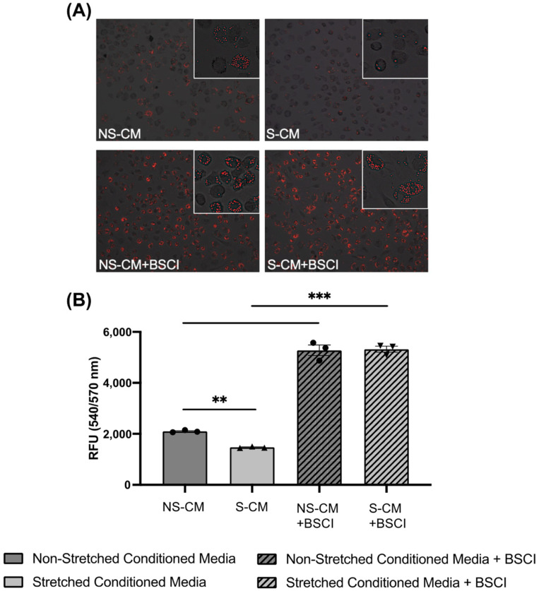
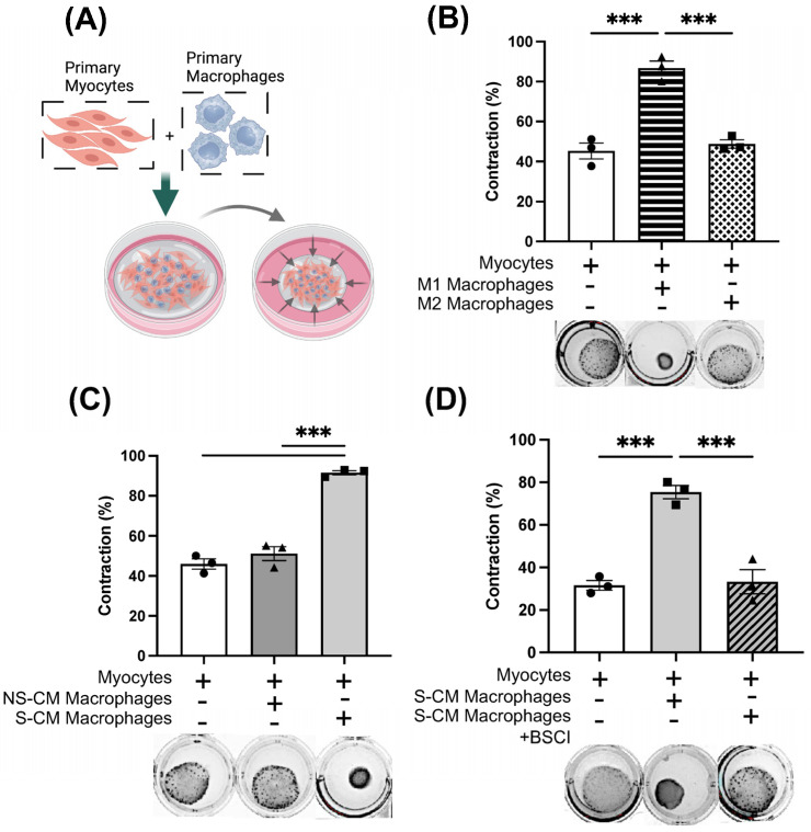
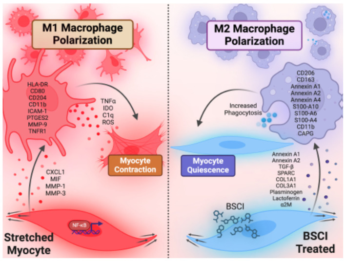
The uterine smooth muscle (myometrium) is an immunomodulatory tissue capable of secreting multiple chemokines during pregnancy. We propose that before term labor, chemokines secreted as a result of mechanical stretch of the uterine walls by the growing fetus(es) induce infiltration of maternal monocytes into myometrium, drive their differentiation into macrophages, and induce pro-inflammatory (M1) polarization, leading to labor contractions. This study used high-throughput proteomic mass-spectrometry to investigate the underlying mechanisms and explored the therapeutic potential of a broad-spectrum chemokine inhibitor (BSCI, FX125L) in modulating these effects. Primary myocytes isolated from the myometrium of term pregnant women were subjected in vitro to static mechanical stretch. Proteomic analysis of stretched myocyte-conditioned media (CM) identified significant upregulation of chemokine-related pathways and ECM degradation proteins. CM induced in vitro differentiation of human monocytes to macrophages and polarization into an M1-like phenotype characterized by elevated ROS production. BSCI treatment altered the myocyte secretome, increasing tissue-remodeling and anti-inflammatory proteins, Annexin A1 and TGF-β. BSCI-treated myocyte secretions induced Annexin A1 expression in macrophages and enhanced their phagocytic activity. We conclude that factors secreted by mechanically stretched myocytes induce pro-inflammatory M1 macrophage polarization, while BSCI modulates myocyte secretome, which reprograms macrophages to a homeostatic M2-like phenotype, thus reducing inflammation. When treated with BSCI, M2-polarized macrophages reduced myocyte-driven collagen gel contraction, whereas M1 macrophages enhanced it. This study reveals novel insights into the myocyte-macrophage interaction and identifies BSCI as a promising drug to modulate myometrial activity. We suggest that uterine macrophages may represent a therapeutic target for preventing preterm labor in women.

Keywords: broad spectrum chemokine inhibitor; cytokine; inflammation; labor; macrophage; mass-spectrometry; mechanical stretch; myometrium; pregnancy; uterus.


Conflict of interest statement

The authors declare no conflicts of interest.

Figures

Figure 1
Figure 1
Proteomic analysis of proteins secreted by stretched primary human myocytes. Myometrial biopsies from pregnant term non-laboring women (N = 4/group) were used to isolate primary uterine myocytes subjected to mechanical stretch (23% elongation, 24 h) using a Flexcell instrument. Conditioned media (CM) from stretched (S-CM) and non-stretched (NS-CM) myocytes were collected, concentrated, and analyzed by TMT-MS to identify differentially secreted proteins. (A) Heatmap displaying secreted proteins significantly modulated by stretch (S-CM vs. NS-CM), with red indicating high abundance and blue indicating low abundance. (B) Volcano plot highlighting differentially secreted proteins, showing significantly increased and decreased proteins (upregulated: red; downregulated: blue) in response to stretch. (C) Gene ontology (GO) enrichment analysis of the top 20 biological processes affected by mechanical stretch, with circle size representing the number of enriched genes and color indicating significance (larger circles indicate higher gene enrichment; higher significance in red). Statistical significance was determined by two-sided unpaired Student’s t-tests.
Figure 2
Figure 2
Differentiation of human peripheral monocytes into macrophages and their polarization in response to conditioned media (CM) from stretched (S-CM) or non-stretched (NS-CM) human uterine myocytes. Monocytes from pregnant term non-laboring women (N = 3–4/group) were cultured for 10 days in CM. Flow cytometry analysis assessed CD14 (monocyte marker), CD68 (macrophage marker), and M1 polarization markers (HLA-DR, CD80). Bar graphs display the percentage of positively stained cells and mean fluorescence intensity (MFI). Statistical significance was determined by one-way ANOVA with Dunnett’s test (* p < 0.05, *** p < 0.001).
Figure 3
Figure 3
Proteomic profiling of macrophages activated by myocyte-secreted factors. Human monocytes (N = 4/group) were treated for 10 days with conditioned media (CM) from stretched (S-CM) or non-stretched (NS-CM) human myocytes, then analyzed by TMT-MS to assess macrophage polarization. (A) Volcano plot showing differentially expressed proteins in S-CM vs. NS-CM-treated macrophages (upregulated: red; downregulated: blue). (B) Bar graph of key protein markers significantly altered by S-CM (mean ± SD; Student’s t-test; * p < 0.05, ** p < 0.01, *** p < 0.001). (C) GO enrichment analysis of the top 20 biological processes affected in S-CM-treated macrophages with circle size representing the number of enriched genes and color indicating significance (larger circles indicate higher gene enrichment; higher significance in red). (D) Fluorescence imaging of S-CM- and NS-CM-treated macrophages stained for ROS production (H2-DCF, green) and Ca2⁺ mobilization (Cal-520 AM, yellow), captured by confocal microscopy.
Figure 4
Figure 4
Proteomic analysis of secreted proteins from BSCI-treated stretched myocytes. Primary human myocytes (N = 4/group) from term pregnant, non-laboring women were pretreated with BSCI (400 nM, 1 h) before undergoing mechanical stretch (23% elongation, 24 h). Conditioned media (CM) from stretched myocytes (S-CM) and BSCI-treated stretched myocytes (S-CM + BSCI) were collected and analyzed by TMT-MS to assess changes in secreted protein composition. (A) Volcano plot showing differentially secreted proteins in S-CM + BSCI vs. S-CM (upregulated: red, downregulated: blue). (B) GO enrichment analysis of the top 20 biological processes modified by BSCI treatment with circle size representing the number of enriched genes and color indicating significance (larger circles indicate higher gene enrichment; higher significance in red). (C) Extracellular matrix (ECM)-associated proteins upregulated in BSCI-treated myocytes, suggesting enhanced tissue remodeling. (D) Key tissue remodeling proteins increased following BSCI treatment, indicating a shift toward an anti-inflammatory, reparative secretome. Statistical significance: two-sided unpaired Student’s t-tests (* p < 0.05, ** p < 0.01, *** p < 0.001).
Figure 4
Figure 4
Proteomic analysis of secreted proteins from BSCI-treated stretched myocytes. Primary human myocytes (N = 4/group) from term pregnant, non-laboring women were pretreated with BSCI (400 nM, 1 h) before undergoing mechanical stretch (23% elongation, 24 h). Conditioned media (CM) from stretched myocytes (S-CM) and BSCI-treated stretched myocytes (S-CM + BSCI) were collected and analyzed by TMT-MS to assess changes in secreted protein composition. (A) Volcano plot showing differentially secreted proteins in S-CM + BSCI vs. S-CM (upregulated: red, downregulated: blue). (B) GO enrichment analysis of the top 20 biological processes modified by BSCI treatment with circle size representing the number of enriched genes and color indicating significance (larger circles indicate higher gene enrichment; higher significance in red). (C) Extracellular matrix (ECM)-associated proteins upregulated in BSCI-treated myocytes, suggesting enhanced tissue remodeling. (D) Key tissue remodeling proteins increased following BSCI treatment, indicating a shift toward an anti-inflammatory, reparative secretome. Statistical significance: two-sided unpaired Student’s t-tests (* p < 0.05, ** p < 0.01, *** p < 0.001).
Figure 5
Figure 5
Proteomic analysis of macrophages exposed to secretome from BSCI-treated myocytes. Primary monocytes (N = 4/group) were cultured for 10 days in conditioned media (CM) from mechanically stretched uterine myocytes (S-CM) pretreated with or without a BSCI (400 nM, 1 h). Total protein content of differentiated macrophages was analyzed using TMT-MS. (A) Volcano plot displays differentially expressed proteins in macrophages treated with S-CM vs. S-CM + BSCI (upregulated: red; downregulated: blue). (B) Gene ontology (GO) enrichment analysis of the top 20 biological processes altered by the BSCI, with circle size representing the number of enriched genes and color indicating significance (larger circles indicate higher gene enrichment; higher significance in red). (C) Bar graph shows fold changes in selected protein markers affected by BSCI treatment. Statistical significance was determined by two-sided unpaired Student’s t-tests (* p < 0.05, ** p < 0.01, *** p < 0.001).
Figure 6
Figure 6
Validation of protein expressed by human macrophages exposed to myocyte secretome and treated with BSCI. Primary human monocytes (N = 3–4/group) were cultured for 10 days in conditioned media (CM) from non-stretched (NS-CM), mechanically stretched (S-CM), and BSCI-treated myocytes (400 nM, 1 h) (NS-CM + BSCI, S-CM + BSCI). Total protein was extracted from differentiated macrophages and analyzed by Western blot. (A) Representative Western blots and densitometric analysis of Annexin A1 and Annexin A2 expression, normalized to ERK2, showing differential expression in response to mechanical stretch and BSCI treatment. (B) Expression of phospho-NF-κBp65 and total NF-κBp65 in macrophages treated with NS-CM, S-CM, or S-CM + BSCI. Densitometric analysis quantifies phospho-NF-κBp65 levels relative to ERK2, indicating changes in NF-κB signaling in response to myocyte-derived factors and the BSCI. Data are presented as mean ± SD, with statistical significance determined by one-way ANOVA followed by Dunnett’s multiple comparisons test (** p < 0.01, *** p < 0.001, “ns”—non significant).
Figure 7
Figure 7
Enhanced phagocytosis in macrophages exposed to BSCI-treated myocyte secretome. Monocytes isolated from pregnant term non-laboring women (N = 3/group) were differentiated into macrophages over 10 days in conditioned media (CM) from four groups: (1) non-stretched myocytes (NS-CM), (2) mechanically stretched myocytes (S-CM), (3) non-stretched myocytes pretreated with BSCI (NS-CM + BSCI), and (4) stretched myocytes pretreated with BSCI (400 nM, 1 h) (S-CM + BSCI). After differentiation, macrophages (5 × 10⁴) were incubated with zymosan particles at 37 °C for 1 h, and phagocytosis was assessed by real-time confocal fluorescence microscopy. (A) Representative immunofluorescent images of zymosan uptake by macrophages. (B) Quantification of relative fluorescence units (RFUs) from duplicate plate readings. Data represent the mean ± SD of three independent macrophage lines (N = 3/group). Statistical significance was determined by one-way ANOVA followed by Dunnett’s multiple comparisons test (** p < 0.01, *** p < 0.001). Original magnification: ×20; inset magnification.
Figure 8
Figure 8
The BSCI inhibits collagen lattice contraction in myocyte–macrophage co-cultures. (A) Schematic of the co-culture treatment protocol. Monocytes and uterine smooth muscle cells were isolated from pregnant term non-laboring women (N = 3/group). Monocytes were differentiated for 10 days before co-culturing with myocytes in a collagen gel lattice, and gel contraction was measured after 48 h. (B) Collagen gel contraction induced by myocyte co-culture with M1 (GM-CSF, LPS, IFNγ) or M2 (M-CSF, IL-13, IL-4) macrophages, compared to myocyte monoculture (negative control). (C) Collagen contraction in co-cultures of myocytes with macrophages treated with conditioned media (CM) from non-stretched (NS-CM) and mechanically stretched (S-CM) myocytes, compared to myocyte monoculture. (D) Effect of the BSCI on collagen contraction. Macrophages treated with CM from stretched myocytes (S-CM) with or without BSCI pretreatment (400 nM, 1 h) were co-cultured with myocytes. Differences in gel surface area were recorded at 48 h. Six wells per condition were used for each primary co-culture (N = 6, technical replicates), with three myometrial cell lines analyzed (N = 3/group). Data are presented as mean ± SD. Statistical significance was determined by one-way ANOVA with Dunnett’s multiple comparisons test and two-sided unpaired Student’s t-tests (*** p < 0.001).
Figure 9
Figure 9
Putative model of myometrial stretch-induced macrophage activation. During late pregnancy, uterine walls are mechanically stretched by growing fetus [52]. Stretched uterine smooth muscle cells (myocytes) secrete multiple factors that induce infiltration of maternal peripheral monocytes [10,11] and promote their differentiation into resident tissue macrophages and polarization to pro-inflammatory M1 phenotype. As a feedback effect, these M1 macrophages can activate uterine myocytes to induce muscle contractions in preparation for the onset of labor [7,27,28,29]. By treating the uterine myocytes with the BSCI, we altered their secretome so they produce anti-inflammatory proteins capable of repolarizing macrophages to a homeostatic M2 phenotype which promotes myocyte quiescence.

Similar articles

References

    1. Quinn J.-A., Munoz F.M., Gonik B., Frau L., Cutland C., Mallett-Moore T., Kissou A., Wittke F., Das M., Nunes T., et al. Preterm Birth: Case Definition & Guidelines for Data Collection, Analysis, and Presentation of Immunisation Safety Data. Vaccine. 2016;34:6047–6056. doi: 10.1016/j.vaccine.2016.03.045. - DOI - PMC - PubMed
    1. Lakshmanan A., Song A.Y., Belfort M.B., Yieh L., Dukhovny D., Friedlich P.S., Gong C.L. The Financial Burden Experienced by Families of Preterm Infants after NICU Discharge. J. Perinatol. 2022;42:223–230. doi: 10.1038/s41372-021-01213-4. - DOI - PMC - PubMed
    1. March of Dimes Report Card. [(accessed on 14 January 2025)]. Available online: https://www.marchofdimes.org/report-card.
    1. Moutquin J.M. Classification and Heterogeneity of Preterm Birth. BJOG Int. J. Obstet. Gynaecol. 2003;110((Suppl. 20)):30–33. doi: 10.1046/j.1471-0528.2003.00021.x. - DOI - PubMed
    1. Sheikh I.A., Ahmad E., Jamal M.S., Rehan M., Assidi M., Tayubi I.A., AlBasri S.F., Bajouh O.S., Turki R.F., Abuzenadah A.M., et al. Spontaneous Preterm Birth and Single Nucleotide Gene Polymorphisms: A Recent Update. BMC Genom. 2016;17:759. doi: 10.1186/s12864-016-3089-0. - DOI - PMC - PubMed

Publication types

Grants and funding

LinkOut - more resources