Review
. 2025 Apr 1;14(7):522.
doi: 10.3390/cells14070522.

Diverse Roles of the Multiple Phosphodiesterases in the Regulation of Cyclic Nucleotide Signaling in Dictyostelium

Affiliations
Review

Diverse Roles of the Multiple Phosphodiesterases in the Regulation of Cyclic Nucleotide Signaling in Dictyostelium

Pundrik Jaiswal et al. Cells. .

Abstract

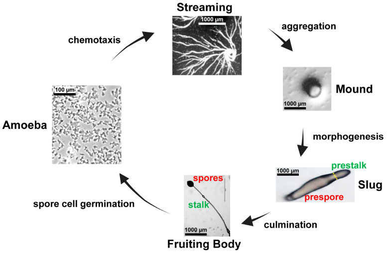
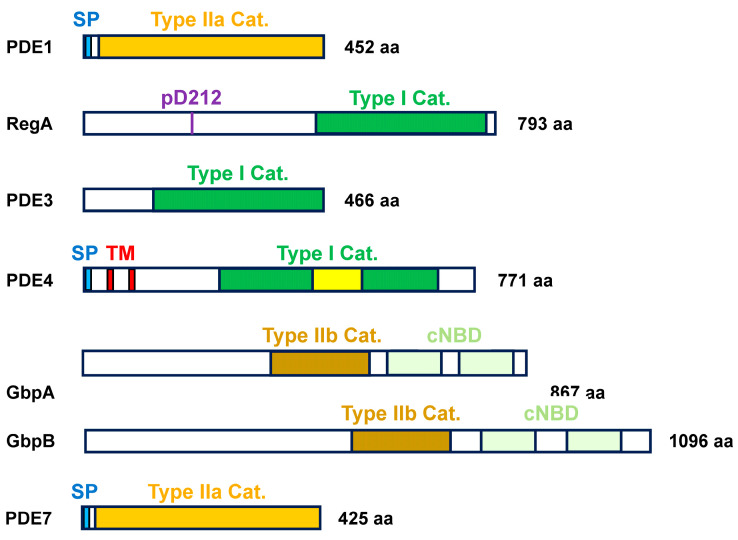
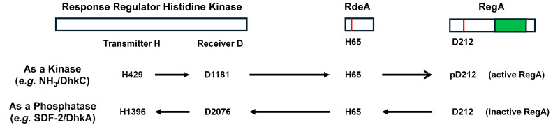
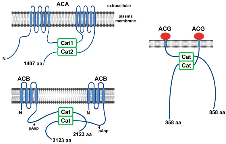
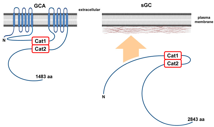
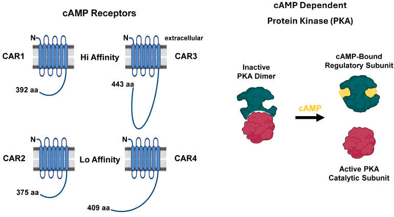
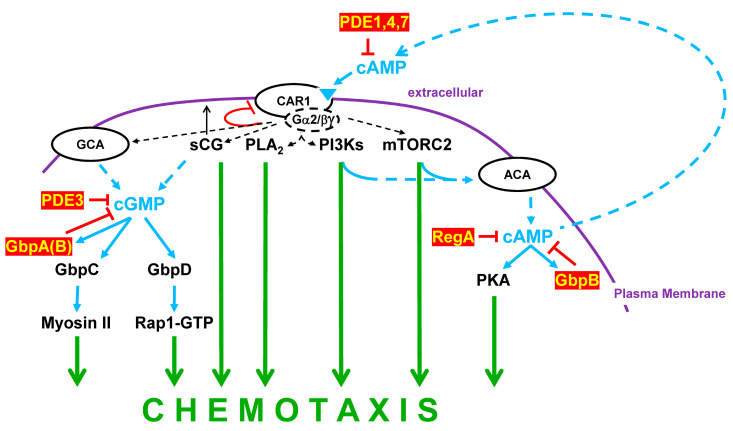
Dictyostelium is a unique model used to study the complex and interactive cyclic nucleotide signaling pathways that regulate multicellular development. Dictyostelium grow as individual single cells, but in the absence of nutrients, they initiate a multicellular developmental program. Central to this is secreted cAMP, a primary GPCR-response signal. Activated cAMP receptors at the cell surface direct a number of downstream signaling pathways, including synthesis of the intracellular second messengers cAMP and cGMP. These, in turn, activate a series of downstream targets that direct chemotaxis within extracellular cAMP gradients, multicellular aggregation, and, ultimately, cell-specific gene expression, morphogenesis, and cytodifferentiation. Extracellular cAMP and intracellular cAMP and cGMP exhibit rapid fluctuations in concentrations and are, thus, subject to exquisite regulation by both synthesis and degradation. The Dictyostelium genome encodes seven phosphodiesterases (PDEs) that degrade cyclic nucleotides to nucleotide 5'-monophosphates. Each PDE has a distinct structure, substrate specificity, regulatory input, cellular localization, and developmentally regulated expression pattern. The intra- or extra-cellular localizations and enzymatic specificities for cAMP or cGMP are essential for degradative precision at different developmental stages. We discuss the diverse PDEs, the nucleotide cyclases, and the target proteins for cAMP and cGMP in Dictyostelium. We further outline the major molecular, cellular, and developmental events regulated by cyclic nucleotide signaling, with emphasis on the input of each PDE and consequence of loss-of-function mutations. Finally, we relate the structures and functions of the Dictyostelium PDEs with those of humans and in the context of potential therapeutic understandings.

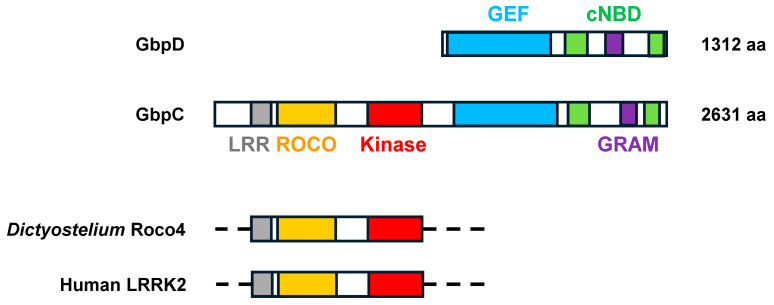
Keywords: GPCRs; LRR/ROCO kinases; PDEs; Protein kinase A; adenylyl cyclases; cAMP; cGMP; guanylyl cyclases.


Conflict of interest statement

The authors declare no conflicts of interests.

Figures

Figure 7
Figure 7
The cGMP targets in Dictyostelium. GbpC and GbpD represent the primary signaling molecules downstream of cGMP, although PDEs GbpA and GbpB are also cyclic nucleotide-binding proteins (Figure 2). Both GbpC and GbpD have GTP exchange factor (GEF) domains, which are activated by cyclic nucleotide-binding and GRAM-mediated interactions. For GbpD, Rap1 serves as the GEF target. For GpbC, the GEF targets the intramolecular bipartite, Ras-like GTPase ROCO domain for activation of the downstream intramolecular kinase and regulation of Myosin II. GbpC was one of the founding members of the LRR/ROCO kinase family, characterized by a leucine-rich repeat (LRR), a ROCO, and a kinase domain. The approximate alignments of these regions within Dictyostelium Roco4 and human LRRK2, relative to GbpC, are shown. Specific mutations in human LRRK2 are associated with Parkinson’s disease, and Dictyostelium Roco4 has proven an effective structural model [87].
Figure 1
Figure 1
Major stages of the Dictyostelium developmental cycle. Dictyostelium grow as single amoeboid cells. When starved for nutrients, Dictyostelium enter a multicellular developmental program. Dictyostelium secrete and become chemotactic to cAMP, and streaming cells aggregate at the centers of cAMP synthesis (see discussion of Figures 8 and 9 regarding PDEs and cyclic nucleotide signaling during chemotaxis). Cells then coalesce into a tight, multicellular aggregation mound. The mound undergoes cytodifferentiation and morphogenesis into the slug, with non-terminally differentiated prestalk cells in the anterior and prespore cells at the posterior (see discussion of Figure 10 regarding PDEs and cyclic nucleotide signaling during cytodifferentiation). Culmination leads to the terminally differentiated fruiting body comprised of mature spore and stalk cells (see discussion of Figure 10 regarding PDEs and cyclic nucleotide signaling during culmination). Under the appropriate physiological conditions, spores will germinate to release amoeba for single-cell growth. Approximate size scales are indicated.
Figure 2
Figure 2
The structural domains of Dictyostelium PDEs. PDE1, PDE4, and PDE7 function to degrade cAMP in the extracellular milieu. PDE1 and PDE7 have high sequence similarity and are primarily secreted; they both may associate with the cell surface by an undefined mechanism. Their catalytic domains are part of the type IIa γ-proteobacteria family. PDE4 is a member of the type I PDE family. PDE4 is membrane-bound and modeled with two transmembrane domains and an extracellular-facing catalytic domain. Its catalytic domain is interrupted by a region of simple sequence amino acids. RegA is an intracellular type I PDE with specificity for cAMP. RegA is part of a two-component regulatory system. It is activated by a phospho-transfer from a histidine kinase to RdeA to Aspartate 212 in the response regulator receiver domain. PDE3 is an intracellular type I PDE with specificity for cGMP. GbpA and GbpB possess two cyclic nucleotide-binding domains, where binding activates their metallo-β-lactamase-type IIb PDE catalytic domain. Both are intracellular. GbpA is cGMP specific. GbpB has a preference for cAMP, but it also has a low affinity for cGMP. SP: signal peptide; TM: transmembrane domain; type IIa Cat: type IIa γ-proteobacteria catalytic family; type IIb Cat: type IIb metallo-β-lactamase catalytic family; type I Cat.: type I PDE catalytic family; cNBD: cyclic nucleotide-binding domain.
Figure 3
Figure 3
Phospho-relay regulation of RegA. Two protein components comprise phospho-relay upstream of RegA, a response regulator histidine kinase (RR-HK) and the phospho-relay RdeA; there are multiple RR-HKs that may integrate into a single RdeA. Multiple targets are likely downstream of RdeA, in addition to RegA. An active RR-HK phosphorylates an intramolecular transmitter histidine, with phospho-transfer to a receiver aspartate. This is followed by intermolecular phospho-transfer to histidine H65 in RdeA and intermolecular phospho-transfer to aspartate D212 in RegA. In a specific example, under high NH3 conditions, RR-HK DhkC is active, leading to the phosphorylation and activation of RegA. Here, intracellular cAMP levels are depleted. RR-HKs can also serve as phosphatases. SDF-2 may inhibit DhKA, with a resulting back phospho-transfer from RegA, leading to its inactivation. Here, intracellular cAMP levels are elevated. The phosphorylation sites on DhkC and DhkA are predicted, as based on comparative sequence modelling [34,36,37].
Figure 4
Figure 4
Structures of the adenylyl cyclases in Dictyostelium. ACA is a classic 12-transmembrane domain adenylyl cyclase. It possesses two intracellular catalytic domains that can be activated when dimerized. ACG is a single-pass, transmembrane protein. Two molecules are suggested to participate in catalytic domain dimerization/activation. The extracellular domain may function as a high osmotic sensor for activation. ACB was defined biochemically by enzymatic assay and as ACR by genetic cloning. ACB is a transmembrane adenylyl cyclase modeled with N- and C-terminal cytoplasmic domains; the precise topologies of the helical transmembrane domains are not defined. ACB is proposed to be part of a two-component regulatory system with activation by a phospho-transfer from an RR-HK (Figure 3). A histidine kinase (HK)-like domain is identified as N-terminal to this RR domain. However, this “HK” lacks a conserved phospho-transmitter histidine, and an active kinase function has not been confirmed. Two molecules of ACB are suggested to participate in intracellular catalytic domain dimerization/activation.
Figure 5
Figure 5
Structures of the guanylyl cyclases in Dictyostelium. GCA is a classic 12-transmembrane domain guanylyl cyclase. It possesses two intracellular catalytic domains that can be activated when dimerized. sGC is structurally analogous to soluble adenylyl cyclases found in mammalian cells. sGC possesses two intracellular catalytic domains that are active when dimerized. Still, sGC is found as both cytosolic and membrane-bound forms. The N-terminal region of sGC allows for association with cortical F-actin. sGC possesses a second function in chemotaxis that is independent of its guanylyl cyclase activity.
Figure 6
Figure 6
The cAMP targets in Dictyostelium. Extracellular cAMP targets four structurally similar cell surface G protein coupled receptors. cAMP receptor 1 (CAR1) has the highest affinity and is primarily responsible for regulating extracellular cAMP signaling and chemotaxis for aggregation. CAR3 also has a high affinity for cAMP and is important for prespore/spore cell fate determination; CAR4 and CAR2 have much lower affinities (>1 M) and are primarily involved in prestalk/stalk cell fate determination. Intracellular cAMP signaling is mediated by the cAMP-dependent protein kinase, PKA. PKA is comprised of a cAMP-binding regulatory subunit and a catalytic subunit. In the schematic, the regulatory and catalytic subunits are bound in the absence of cAMP, and PKA is inactive. In the presence of cAMP, the dimer dissociates, and the catalytic subunit becomes activated.
Figure 8
Figure 8
The roles of cAMP PDEs in cAMP oscillations. Quiescent, basal cells have minimal levels of intracellular cAMP, as the result of an inactive ACA and an active intracellular cAMP PDE RegA, mediated by phospho-relay from a RR-HK to RedA to RegA (Figure 3). Upon cAMP receptor (CAR1) stimulation by extracellular cAMP, ACA is activated by a complex interplay involving multiple RAS proteins, CRAC, mTORC2, and other factors. RegA is also inhibited by the phospho-activation of ERK2. Thus, intracellular cAMP accumulates. cAMP is also secreted, recruiting additional cells in response signaling. To allow cAMP levels to oscillate, stimulated cells must return to their quiescent state. This occurs in several phases. Activated CAR1 becomes de-sensitized to further stimulation. Extracellular cAMP is degraded by PDEs 1, 7, and 4; PDE1 and 7 are subject to additional regulatory inhibition by secreted PDI. Active ERK2 leads to active ERK1, promoting the de-phosphorylation and de-activation of ERK2. In the absence of CAR1 stimulation, RegA, ACA, and ERK2 return to their basal state. Finally, ERK1 is inactivated through the action of PKA. CAR1 becomes resensitized. Cells are now-re-set for another cycle of cAMP signaling. Oscillation time between on/off responses occurs with intervals of ~6 min.
Figure 9
Figure 9
The Roles of PDEs in chemotaxis. Multiple pathway arms are activated downstream of cAMP receptor signaling, which collectively direct chemotaxis during developmental aggregation. The primary cyclases activated downstream of CAR1 during aggregation are GCA, sGC, and ACA. GCA and sGC cooperate to promote cGMP accumulation. cGMP activates the GEF domains in proteins GbpC and GbpD. The GbpD GEF domain promotes Rap1-GTP formation, with the consequent pathway signaling driving chemotaxis. The GbpC GEF domain then activates the intramolecular ROCO domain (Figure 7), whose target is the GbpC kinase in the myosin II pathway. cGMP will also activate GbpA, which, in conjunction with PDE3, degrades intracellular cGMP. GbpB plays an additional minor role in cGMP degradation. ACA promotes cAMP accumulation, with the primary intracellular target PKA (Figure 6). cAMP will also activate GbpB, which, in conjunction with RegA, degrades intracellular cAMP. Fundamentally, chemotaxis is dependent on the collective signaling pathways downstream of CAR1, which regulate and coordinate cellular structural parameters that drive directional movement within a cAMP gradient. Other functions proximate to CAR1, but independent of cyclic nucleotide signaling, include PI3Ks, mTORC2, PLC, PLA2, and the non-catalytic activity of sGC. Many are immediately responsive to CAR1-dependent RAS activations, downstream of G protein signaling. It should also be evident that, with the oscillation of extracellular cAMP and the on/off cycling of CAR1 activation (Figure 8), intracellular levels of cAMP and cGMP, and other downstream pathway activities, will also oscillate. This serves to re-enforce directed cell migration toward the highest concentrations of cAMP.
Figure 10
Figure 10
The role of RegA in cell-fate specification. Prestalk/stalk and prespore/spore differentiations are complex and cannot simply be described as a binary choice. There are subtypes within each major class, but cell development is also a continuum, from precursor to terminal differentiation. Nonetheless, prestalk/stalk and prespore/spore differentiations are stylized and compressed into a single representation for each. Prespore cells express CAR3, and prestalk cells express CAR4. In multicellular structures, extracellular cAMP-stimulated CAR3 drives PKA (via ACA) and GSK3 activations, which are required for prespore differentiation; extracellular cAMP-stimulated CAR4 drives PKA (via ACA) activation but inhibits GSK3, as required for prestalk differentiation. Prespore and prestalk cells are mostly sorted from one another. First, prespore cells form the base of the aggregation mound. The prestalk cells extend to the tip of the mound and then further to an elongated migrating slug. In general, within the slug, prestalk cells form the anterior 20%, and prespore cells the posterior 80%. However, there are subtypes, substructures, and even intermingled cell-types that are not further detailed. Morphogenetic and cell developmental progression for culmination beyond the slug stage requires continued intracellular cAMP accumulation in pretalk cells. Environmental NH3 is one factor that suppresses cAMP accumulation. RR-HK DhkC becomes activated, which maintains the high phospho-activity of RegA, which limits cAMP accumulation, culmination, and terminal differentiation of both the stalk and spores. Whence the slug moves to the open area where NH3 is dispersed, RegA activation is attenuated. Several processes then facilitate intracellular accumulation in prestalk and prespore cells. Limited RegA activity, coupled with the actions of ACA and ACB, leads to an increase in intracellular cAMP, which promotes culmination. Prestalk lineage cells produce two factors that further enhance cAMP levels. DgcA is a dinucleotide cyclase, and c-di-GMP is a presumed activator of ACA in prestalk cells. TagC in prestalk cells mediates the release of SDF-2, which inhibits RegA in prespore cells. Functionally, SDF-2 acts to de-phosphorylate/inactivate RegA. In the presence of SDF-2, the RR-HK DhkA is a phosphatase of RegA (Figure 3), although the precise mechanism is not determined. This is depicted as HK inhibition (*). Two other inputs promote intracellular cAMP accumulation. Discadenine functions through RR-HK DhkB to activate ACB, independently of RdeA (Figure 3 and Figure 4). High osmolarity activates ACG (Figure 4). Interactions of NH3, c-di-GMP, and discadenine with their target proteins are not meant to indicate direct binding. The mechanistic interactions are not known.

Similar articles

References

    1. Jaiswal P., Kimmel A.R. mTORC1/AMPK responses define a core gene set for developmental cell fate switching. BMC Biol. 2019;17:58. doi: 10.1186/s12915-019-0673-1. - DOI - PMC - PubMed
    1. Jaiswal P., Majithia A.R., Rosel D., Liao X.H., Khurana T., Kimmel A.R. Integrated actions of mTOR complexes 1 and 2 for growth and development of Dictyostelium. Int. J. Dev. Biol. 2019;63:521–527. doi: 10.1387/ijdb.190245ak. - DOI - PubMed
    1. van Haastert P.J.M., Keizer-Gunnink I., Pots H., Ortiz-Mateos C., Veltman D., van Egmond W., Kortholt A. Forty-five years of cGMP research in Dictyostelium: Understanding the regulation and function of the cGMP pathway for cell movement and chemotaxis. Mol. Biol. Cell. 2021;32:ar8. doi: 10.1091/mbc.E21-04-0171. - DOI - PMC - PubMed
    1. Jaiswal P., Meena N.P., Chang F.S., Liao X.H., Kim L., Kimmel A.R. An integrated, cross-regulation pathway model involving activating/adaptive and feed-forward/feed-back loops for directed oscillatory cAMP signal-relay/response during the development of Dictyostelium. Front. Cell Dev. Biol. 2023;11:1263316. doi: 10.3389/fcell.2023.1263316. - DOI - PMC - PubMed
    1. McMains V.C., Liao X.H., Kimmel A.R. Oscillatory signaling and network responses during the development of Dictyostelium discoideum. Ageing Res. Rev. 2008;7:234–248. doi: 10.1016/j.arr.2008.04.003. - DOI - PMC - PubMed

Publication types

MeSH terms

LinkOut - more resources