. 2025 Apr 1;14(7):521.
doi: 10.3390/cells14070521.

ALKBH5 Improves the Epithelial Cell Tight Junctions to Inhibit Escherichia coli-Induced Mastitis

Affiliations

ALKBH5 Improves the Epithelial Cell Tight Junctions to Inhibit Escherichia coli-Induced Mastitis

Xuan Wu et al. Cells. .

Abstract

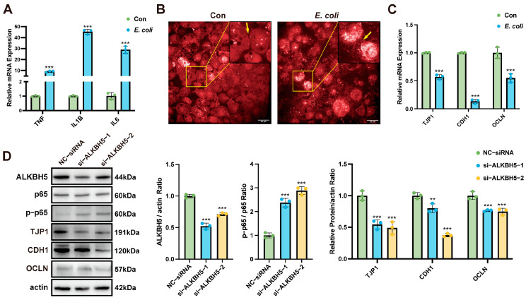
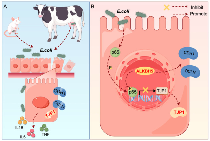
Mastitis poses a severe threat to the global cattle industry, causing huge economic losses. Environmental mastitis is mainly induced by Escherichia coli (E. coli), and the current treatment is still using antibiotics, with problems such as drug resistance and food safety. ALKBH5 is an RNA m6A demethylase that plays an important role in various biological processes, while p65 is a key regulator of inflammatory responses. Therefore, studying the interaction between ALKBH5 and p65 in protecting the mammary epithelial barrier provides new insights into the pathogenesis of mastitis. This study revealed that E. coli-induced acute inflammation activated the NF-κB/p65 signaling pathway and disrupted mammary epithelial cell tight junctions. Knockdown of ALKBH5 promoted p65 phosphorylation and inhibited the expressions of the tight junction proteins TJP1, CDH1, and OCLN. Furthermore, motif analysis, CHIP-PCR, and dual luciferase assay confirmed that phosphorylated p65 inhibited TJP1 promoter activity, thereby inhibiting TJP1 expression. In addition, the mouse experiment further demonstrated that knockdown of ALKBH5 aggravated E. coli-induced acute mastitis and epithelial cell tight junction disruption, and promoted E. coli invasion and proliferation. Significantly, this study is the first to demonstrate the details of the interaction between p65 and TJP1 and to declare the molecular mechanism of ALKBH5 in improving the cell tight junction, which lays a potential target and theoretical foundation for the treatment of mastitis and other infectious diseases.

Keywords: ALKBH5; E. coli; TJP1; mastitis; p65; tight junction.


Conflict of interest statement

The authors declare no conflicts of interest.

Figures

Figure 1
Figure 1
ALKBH5 inhibits E. coli-induced MAC-T cell tight junction disruption. (A) E. coli induced upregulation of TNF, IL1B, and IL6 mRNA expressions in MAC-T cells (24 h infection). Measured by RT-qPCR, normalized to ACTB; (B) E. coli induced tight junction disruption in MAC-T cells (24 h infection), yellow arrows indicate inter-cellular junction. Scale bar = 20 µm; (C) E. coli-induced down-regulation of TJP1, CDH1, and OCLN mRNA expressions in MAC-T cells (24 h infection). Measured by RT-qPCR, normalized to ACTB; (D) ALKBH5 knockdown induced p65 phosphorylation and inhibited TJP1, CDH1, and OCLN protein expressions. MAC-T cells transfected with ALKBH5 siRNA (24 h) followed by E. coli infection (24 h). Analyzed by Western blotting, with actin as loading control. Data are mean ± SD (n = 3 replicates per group, 3 independent experiments). ** p < 0.01, *** p < 0.001.
Figure 2
Figure 2
ALKBH5 mediates p65 to promote TJP1 expression. (A) Molecular docking analysis indicates that ALKBH5 binds to the p65 protein; (B) The DNA recognition motif of p65 perfectly matches part of the TJP1 promoter; (C) CHIP-PCR/qPCR confirmed that p65 binds to the TJP1 promoter; (D) Dual luciferase assays demonstrated that E. coli mediates p65 phosphorylation and translocation into the nucleus to inhibit the promoter activity of TJP1; (E) p65 inhibits TJP1 expression in MAC-T cells infected with E. coli for 24 h. Immunofluorescence staining shows p65 (yellow) and TJP1 (green) with DAPI (blue) indicating nuclei. Scale bar = 20 µm. Data are presented as mean ± SD (n = 3 replicates per group, 3 independent experiments), ns = no significant difference, *** p < 0.001.
Figure 3
Figure 3
E. coli induces mice mastitis and disrupts epithelial cell tight junctions. (A) Small animal live imaging demonstrating the proliferation and spread of bioluminescent E. coli in the mammary glands of mouse; (B) Appearance, dissection, and mammary tissue of control and E. coli-infected mouse; (C) Bacterial load measurement in mammary tissue of control and E. coli-infected mouse. Data are presented as median bacterial load (CFU/g); (D) TEM observation of the damage of mouse mammary gland epithelial cell junction. Blue arrows indicate mitochondria, green arrows indicate nucleus, and red arrows indicate epithelial junction structures. Scale bar = 2 µm; (E) HE and IHC detection of mammary tissue of control and E. coli-infected mouse, and quantification of p65 nuclear translocation by IHC. Scale bar = 50 µm. Data are presented as mean ± SD (n = 6 mice per group), *** p < 0.001.
Figure 4
Figure 4
Inhibition of ALKBH5 aggravates mastitis. (A) Construction and grouping of the mouse mastitis model. Mice were intraperitoneally injected with ALKBH5 inhibitor IOX1 or controls (5%DMSO or PBS). Mammary glands were injected with bioluminescent E. coli after 30 min; (B) RT-qPCR showed that IOX1 inhibited Alkbh5 expression and promoted E. coli-induced inflammatory cytokine expressions (Il1b, Il6, Tnf) in mouse mammary tissues. Data are normalized to Actb; (C) Dissection of mammary glands showing exacerbated tissue injury in mice treated with IOX1 + E. coli compared to E. coli alone; (D) Bacterial load measurement in mammary tissue showing higher E. coli concentrations in the IOX1 + E. coli group. Data are presented as median bacterial load (CFU/g); (E) Small animal live imaging demonstrating that IOX1 enhanced E. coli proliferation and spread in the mouse mammary glands. Luminescence signal intensity indicated bacterial load; (F) Three-dimensional modeling showed that IOX1 aggravated E. coli proliferation. Data are presented as mean ± SD (n = 6 mice per group), *** p < 0.001.
Figure 5
Figure 5
Inhibition of ALKBH5 aggravates epithelial cell tight junction disruption. (A) WB detected up-regulation of ALKBH5 and p-p65, and down-regulation of TJP1 in mouse mammary tissues during IOX1 induction. β-actin was used as a loading control; (B) TEM imaging of mammary glands of mice in the IOX1 group, the E. coli group, and the IOX1 + E. coli group. Blue arrows indicate mitochondria, green arrows indicate nucleus, and red arrows indicate epithelial junction structures. Scale bar = 2 µm; (C) HE and detection of mouse mammary tissue from the IOX1, E. coli, and IOX1 + E. coli groups, and quantification of p65 nuclear translocation by IHC. Scale bar = 50 µm. Data are presented as mean ± SD (n = 6 mice per group), ** p < 0.01, *** p < 0.001.
Figure 6
Figure 6
Schematic diagram of ALKBH5 protecting cell tight junction. (A) E. coli-induced mastitis disrupts epithelial cell tight junction; (B) ALKBH5 protects epithelial cell tight junction.

References

    1. Huma Z.I., Sharma N., Kour S., Lee S.J. Phenotypic and Molecular Characterization of Bovine Mastitis Milk Origin Bacteria and Linkage of Intramammary Infection with Milk Quality. Front. Vet. Sci. 2022;9:885134. doi: 10.3389/fvets.2022.885134. - DOI - PMC - PubMed
    1. Sharun K., Dhama K., Tiwari R., Gugjoo M.B., Yatoo M.I., Patel S.K., Pathak M., Karthik K., Khurana S.K., Singh R., et al. Advances in therapeutic and managemental approaches of bovine mastitis: A comprehensive review. Vet. Q. 2021;41:107–136. doi: 10.1080/01652176.2021.1882713. - DOI - PMC - PubMed
    1. Morales-Ubaldo A.L., Rivero-Perez N., Valladares-Carranza B., Velázquez-Ordoñez V., Delgadillo-Ruiz L., Zaragoza-Bastida A. Bovine mastitis, a worldwide impact disease: Prevalence, antimicrobial resistance, and viable alternative approaches. Vet. Anim. Sci. 2023;21:100306. doi: 10.1016/j.vas.2023.100306. - DOI - PMC - PubMed
    1. Xu H., Wu X., Yang Z., Shi X., Guo A., Hu C. N6-methyladenosine-modified lncRNA in Staphylococcus aureus-injured bovine mammary epithelial cells. Arch. Microbiol. 2024;206:431. doi: 10.1007/s00203-024-04156-4. - DOI - PubMed
    1. Saini V., McClure J., Léger D., Dufour S., Sheldon A., Scholl D., Barkema H. Antimicrobial use on Canadian dairy farms. J. Dairy Sci. 2012;95:1209–1221. doi: 10.3168/jds.2011-4527. - DOI - PubMed

Publication types

MeSH terms

Substances

LinkOut - more resources