. 2025 Apr 6;14(7):552.
doi: 10.3390/cells14070552.

Effects of Anti-CD20 Antibody Therapy on Immune Cell Dynamics in Relapsing-Remitting Multiple Sclerosis

Affiliations

Effects of Anti-CD20 Antibody Therapy on Immune Cell Dynamics in Relapsing-Remitting Multiple Sclerosis

Alice G Willison et al. Cells. .

Abstract

Introduction: The efficacy of anti-CD20 antibodies has significantly contributed to advancing our understanding of disease pathogenesis and improved treatment outcomes in relapsing-remitting multiple sclerosis (RRMS). A comprehensive analysis of the peripheral immune cell profile, combined with prospective clinical characterization, of RRMS patients treated with ocrelizumab (OCR) or ofatumumab (OFA) was performed to further understand immune reconstitution following B-cell depletion.

Methods: REBELLION-MS is a longitudinal analysis of RRMS patients treated with either OCR (n = 34) or OFA (n = 25). Analysis of B, T, natural killer (NK) and natural killer T (NKT) cells at baseline, month 1, and 12 was performed by multidimensional flow cytometry. Data were analyzed by conventional gating and unsupervised computational approaches. In parallel, different clinical parameters were longitudinally assessed. Twenty treatment-naïve age/sex-matched RRMS patients were included as the control cohort.

Results: B-cell depletion by OCR and OFA resulted in significant reductions in CD20+ T and B cells as well as B-cell subsets, alongside an expansion of CD5+CD19+CD20- B cells, while also elevating exhaustion markers (CTLA-4, PD-1, TIGIT, TIM-3) across T, B, NK, and NKT cells. Additionally, regulatory T-cell (TREG) numbers increased, especially in OCR-treated patients, and reductions in double-negative (CD3+CD4-CD8-) T cells (DN T cells) were observed, with these DN T cells having higher CD20 expression compared to CD4 or CD8 positive T cells. These immune profile changes correlated with clinical parameters, suggesting pathophysiological relevance in RRMS.

Conclusions: Our interim data add weight to the argumentation that the exhaustion/activation markers, notably TIGIT, may be relevant to the pathogenesis of MS. In addition, we identify a potentially interesting increase in the expression of CD5+ on B cells. Finally, we identified a population of double-negative T cells (KLRG1+HLADR+, in particular) that is associated with MS activity and decreased with CD20 depletion.

Trial registration: ClinicalTrials.gov NCT06586177.

Keywords: autoimmunity; immune reconstitution; multiple sclerosis; ocrelizumab; ofatumumab.


Conflict of interest statement

AGW reports personal fees from Merck, travel reimbursements and meeting attendance fees from Novartin, Merck, and Sanofi; RH declares no relevant competing interests; MW declares no relevant competing interests; SE declares no relevant competing interests; NH reports personal fees from ArgenX, Merck, Novartis, and Viatris, travel reimbursements and meeting attendance fees from Alexion, ArgenX, Merck, and Novartis, research support by the UKD FUTURE program of the Deutsche Forschungsgesellschaft outside the scope of this study; LM reports no conflicts of interest related to this study; he has received honoraria for lecturing, consulting, and travel expenses for attending meetings from Biogen, Merck, Sanofi, argenX, Roche, Alexion, and Novartis, all outside the scope of this work, his research is funded by the German Multiple Sclerosis Foundation (DMSG) and the Deutsche Forschungsgemeinschaft (DFG, German Research Foundation)-493659010; SP received travel grants from Sanofi–Aventis, Alexion, Novartis, and Merck, lecturing honoraria or compensation for consulting from Alexion, Hexal, Merck, Novartis, Sanofi–Aventis, Mylan Healthcare, Roche, and Biogen, and research support from Diamed, Merck, Biogen, Novartis, and the German Multiple Sclerosis Society North-Rhine-Westphalia; SL declares no relevant competing interests; LMP declares no relevant competing interests; LR received travel reimbursements from Merck Serono and Sanofi–Aventis; MÖ declares no relevant competing interests; TR reports grants from German Ministry of Education, Science, Research and Technology, grants from the German Research Foundation (DFG), grants and personal fees from Sanofi, Argenx, and Alexion, personal fees from Biogen, Roche, UCB, Novartis, and Teva, personal fees and nonfinancial support from Merck, outside the submitted work; NM has received honoraria for lecturing and travel expenses for attending meetings from Biogen Idec, GlaxoSmith Kline, Teva, Novartis Pharma, Bayer Healthcare, Genzyme, Alexion Pharmaceuticals, Fresenius Medical Care, Diamed, UCB Pharma, AngeliniPharma, Bial, and Sanofi–Aventis, has received royalties for consulting from UCB Pharma, Alexion Pharmaceuticals, and Sanofi–Aventis, and has received financial research support from Euroimmun, Fresenius Medical Care, Diamed, Alexion Pharmaceuticals, and Novartis Pharma; MK received honoraria for lecturing and consulting and travel expenses for attending meetings from Biogen, Hexal, Merck Healthcare Germany, Neuraxpharm, Novartis, and Sanofi, and research support from Merck Healthcare Germany, Novartis, Viatris, and the German Multiple Sclerosis Society; SLH currently serves on the scientific advisory board of Accure, Alector, Annexon, and Hinge Therapeutics, he has previously consulted for BD, Moderna, NGM Bio, Nurix Therapeutics, Pheno Therapeutics, and previously served on the Board of Directors of Neurona, Dr. Hauser also has received travel reimbursement and writing support from F. Hoffmann–La Roche and Novartis AG for anti-CD20-therapy-related meetings and presentations; HPH received fees for serving on steering and data monitoring committees from Aurinia Pharma, BMS Celgene, Boehringer Ingelheim, Hoffmann–La Roche, Merck, Novartis, Octapharma, TG Therapeutics; PAL contributes to the development of LCMV for clinical application in oncology in cooperation with, as founder of and as advisor to, Abalos Therapeutics GmbH; MP received honoraria for lecturing and travel expenses for attending meetings from Alexion, ArgenX, Bayer Health Care, Biogen, Hexal, Merck Serono, Novartis, Roche, Sanofi–Aventis, Takeda, and Teva, his research is funded by ArgenX, Biogen, Demecan, Hexal, Horizon Merck Serono, Novartis, Roche, Viatris, Takeda, and Teva; SR received travel grants from Merck Healthcare Germany GmbH, Alexion Pharmaceuticals, and Bristol Myers Squibb, she served on a scientific advisory board from Merck Healthcare Germany GmbH and received honoraria for lecturing from Roche and Merck Healthcare Germany GmbH, her research was supported by Novartis, ’Stiftung zur Förderung junger Neurowissenschaftler’, and ’Else Kröner-Fresenius-Stiftung’; SGM receives honoraria for lecturing, and travel expenses for attending meetings from Academy 2, Argenx, Alexion, Almirall, Amicus Therapeutics Germany, Bayer Health Care, Biogen, BioNtech, BMS, Celgene, Datamed, Demecan, Desitin, Diamed, Diaplan, DIU Dresden, DPmed, Gen Medicine and Healthcare products, Genzyme, Hexal AG, IGES, Impulze GmbH, Janssen Cilag, KW Medipoint, MedDay Pharmaceuticals, Merck Serono, MICE, Mylan, Neuraxpharm, Neuropoint, Novartis, Novo Nordisk, ONO Pharma, Oxford PharmaGenesis, QuintilesIMS, Roche, Sanofi–Aventis, Springer Medizin Verlag, STADA, Chugai Pharma, Teva, UCB, Viatris, Wings for Life international and Xcenda, his research is funded by the German Ministry for Education and Research (BMBF), Bundesinstitut für Risikobewertung (BfR), Deutsche Forschungsgemeinschaft (DFG), Else Kröner Fresenius Foundation, Gemeinsamer Bundesausschuss (G-BA), German Academic Exchange Service, Hertie Foundation, Interdisciplinary Center for Clinical Studies (IZKF) Muenster, German Foundation Neurology and Alexion, Almirall, Amicus Therapeutics Germany, Biogen, Diamed, DGM e.v., Fresenius Medical Care, Genzyme, Gesellschaft von Freunden und Förderern der Heinrich-Heine-Universität Düsseldorf e.V., HERZ Burgdorf, Merck Serono, Novartis, ONO Pharma, Roche, and Teva.

Figures

Figure 1
Figure 1
Study design for evaluating OCR and OFA therapies in RRMS patients. The cohort was assessed at months 0, 1, and 12 for the following parameters: basic disease characteristics (only month 0), EDSS, relapse documentation, and immunological analyses using PBMCs. Figure 1 was created in BioRender (https://biorender.com/, last accessed on 27 March 2025) Willison, A. (2024) BioRender.com/n12e680 [6]. EDSS—expanded disability status scale; OCR—ocrelizumab; OFA—ofatumumab; PBMCs—peripheral blood mononuclear cells.
Figure 2
Figure 2
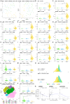
Reduction in CD20+ B and T cells, while CD5 expression was increased on B cells of OCR- and OFA-treated compared to treatment-naïve RRMS patients. (A) UMAP analysis including the PB immune cell subsets assessed by mFC. Each colored symbol presents an individual patient. (BM) Violin plots with overlaying boxplots illustrating CD20⁺ B cells, CD20+ T cells, and various B cell subsets (% of living cells) in the PB of OCR-treated, OFA-treated, and treatment-naïve RRMS patients. Boxes show the median and the 25th as well as the 75th percentiles. The whiskers extend from the hinge to the largest and smallest values, respectively, but no further than 1.5 * IQR from the hinge. p-values were calculated using analysis of variance (ANOVA) with post hoc Tukey Honestly Significant Difference (HSD), if normality could be assumed based on the Shapiro–Wilk test, otherwise the Kruskal–Wallis test was used with the Dunn post hoc test (p-adjustment method: Benjamini–Hochberg). (N) Ridge plots illustrating the MFI of CD5 on B cells of OCR- and OFA-treated compared to treatment-naïve RRMS patients. p-values were calculated using the Kruskal–Wallis test with the Dunn post hoc test (p-adjustment method: Benjamini–Hochberg) as normality could not be assumed based on the Shapiro–Wilk test. (O) Opt-SNE plots with B-cell clusters identified by FlowSOM. Opt-SNE plot including mFC data from OCR-treated, OFA-treated, and treatment-naïve RRMS patients were created with the platform OMIQ from Dotmatics (www.omiq.ai, www.dotmatics.com, last accessed on 27 March 2025) [8] using the default parameters (max iterations = 1000, opt-SNE end = 5000, perplexity = 30, theta = 0.5, components = 2, random seed = 1759, verbosity = 25). The algorithm FlowSOM (xdim = 12, ydim = 12, rlen = 10, distance metric euclidean) was used for cluster identification. (P) Violin plots with overlaying box plots depicting CD19+CD20CD5+ B cells (% of living cells) identified by FlowSOM. Boxes show the median and the 25th as well as the 75th percentiles. The whiskers extend from the hinge to the largest and smallest values, respectively, but no further than 1.5 * IQR from the hinge. p-values were calculated using the Kruskal–Wallis test with the Dunn post hoc test (p-adjustment method: Benjamini–Hochberg) as normality could not be assumed based on the Shapiro–Wilk test. (Q) Correlation analysis between the CD5 MFI of B cells and EDSS as well as time since last relapse. Spearman correlation coefficients were calculated as normality of data could not be assumed. Bc—B cell; Breg—regulatory B cells; EDSS—expanded disability status scale; m—month(s); MFI—mean fluorescent intensity; OCR—ocrelizumab; OFA—ofatumumab; PB—peripheral blood; tnRRMS—treatment-naïve relapsing-remitting multiple sclerosis; tnOCR/OFA—OCR- and OFA-treated RRMS patients who were treatment-naïve at m0, UMAP: Uniform Manifold Approximation and Projection for Dimension Reduction. Statistical significance: p ≤ 0.05 (*), p ≤ 0.001 (***), p ≤ 0.0001 (****).
Figure 3
Figure 3
Increased expression of exhaustion/activation markers on T cells of OCR- and OFA-treated RRMS patients. Ridge plots illustrating the MFIs of different exhaustion/activation markers on T cells (AK) of OCR- and OFA-treated compared to treatment-naïve RRMS patients. p-values were calculated using analysis of variance (ANOVA) with post-hoc Tukey Honestly Significant Difference (HSD), if normality could be assumed based on the Shapiro–Wilk test, otherwise the Kruskal–Wallis test was used with the Dunn post hoc test (p-adjustment method: Benjamini–Hochberg). m—month(s); MFI—mean fluorescent intensity; OCR—ocrelizumab; OFA—ofatumumab; RRMS—relapsing-remitting multiple sclerosis; tnRRMS—treatment-naïve relapsing-remitting multiple sclerosis. Statistical significance: p ≤ 0.05 (*), p ≤ 0.01 (**), p ≤ 0.001 (***), p ≤ 0.0001 (****).
Figure 4
Figure 4
Expression of exhaustion/activation markers on immune cells correlates with clinical parameters of RRMS patients. Correlation analysis between different exhaustion/activation markers on T cells (AH), B cells (IL), NKT cells (MP), and NK cells (Q) and EDSS as well as time since disease manifestation. Spearman correlation coefficients were calculated as normality of data could not be assumed. EDSS—expanded disability status scale; m—month(s); MFI—mean fluorescent intensity; NK—natural killer cells; OCR—ocrelizumab; OFA—ofatumumab; RRMS—relapsing-remitting multiple sclerosis; tnOCR/OFA—OCR- and OFA-treated RRMS patients who were treatment- naïve at m0; tnRRMS—treatment-naïve relapsing-remitting multiple sclerosis.
Figure 5
Figure 5
Higher expression of exhaustion/activation markers on B, NK, and NKT cells of OCR- and OFA-treated RRMS patients. Ridge plots illustrating the MFIs of different exhaustion/activation markers on B, NK, and NKT cells (AM) of OCR- and OFA-treated compared to treatment-naïve RRMS patients. p-values were calculated using analysis of variance (ANOVA) with post hoc Tukey Honestly Significant Difference (HSD), if normality could be assumed based on the Shapiro–Wilk test, otherwise the Kruskal–Wallis test was used with the Dunn post hoc test (p-adjustment method: Benjamini–Hochberg). m—month(s); MFI—mean fluorescent intensity; NK—natural killer cells; OCR—ocrelizumab; OFA—ofatumumab; RRMS—relapsing-remitting multiple sclerosis; tnRRMS—treatment-naïve relapsing-remitting multiple sclerosis. Statistical significance: p ≤ 0.05 (*), p ≤ 0.001 (***), p ≤ 0.0001 (****).
Figure 6
Figure 6
Increase in regulatory T cells and reduction in double negative T cells in OCR- and OFA-treated RRMS patients. (A) Violin plots with overlaying boxplots showing TREGs (% of living cells) in the PB of OCR-treated, OFA-treated, and treatment-naïve RRMS patients. Boxes show the median and the 25th as well as the 75th percentiles. The whiskers extend from the hinge to the largest and smallest values, respectively, but no further than 1.5 * IQR from the hinge. p-values were calculated using the Kruskal–Wallis test with the Dunn post hoc test (p-adjustment method: Benjamini–Hochberg) as normality could not be assumed based on the Shapiro–Wilk test. (B) Opt-SNE plots with T cell clusters identified by FlowSOM. Opt-SNE plot including mFC data from OCR-treated, OFA-treated, and treatment-naïve RRMS patients were created with the platform OMIQ from Dotmatics (www.omiq.ai, www.dotmatics.com) using the default parameters (max iterations = 1000, opt-SNE end = 5000, perplexity = 30, theta = 0.5, components = 2, random seed = 1759, verbosity = 25). The algorithm FlowSOM (xdim = 12, ydim = 12, rlen = 10, distance metric euclidean) was used for cluster identification. (CE) Violin plots with overlaying box plots depicting KLRG1HLADR+ DN T cells (C), KLRG1+HLADR DN T cells (D), and KLRG1+HLADR+ DN T cells (E). Boxes show the median and the 25th as well as the 75th percentiles. The whiskers extend from the hinge to the largest and smallest values, respectively, but no further than 1.5 * IQR from the hinge. p-values were calculated using analysis of variance (ANOVA) with post hoc Tukey Honestly Significant Difference (HSD), if normality could be assumed based on the Shapiro–Wilk test, otherwise the Kruskal–Wallis test was used with the Dunn post hoc test (p-adjustment method: Benjamini–Hochberg). (F) Violin plots with overlaying boxplots illustrating the percentage of CD20+ T cells among all DN T cells compared to the percentage of CD20+ T cells among all CD4+ and/or CD8+ T cells. Boxes show the median and the 25th as well as the 75th percentiles. The whiskers extend from the hinge to the largest and smallest values, respectively, but no further than 1.5 * IQR from the hinge. p-values were calculated with paired Wilcoxon’s rank-sum test. (GI) Correlation analysis between TREGs and EDSS at m1 (G), KLRG1HLADR+ DN T cells and relapse quantity at m0 (H), as well as KLRG1HLADR+ DN T cells and EDSS at m12 (I). Spearman correlation coefficients were calculated as normality of data could not be assumed. DN Tc—CD4CD8 T cells; EDSS—expanded disability status scale; m—month(s); OCR—ocrelizumab; OFA—ofatumumab; pos. Tc—CD4+ and/or CD8+ T cells; PB—peripheral blood; RRMS—relapsing-remitting multiple sclerosis; tnRRMS—treatment-naïve relapsing-remitting multiple sclerosis; tnOCR/OFA—OCR- and OFA-treated RRMS patients who were treatment- naïve at m0; Tc—T cells; Treg—regulatory T cells. Statistical significance: p ≤ 0.05 (*), p ≤ 0.01 (**), p ≤ 0.001 (***), p ≤ 0.0001 (****).

References

    1. Gelfand J.M., Cree B.A.C., Hauser S.L. Ocrelizumab and Other CD20(+) B-Cell-Depleting Therapies in Multiple Sclerosis. Neurotherapeutics. 2017;14:835–841. doi: 10.1007/s13311-017-0557-4. - DOI - PMC - PubMed
    1. Hauser S.L., Bar-Or A., Cohen J.A., Comi G., Correale J., Coyle P.K., Cross A.H., de Seze J., Leppert D., Montalban X., et al. Ofatumumab versus Teriflunomide in Multiple Sclerosis. N. Engl. J. Med. 2020;383:546–557. doi: 10.1056/NEJMoa1917246. - DOI - PubMed
    1. Hauser S.L., Bar-Or A., Comi G., Giovannoni G., Hartung H.-P., Hemmer B., Lublin F., Montalban X., Rammohan K.W., Selmaj K., et al. Ocrelizumab versus Interferon Beta-1a in Relapsing Multiple Sclerosis. N. Engl. J. Med. 2017;376:221–234. doi: 10.1056/NEJMoa1601277. - DOI - PubMed
    1. Frisch E.S., Pretzsch R., Weber M.S. A Milestone in Multiple Sclerosis Therapy: Monoclonal Antibodies Against CD20—Yet Progress Continues. Neurotherapeutics. 2021;18:1602–1622. doi: 10.1007/s13311-021-01048-z. - DOI - PMC - PubMed
    1. Thompson A.J., Banwell B.L., Barkhof F., Carroll W.M., Coetzee T., Comi G., Correale J., Fazekas F., Filippi M., Freedman M.S., et al. Diagnosis of multiple sclerosis: 2017 revisions of the McDonald criteria. Lancet Neurol. 2018;17:162–173. doi: 10.1016/S1474-4422(17)30470-2. - DOI - PubMed

Publication types

MeSH terms

Associated data

LinkOut - more resources