. 2025 Mar 21;15(7):475.
doi: 10.3390/nano15070475.

A New Method for Accelerated Aging of Nanoparticles to Assess the Colloidal Stability of Albumin-Coated Magnetic Nanoparticles

Affiliations

A New Method for Accelerated Aging of Nanoparticles to Assess the Colloidal Stability of Albumin-Coated Magnetic Nanoparticles

Boris Nikolaev et al. Nanomaterials (Basel). .

Abstract

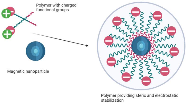
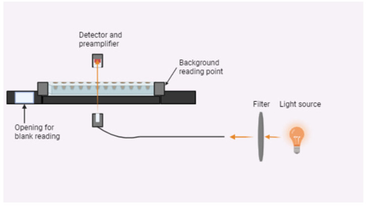
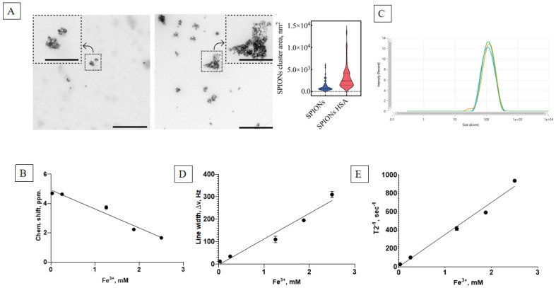
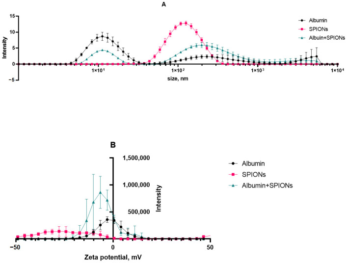
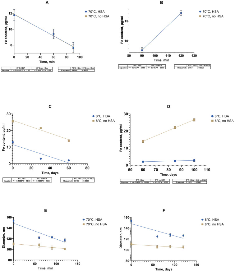
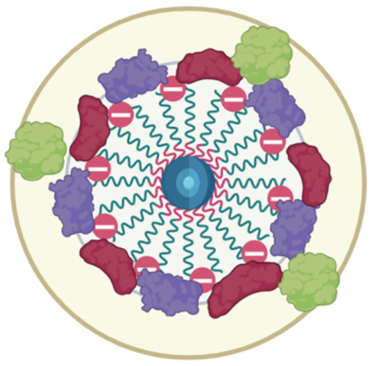
The colloidal long-storage stability of nanosized drugs is a crucial factor for pharmacology, as they require much time for robust estimation. The application of bioavailable magnetic nanosuspensions in theranostics is limited by incomplete information about their colloidal stability in the internal media of human organisms. A method for the accelerated temperature stress "aging" of magnetic nanosized suspensions is proposed for the rapid assessment and prediction of the colloidal stability over time of nanosized iron oxide suspensions stabilized by albumin HSA. Colloidal stability is assessed using dynamic light scattering (DLS), fluorescence spectroscopy, electrophoresis, and ion monitoring methods during short- and long-term storage. Rapid assessment is achieved by short high-temperature (70 °C) processing of carboxymethyl-dextran-coated nanosol in the presence of albumin. The role of albumin in the sustained stability of superparamagnetic iron oxide particles (SPIONs) was studied under conditions mimicking blood plasma (pH = 7.4) and endolysosomal cell compartments (pH = 5.5). According to the fluorescence quenching and DLS data, colloidal stability is ensured by the formation of an HSA corona on carboxymethyl-dextran-coated SPIONs and their process of clustering. In the presence of albumin, the colloidal stability of nanoparticles is shown to increase from 80 to 121 days at a storage temperature of 8 °C The prognostic shelf life of magnetic nanosol is estimated by calculating the Van't Hoff's relation for the rate of chemical reactions. The validity of using the Van't Hoff's rule is confirmed by the agreement of the calculated activation energy at 8 °C and 70 °C. The developed method of the accelerated aging of nanoparticles can not only be employed for the estimation of the shelf life of magnetic nanoparticles coated with HSA in vitro but also for assessing the stability of SPIONs applied in vivo.

Keywords: SPIONs; metal oxide nanoparticles; method of accelerated aging; nanoparticle aggregation; nanoparticle stability; protein corona; superparamagnetic iron oxide nanoparticles.


Conflict of interest statement

All authors declare no conflicts of interest.

Figures

Figure 1
Figure 1
Electrosteric coating of nanoparticles.
Figure 2
Figure 2
Turbidimetry experiment scheme.
Figure 3
Figure 3
Physicochemical characterization of the magnetic nanoparticles. (A) TEM images of CMDx-SPION clusters without (left panel) and with HSA (center panel). The right panel presents the distribution of cluster areas (scaled by ×10⁻3 nm2 for graphical clarity). Scale bars: 500 nm for the main images; 200 nm for insets. The violin plot shows the median cluster areas (dashed lines) and interquartile ranges (25% to 75%, dotted lines), illustrating the central tendency and variability in cluster sizes. The width of each distribution in the violin plot reflects the density of data points in different cluster areas, with broader sections indicating a higher concentration of values. (B) Chemical shift dependence on the SPION concentration in water. (C) Size distribution of the nanoparticles. (D) Line width dependence on the SPION concentration in water. (E) Relaxation time of the SPION water suspensions.
Figure 4
Figure 4
Analysis of the albumin–nanoparticle interactions according to the dependence of the albumin fluorescence intensity (340 nm) on the Fe+3 content in SPIONs: (A,B) fluorescence intensity of the SPIONs with BSA and HSA, respectively, in PBS; (C,D) fluorescence intensity of the SPIONs with BSA and HSA, respectively, at a set pH; (E) calculated quenching constants.
Figure 5
Figure 5
DLS results for SPIONs upon incubation with albumin: (A) size distribution; (B) zeta-potential distribution.
Figure 6
Figure 6
Turbidity of the magnetic suspension of SPION/albumin at a light absorbance of 405 nm after a 2 h treatment at an ambient temperature of 22–25 °C after 5 days of storage.
Figure 7
Figure 7
Physicochemical parameters of the SPIONs in the stress test: (A) Fe3+ content; (B) hydrodynamic diameter; (C) polydispersity index. n = 3 (for each parameter).
Figure 8
Figure 8
Iron content in the SPION sedimentation test after heating with HSA. ****—p < 0.0001; ns—non-significant.
Figure 9
Figure 9
Properties of SPIONs during prolonged storage: (A) Fe3+ content; (B) hydrodynamic diameter; (C) polydispersity index. n = 3 (for each parameter).
Figure 10
Figure 10
Storage stability of SPIONs in the dispersion during long cold storage (8 °C) and after short temperature stress action (70 °C) in the presence albumin: (A,B) Fe3+ release in degradation after temperature stress; (C,D) Fe3+ release in degradation during long storage at 8 °C; (E,F) nanocluster size dynamics for short stress and long storage, respectively. Control sample—no albumin added.
Figure 11
Figure 11
Schematic view of the sera protein (including albumin) corona around magnetic iron oxide nanoparticles stabilized by carboxymethyl dextran.

Similar articles

References

    1. Gareev K.G., Grouzdev D.S., Koziaeva V.V., Sitkov N.O., Gao H., Zimina T.M., Shevtsov M. Biomimetic Nanomaterials: Diversity, Technology, and Biomedical Applications. Nanomaterials. 2022;12:2485. doi: 10.3390/nano12142485. - DOI - PMC - PubMed
    1. Zhou Y., Liu R., Shevtsov M., Gao H. When imaging meets size-transformable nanosystems. Adv. Drug Deliv. Rev. 2022;183:114176. doi: 10.1016/j.addr.2022.114176. - DOI - PubMed
    1. Shevtsov M., Kaesler S., Posch C., Multhoff G., Biedermann T. Magnetic nanoparticles in theranostics of malignant melanoma. EJNMMI Res. 2021;11:127. doi: 10.1186/s13550-021-00868-6. - DOI - PMC - PubMed
    1. Han H., Yao Y., Robinson R.D. Interplay between Chemical Transformations and Atomic Structure in Nanocrystals and Nanoclusters. Acc. Chem. Res. 2021;54:509–519. doi: 10.1021/acs.accounts.0c00704. - DOI - PubMed
    1. Chatterjee K., Sarkar S., Jagajjanani Rao K., Paria S. Core/shell nanoparticles in biomedical applications. Adv. Colloid Interface Sci. 2014;209:8–39. doi: 10.1016/j.cis.2013.12.008. - DOI - PubMed

LinkOut - more resources