

Pre-treatment and post-treatment nasopharyngeal carcinoma imaging: imaging updates, pearls and pitfalls
- PMID: 40214770
- PMCID: PMC12041163
- DOI: 10.1007/s00234-025-03596-z
Pre-treatment and post-treatment nasopharyngeal carcinoma imaging: imaging updates, pearls and pitfalls
Abstract
Purpose: Nasopharyngeal carcinoma (NPC) is endemic in Southeast Asia, requiring precise imaging for personalized treatment. This review highlights key imaging challenges and updates from recent literature, emphasizing findings that impact oncological management.
Methods: We discuss common and uncommon clinical entities, detailing salient imaging features and diagnostic distinctions to aid accurate interpretation.
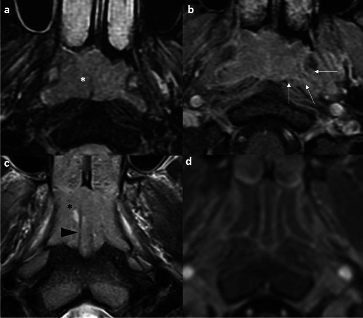
Results: In the pre-treatment setting, leveraging the characteristic MR signals and spread patterns of NPC aids in defining the tumor volume for accurate staging and radiotherapy contouring. Key diagnostic challenges include differentiating tumor from benign hyperplasia, skull base osteomyelitis, and other skull base tumors. Perineural tumor spread, radiological extranodal extension and nodal necrosis further refine primary tumor and nodal assessment. In the post-treatment setting, the key question is whether tumor recurrence exists. Diagnostic challenges involve distinguishing tumor recurrence from scar tissue, post-radiation nasopharyngeal necrosis, or hypertrophied cervical ganglia. For recurrences, endoscopic nasopharyngectomy has emerged as the preferred approach over open surgery or re-irradiation. The text highlights characteristic post-treatment appearances and emphasizes recognizing these patterns to avoid misinterpretation and guide appropriate management.
Conclusion: Imaging plays a pivotal role in NPC precision oncology. Mastering imaging pearls and pitfalls empowers radiologists to provide clinicians with reliable, actionable guidance.
Keywords: Computed tomography; Diffusion-weighted imaging; Imaging; Magnetic resonance imaging; Nasopharyngeal carcinoma; Positron emission tomography.
© 2025. The Author(s).
Conflict of interest statement
Declarations. Ethical approval: The study was approved by the Central Institutional Review Board (CIRB-2024–441-2) and complied with the Declaration of Helsinki. Informed consent was waived, given the retrospective nature of the study. Conflict of interest: The authors declare no competing interests.
Figures


Similar articles
-
Image-based diagnosis of residual or recurrent nasopharyngeal carcinoma may be a phantom tumor phenomenon.Medicine (Baltimore). 2021 Feb 26;100(8):e24555. doi: 10.1097/MD.0000000000024555. Medicine (Baltimore). 2021. PMID: 33663063 Free PMC article.
-
Management of locally recurrent nasopharyngeal carcinoma.Cancer Treat Rev. 2019 Sep;79:101890. doi: 10.1016/j.ctrv.2019.101890. Epub 2019 Aug 21. Cancer Treat Rev. 2019. PMID: 31470314 Review.
-
PET/CT: Nasopharyngeal Cancers.PET Clin. 2022 Apr;17(2):285-296. doi: 10.1016/j.cpet.2021.12.006. Epub 2022 Mar 4. PET Clin. 2022. PMID: 35256301 Review.
-
Machine Learning Methods for Optimal Radiomics-Based Differentiation Between Recurrence and Inflammation: Application to Nasopharyngeal Carcinoma Post-therapy PET/CT Images.Mol Imaging Biol. 2020 Jun;22(3):730-738. doi: 10.1007/s11307-019-01411-9. Mol Imaging Biol. 2020. PMID: 31338709
-
Pitfalls in the staging of cancer of nasopharyngeal carcinoma.Neuroimaging Clin N Am. 2013 Feb;23(1):9-25. doi: 10.1016/j.nic.2012.08.006. Neuroimaging Clin N Am. 2013. PMID: 23199659 Review.
References
-
- King AD (2022) MR imaging of nasopharyngeal carcinoma. Magn Reson Imaging Clin N Am 30(1):19–33. 10.1016/j.mric.2021.06.015 - PubMed
-
- Liang SB, Sun Y, Liu LZ, Chen Y, Chen L, Mao YP, Tang LL, Tian L, Lin AH, Liu MZ, Li L, Ma J (2009) Extension of local disease in nasopharyngeal carcinoma detected by magnetic resonance imaging: improvement of clinical target volume delineation. Int J Radiat Oncol Biol Phys 75(3):742–750. 10.1016/j.ijrobp.2008.11.053 - PubMed
Publication types
MeSH terms
LinkOut - more resources
Full Text Sources
Medical
