. 2025 Apr 11;48(1):359.
doi: 10.1007/s10143-025-03511-3.

Beyond the blood-brain barrier: feasibility and technical validation of dual-compartment circulating tumor cells detection in high-grade glioma patients

Affiliations

Beyond the blood-brain barrier: feasibility and technical validation of dual-compartment circulating tumor cells detection in high-grade glioma patients

Yu-Chung Juan et al. Neurosurg Rev. .

Abstract

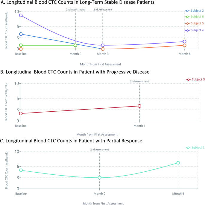
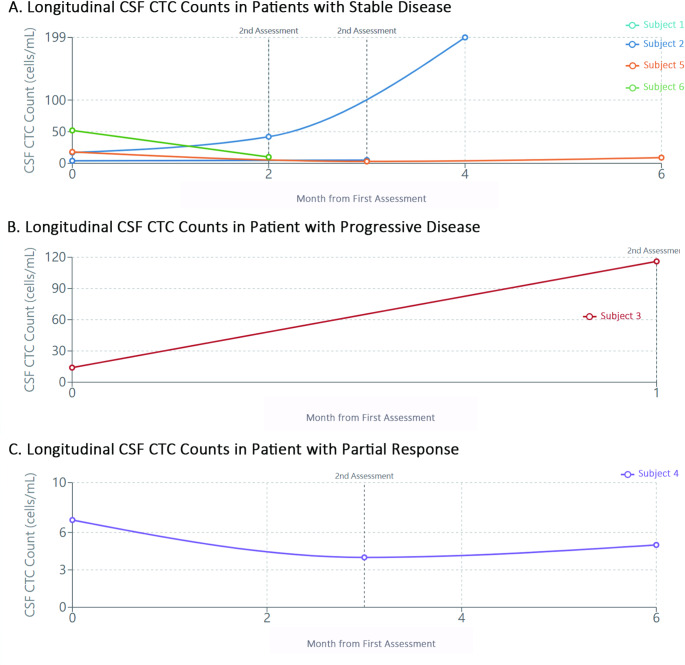
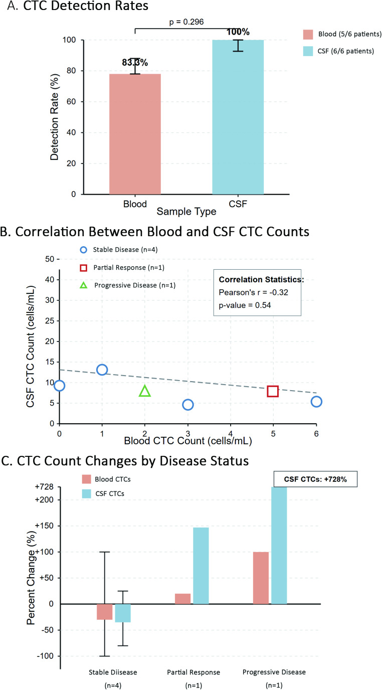
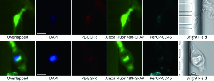
The elusive nature of brain tumor progression, hidden behind the blood-brain barrier, presents significant challenges for treatment monitoring in high-grade gliomas. In this feasibility study, we evaluate a novel approach to tracking glioblastoma through liquid biopsy, assessing whether tumor cells leave detectable molecular footprints in both blood and cerebrospinal fluid (CSF). Using the MiSelect R II System with specialized microfluidic technology, we analyzed paired blood and CSF samples from six glioblastoma patients, revealing a striking presence of circulating tumor cells (CTCs)- with higher abundance in CSF, where detection rates reached 100% compared to 83.3% in blood. Our technical validation demonstrates the system's capability to identify CTCs through multi-marker analysis (EGFR+/GFAP+/CD45-). Preliminary observations revealed higher CTC counts in CSF (median 15.5 cells/mL) compared to blood (median 3.0 cells/mL), with notable differences between compartments suggesting they may reflect distinct aspects of disease biology. In a patient who developed progressive disease, we observed a substantial increase in CSF CTCs from 14 to 116 cells/mL, warranting further investigation in larger cohorts. Additionally, we detected CTC clusters in both compartments, an intriguing finding with potential biological significance. While our interim analysis provides technical proof-of-concept for CTC detection in glioblastoma patients, the limited sample size precludes definitive conclusions regarding clinical utility. These findings establish a methodological foundation for future comprehensive studies exploring the relationship between CTC dynamics and clinical outcomes in high-grade gliomas.

Keywords: Biomarkers; Cerebrospinal fluid; Circulating tumor cells (CTCs); Glioblastoma; High-grade glioma; Liquid biopsy; MiSelect R II system; Microfluidic technology.


Conflict of interest statement

Declarations. Ethics approval and consent to participate: This study was approved by the Institutional Review Board of China Medical University and Hospital in Taiwan (approval No. CMUH112-REC2-103). The trial is registered at ClinicalTrials.gov (registration number pending). The research in accordance with local regulations and international standards, including the Declaration of Helsinki. Consent to participate: Informed consent was obtained from all individual participants included in the study. Competing interests: The authors declare no competing interests.

Figures

Fig. 1
Fig. 1
Longitudinal Analysis of Circulating Tumor Cells in Blood Samples from Glioblastoma Patients. Figure 1 shows the longitudinal monitoring of circulating tumor cell (CTC) counts in peripheral blood samples from six patients with IDH-wild type glioblastoma. Patients are grouped according to their clinical disease status at follow-up. (A) Blood CTC counts in patients with stable disease (n = 4), showing relatively consistent or decreasing counts over time. (B) Blood CTC dynamics in a patient with progressive disease (Subject 3), demonstrating an increase in CTC count from 2 to 4 cells/mL coinciding with clinical progression. (C) Blood CTC pattern in a patient with partial response followed by progression (Subject 1), showing an initial decrease followed by an increase in CTC count corresponding to disease progression. These preliminary findings suggest potential utility of blood CTC monitoring as a complementary approach to conventional imaging in assessing treatment response. CTC counts are presented as cells per mL of blood. Time points represent baseline (post-surgery) and follow-up assessments. Dotted vertical lines indicate clinical assessment timepoints. Error bars are not included as measurements represent single determinations at each timepoint
Fig. 2
Fig. 2
Longitudinal Analysis of Circulating Tumor Cells in Cerebrospinal Fluid Samples from Glioblastoma Patients. Figure 2 illustrates the longitudinal monitoring of circulating tumor cell (CTC) counts in cerebrospinal fluid (CSF) samples from the same six patients with IDH-wild type glioblastoma. (A) CSF CTC dynamics in patients with stable disease (n = 4), showing relatively consistent counts over the observation period. (B) CSF CTC pattern in a patient with progressive disease (Subject 3), demonstrating a substantial increase from 14 to 116 cells/mL, preceding clinical evidence of disease progression. (C) CSF CTC counts in a patient with partial response followed by progression (Subject 1), showing a pattern of increase corresponding to disease status changes. These preliminary observations suggest that CSF CTCs may be a more sensitive indicator of disease status compared to blood CTCs in high-grade glioma patients. CTC counts are presented as cells per mL of CSF. Time points represent baseline (post-surgery) and follow-up assessments. Dotted vertical lines indicate clinical assessment timepoints. Error bars are not included as measurements represent single determinations at each timepoint
Fig. 3
Fig. 3
Comparison of CTC Detection in Blood and CSF Samples and Relationship to Disease Status. Figure 3 presents a comprehensive analysis of circulating tumor cell (CTC) detection in matched blood and CSF samples from glioblastoma patients. (A) Detection rates of CTCs in blood versus CSF samples across all patients (n = 6), demonstrating the higher sensitivity of CSF sampling (100%) compared to blood sampling (83.3%). (B) Correlation analysis between blood and CSF CTC counts at baseline assessment, showing a negative correlation (r = -0.525) that suggests these two compartments may reflect different aspects of disease biology. (C) Percent change in CTC counts between baseline and second assessment grouped by disease status, highlighting the substantial increase in CSF CTC count in the progressive disease case compared to stable disease and partial response cases. These findings support the potential complementary role of dual-compartment CTC monitoring in assessing treatment response in glioblastoma patients. In Panel A, detection rates were compared using Fisher’s exact test with 95% confidence intervals calculated using Wilson methods. In Panel B, different symbols represent disease status at second assessment. In Panel C, error bars represent standard error of the mean; for Progressive Disease and Partial Response (n = 1 each), no error bars are shown as these represent single patient observations
Fig. 4
Fig. 4
Identification of Circulating Tumor Cells in High-Grade Glioma Patients Using Multi-Marker Immunofluorescence. Figure 4 demonstrates the identification and characterization of circulating tumor cells (CTCs) in cerebrospinal fluid from a high-grade glioma patient using the MiSelect R II System. Representative immunofluorescence images show a CTC identified based on multiple marker expression: EGFR positivity (red), GFAP positivity (green), nuclear staining with DAPI (blue), and absence of the leukocyte marker CD45. The bright field image reveals the morphological features of the isolated CTC and CTC cluster. This multi-parametric approach enables reliable identification of CTCs originating from high-grade gliomas, distinguishing them from other cellular components in the CSF. CTCs were identified based on the following criteria: EGFR+ (red), GFAP+ (green), DAPI + nuclear staining (blue), and CD45- (absence of leukocyte marker). Scale bar represents 10 μm. Images were acquired using the MiSelect R II System with standardized exposure parameters. CTC cluster showed the same immunophenotypes with CTCs and defined as aggregates of two or more CTCs

Similar articles

References

    1. Fabro F, Lamfers MLM, Leenstra S (2022) Advancements, challenges, and future directions in tackling glioblastoma resistance to small kinase inhibitors. Cancers (Basel), 14(3) - PMC - PubMed
    1. Loya J et al (2019) Biological intratumoral therapy for the high-grade glioma part I: intratumoral delivery and immunotoxins. CNS Oncol 8(3):CNS38 - PMC - PubMed
    1. Birk HS, Han SJ, Butowski NA (2017) Treatment options for recurrent high-grade gliomas. CNS Oncol 6(1):61–70 - PMC - PubMed
    1. Fisher JP, Adamson DC (2021) Current FDA-Approved therapies for High-Grade malignant gliomas. Biomedicines, 9(3) - PMC - PubMed
    1. Mohammed S, Dinesan M, Ajayakumar T (2022) Survival and quality of life analysis in glioblastoma multiforme with adjuvant chemoradiotherapy: a retrospective study. Rep Pract Oncol Radiother 27(6):1026–1036 - PMC - PubMed

MeSH terms

Substances