Review
. 2025 Aug;43(8):1239-1260.
doi: 10.1007/s11604-025-01777-7. Epub 2025 Apr 11.

Rare pancreatic ductal adenocarcinoma variants and other malignant epithelial tumors: a comprehensive clinical and radiologic review

Affiliations
Review

Rare pancreatic ductal adenocarcinoma variants and other malignant epithelial tumors: a comprehensive clinical and radiologic review

Yukihisa Takayama et al. Jpn J Radiol. 2025 Aug.

Abstract

Over 95% of pancreatic carcinomas are classified as conventional pancreatic ductal adenocarcinoma (cPDAC), while less than 5% consist of rare histological subtypes. Some of these rare histological subtypes, such as colloid carcinoma, medullary carcinoma, and undifferentiated carcinoma with osteoclast-like giant cells, are associated with a relatively better prognosis compared to cPDAC, whereas others, including signet ring cell carcinoma/poorly cohesive carcinoma, adenosquamous carcinoma, large cell carcinoma with rhabdoid phenotype, and undifferentiated carcinoma, have a worse prognosis. Other malignant pancreatic epithelial tumors (MPET) include acinar cell carcinoma, pancreatoblastoma, and solid-pseudopapillary neoplasm that should also be differentiate from PDACs. Accurate differentiation among PDAC subtypes and other MPETs is essential for precise survival predictions and effective therapeutic planning. However, cPDAC, rare histological subtypes of PDAC and MPETs often exhibit similar imaging findings, making it challenging to establish a diagnosis based solely on imaging. Thus, needle biopsy or surgical resection is generally required for the final diagnosis. We herein present a review article based on case studies and literature reviews of rare histological subtypes of PDAC and other MPET, with particular focus on their imaging characteristics, referencing the 5th edition of the World Health Organization classification.

Keywords: 1⁸F-FDG-PET; CT; MRI; Pancreatic ductal adenocarcinoma; Radiological features; Rare histologic subtype.


Conflict of interest statement

Declarations. Conflict of interest: The authors declare no conflict of interest associated with this manuscript. Ethical approval: Ethical approval for this manuscript was waived by the institutional review board of our institution as it is a review article based on published literature and case presentations.

Figures

Fig. 1
Fig. 1
An 80-year-old female patient with conventional pancreatic ductal adenocarcinoma (cPDAC). a Fat-suppressed T1-weighted imaging (FS-T1WI), b T2-weighted imaging (T2WI), c diffusion-weighted imaging (DWI) with b-value of 800 s/mm2, d apparent diffusion coefficient (ADC) map, and magnetic resonance cholangiopancreatography (MRCP). The tumor is located in the pancreatic uncinate process (arrow), measuring 3.0 cm in diameter, and exhibits infiltrative growth with partially ill-defined borders. The tumor exhibits heterogeneous isointensity on FS-T1WI and T2WI, heterogeneous hyperintensity on DWI, and slightly lower ADC values (1.02 × 10−3 mm2/s) relative to the pancreas parenchyma. MRCP shows that the tumor-induced obstruction of the pancreatic and/or bile ducts results in upstream ductal dilation and retention cyst formation
Fig. 2
Fig. 2
An 80-year-old female patient with conventional pancreatic ductal adenocarcinoma (cPDAC), the same patient as in Fig. 1. a Non-contrast-enhanced computed tomography (non-CECT) and dynamic contrast-enhanced CT in the b pancreatic phase, c portal phase, and d delayed phase, and e 18F-fluoro-2-deoxy-D-glucose positron emission tomography with CT (18F-FDG-PET/CT). The tumor is located in the pancreatic uncinate process (arrow), measuring 3.0 cm in diameter, and exhibits infiltrative growth with partially ill-defined borders. It appears isoattenuation on non-CECT and demonstrates gradual delayed enhancement during the portal and delayed phases, suggesting intratumoral fibrotic stroma, a characteristic finding of cPDAC. On 18F-FDG-PET/CT, the tumor shows mildly increased 18F-FDG uptake, with a maximum standardized uptake value of 2.9
Fig. 3
Fig. 3
A 79-year-old female patient with colloid carcinoma (CC). a Fat-suppressed T1-weighted imaging (FS-T1WI), b T2-weighted imaging (T2WI), c diffusion-weighted imaging (DWI) with b value of 800 s/mm2, d apparent diffusion coefficient (ADC) map, e non-contrast-enhanced computed tomography (non-CECT) and f 18F-fluoro-2-deoxy-D-glucose positron emission tomography with CT (18F-FDG-PET/CT). The tumor is located in the pancreatic tail (arrow), measuring 3.0 cm in diameter, and demonstrates an ill-defined hypointensity on FS-T1WI, "mesh-like" mild hyperintensity on T2WI and DWI, higher ADC values (1.74 × 10−3 mm2/s), and hypoattenuation on non-CECT relative to the pancreatic parenchyma. On 18F-FDG PET/CT, the tumor exhibits increased 18F-FDG uptake, with a maximum standardized uptake value max of 4.30. A well-defined adjacent cyst, identified as a retention cyst, coexists with the CC but is not a pathologically neoplastic lesion
Fig. 4
Fig. 4
A 67-year-old male patient with adenosquamous carcinoma. a Fat-suppressed T1-weighted imaging (FS-T1WI), b T2-weighted imaging (T2WI), c diffusion-weighted imaging (DWI) with b value of 800 s/mm2, d apparent diffusion coefficient (ADC) map. The tumor is located in the pancreatic tail (arrow), measuring 8.0 cm in diameter, and demonstrates an expansile growth with well-defined margins. The tumor shows heterogeneous hypointensity on FS-T1WI and heterogeneous iso-to-hyperintensity on T2WI and DWI, and lower ADC values (1.09 × 10−3 mm2/s) relative to the pancreatic parenchyma
Fig. 5
Fig. 5
A 67-years-old male patient with adenosquamous carcinoma, the same patient as in Fig. 4. a Non-contrast-enhanced computed tomography (non-CECT) and dynamic contrast-enhanced CT in the b pancreatic phase, c portal phase, and d delayed phase, and e 18F-fluoro-2-deoxy-D-glucose positron emission tomography with CT (18F-FDG-PET/CT). The tumor is located in the pancreatic tail (arrow), measuring 8.0 cm in diameter. On non-CECT, the tumor exhibits a well-defined heterogeneous hypo – to isoattenuation. The central necrosis displays heterogeneous hypoattenuation without enhancement, consistent with central necrosis. In contrast, the solid peripheral region shows hypo- to isoattenuation on non-CECT and persistent enhancement on the delayed phase, often described as 'ring-like enhancement,' which is suggestive of a fibrous capsule and may serve as a useful predictive factor for ASqC. On 18F-FDG-PET/CT, the solid peripheral tumor shows increased 18F-FDG uptake, with a maximum standardized uptake value of 13.40 in the peripheral solid region of the tumor
Fig. 6
Fig. 6
A 61-year-old female patient with undifferentiated carcinoma with osteoclast-like giant cells. a Fat-suppressed T1-weighted imaging (FS-T1WI), b T2-weighted imaging (T2WI), c diffusion-weighted imaging (DWI) with b value of 800 s/mm2, d apparent diffusion coefficient (ADC) map, e R2* map, and f magnetic resonance cholangiopancreatography (MRCP). The tumor is located in the pancreatic body (arrow), measuring 1.7 cm in diameter, and demonstrates an expansile growth with well-defined borders. The tumor shows heterogeneous hypointensity on FS-T1WI, T2WI, and DWI, and lower ADC values (0.86 × 10−3 mm2/s) relative to the pancreas parenchyma. R2* value is 300 per second which clearly revealed intratumoral hemorrhage. These findings are characteristic of the tumor and allowed for a preoperative differential diagnosis. MRCP shows that the tumor-induced obstruction of the pancreatic ducts results in upstream ductal dilation. These images are reproduced with permission from Sato et al., 'Undifferentiated carcinoma of the pancreas with osteoclast-like giant cells showing intraductal growth and intratumoral hemorrhage: MRI features'. Radiol Case Rep. 2019 Aug 14;14(10):1283–1287 © 2019 by Elsevier
Fig. 7
Fig. 7
A 61-year-old female patient with undifferentiated carcinoma with osteoclast-like giant cells (UCOGC), the same patient as in Fig. 6. a Non-contrast-enhanced computed tomography (non-CECT) and dynamic contrast-enhanced CT in the b pancreatic phase, c portal phase, and d delayed phase, and e 18F-fluoro-2-deoxy-D-glucose positron emission tomography with CT (18F-FDG-PET/CT). The tumor is located in the pancreatic body (arrow), measuring 1.7 cm in diameter. The tumor demonstrates an expansile growth with well-defined margins. The tumor shows isoattenuation on non-CECT, while DCE-CT demonstrates that the tumor shows a homogenous enhancement on pancreatic and portal phases but slight hypoattenuation on the delayed phase. This enhancement pattern is not typical for large-sized UCOGCs. On 18F-FDG-PET/CT, the tumor shows increased 18F-FDG uptake, with a maximum standardized uptake value of 4.03. These images are reproduced with permission from Sato et al., 'Undifferentiated carcinoma of the pancreas with osteoclast-like giant cells showing intraductal growth and intratumoral hemorrhage: MRI features'. Radiol Case Rep. 2019 Aug 14;14(10):1283–1287 © 2019 by Elsevier
Fig. 8
Fig. 8
A 59-year-old male patient was diagnosed with acinar cell carcinoma (ACC). a Fat-suppressed T1-weighted imaging (FS-T1WI), b fat-suppressed T2-weighted imaging (FS-T2WI), c diffusion-weighted imaging (DWI) with b-value of 800 s/mm2, and d an apparent diffusion coefficient (ADC) map. The tumor is located in the pancreatic tail (arrow), measuring 15.0 cm in diameter, and exhibits expansile growth with well-defined margins. The tumor demonstrates heterogeneous hypointensity on FS-T1WI and heterogeneous iso- to hyperintensity on FS-T2WI and DWI. The solid region of the tumor shows lower ADC values (0.64 × 10⁻3 mm2/s), while the necrotic region exhibits higher ADC values (3.29 × 10⁻3 mm2/s) compared to the pancreatic parenchyma
Fig. 9
Fig. 9
A 59-year-old male patient with acinar cell carcinoma, the same patient as in Fig. 8. a Non-contrast-enhanced computed tomography (non-CECT), b dynamic contrast-enhanced CT in the pancreatic phase, c portal phase, d delayed phase, and e 1⁸F-fluoro-2-deoxy-D-glucose positron emission tomography with CT (1⁸F-FDG-PET/CT). The tumor is located in the pancreatic tail (arrow), measuring 15.0 cm in diameter, and exhibits expansile growth with well-defined margins. The solid region of the tumor shows heterogeneous isoattenuation relative to the pancreatic parenchyma on non-CECT. In addition, it shows heterogeneous enhancement in the pancreatic phase with slight subsequent washout in the portal and delayed phases. Necrotic regions exhibit no contrast enhancement. On 1⁸F-FDG-PET/CT, the tumor shows increased 18F-FDG uptake, with a maximum standardized uptake value of 7.42 in the solid regions of the tumor
Fig. 10
Fig. 10
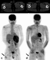
A 59-year-old male patient with acinar cell carcinoma (ACC), the same patient as in Figs. 8 and 9. a and b Dynamic contrast-enhanced CT (DCE-CT) in the pancreatic phase, c and d maximum intensity projection images of 1⁸F-fluoro-2-deoxy-D-glucose positron emission tomography with CT (1⁸F-FDG-PET/CT). Images (b) and (d) are obtained 7 months after the examinations shown in (a) and (b), respectively. The tumor is located in the pancreatic tail, measuring 15 cm in diameter, and exhibits expansile growth with well-defined margins. DCE-CT reveals an ill-defined, hazy appearance of the subcutaneous fat in the right thigh (arrow). 1⁸F-FDG-PET/CT demonstrates slightly increased 1⁸F-FDG uptake, with a maximum standardized uptake value of 2.14 in this region. These findings are consistent with pancreatic panniculitis (PP), characterized by subcutaneous fat necrosis associated with ACC. Following the surgical resection of the ACC, the ill-defined hazy appearance and 1⁸F-FDG uptake resolved, indicating an improvement in PP associated with the ACC
Fig. 11
Fig. 11
A 43-year-old male patient with a solid-pseudopapillary neoplasm. a Fat-suppressed T1-weighted imaging (FS-T1WI), b fat-suppressed T2-weighted imaging (FS-T2WI), c diffusion-weighted imaging (DWI) with b value of 800 s/mm2, and d an apparent diffusion coefficient (ADC) map. The tumor is located in the pancreatic head (arrow), measuring 2.0 cm in diameter, and exhibits expansile growth with well-defined margins. The central region of the tumor exhibits heterogeneous hyperintensity on FS-T1WI, FS-T2WI, and DWI, with an equivalent ADC values (1.46 × 10⁻3 mm2/s) relative to the pancreatic parenchyma. Additionally, the tumor demonstrates a hypointense rim on FS-T1WI, FS-T2WI, and DWI, corresponding to the peripheral rim calcification observed on CT (shown in Fig. 13)
Fig. 12
Fig. 12
A 45-year-old male patient with solid-pseudopapillary neoplasm. a Fat-suppressed T1-weighted imaging (FS-T1WI), b fat-suppressed T2-weighted imaging (FS-T2WI), c diffusion-weighted imaging (DWI) with b value of 800 s/mm2, and d apparent diffusion coefficient (ADC) map. The tumor is located in the pancreatic head (arrow), measuring 1.5 cm in diameter, and exhibits expansile growth with well-defined margins. The tumor exhibits heterogeneous hypointensity on FS-T1WI, heterogeneous hyperintensity on FS-T2WI, and DWI, with higher ADC values (3.10 × 10⁻3 mm2/s), relative to the pancreatic parenchyma. Additionally, the tumor demonstrates a small cystic component on the right side of the tumor on FS-T2WI
Fig. 13
Fig. 13
A 43-year-old female patient was diagnosed with a solid-pseudopapillary neoplasm, the same patient as in Fig. 11. a Non-contrast-enhanced computed tomography (CT), b dynamic contrast-enhanced CT in the pancreatic phase, c portal venous phase, d delayed phase, and e 1⁸F-fluoro-2-deoxy-D-glucose positron emission tomography with CT (1⁸F-FDG-PET/CT). The tumor is located in the pancreatic head (arrow), measuring 2.0 cm in diameter, and exhibits expansile growth with well-defined margins with peripheral rim calcification. The central region of the tumor demonstrates poor enhancement during the pancreatic phase, with slightly persistent enhancement observed in the portal venous and delayed phases. On 1⁸F-FDG-PET/CT, the tumor shows mildly increased 18F-FDG uptake, with a maximum standardized uptake value of 2.51
Fig. 14
Fig. 14
A 45-year-old female patient with solid-pseudopapillary neoplasm, the same patient as in Fig. 10. a Non-contrast-enhanced computed tomography (CT), b dynamic contrast-enhanced CT in the pancreatic phase, c portal phase, d delayed phase, and e 1⁸F-fluoro-2-deoxy-D-glucose positron emission tomography with CT (1⁸F-FDG-PET/CT). The tumor is located in the pancreatic head (arrow), measuring 1.5 cm in diameter, and exhibits expansile growth with well-defined margins. The tumor shows poor enhancement in the pancreatic phase with persistent enhancement in the portal and delayed phases. Tumor necrosis and calcification are not evident. On 1⁸F-FDG-PET/CT, the tumor shows mildly increased 18F-FDG uptake, with a maximum standardized uptake value of 2.44

Similar articles

References

    1. Vital Statistics Japan reported by the Ministry of Health, Labour and Welfare (2022) [Internet]. Available from: https://www.mhlw.go.jp/toukei/saikin/hw/jinkou/geppo/nengai22/index.html.
    1. WHO Classification of Tumours Editorial Board: WHO Classification of Tumors, 5th ed, vol. 1 (2019) Digestive system tumours. Chapter 10. Tumours of the pancreas. IARC Press Lyon.
    1. Bengtsson A, Andersson R, Ansari D. Histological variants of pancreatic ductal adenocarcinoma: a survival analysis. Langenbecks Arch Surg. 2024;409(1):312. 10.1007/s00423-024-03506-6. - PMC - PubMed
    1. Japanese Radiological Society ed. Diagnostic imaging guidelines 2021: English version. 2021. Available from: https://www.radiology.jp/content/files/gl2021/diagnostic_imaging_guideli...
    1. Schawkat K, Manning MA, Glickman JN, Mortele KJ. Pancreatic ductal adenocarcinoma and its variants: pearls and perils. Radiographics. 2020;40(5):1219–39. 10.1148/rg.2020190184. - PubMed

MeSH terms