. 2025 Apr 11;11(15):eadr6211.
doi: 10.1126/sciadv.adr6211. Epub 2025 Apr 11.

Female sex hormones exacerbate retinal neurodegeneration

Affiliations

Female sex hormones exacerbate retinal neurodegeneration

Ashley A Rowe et al. Sci Adv. .

Abstract

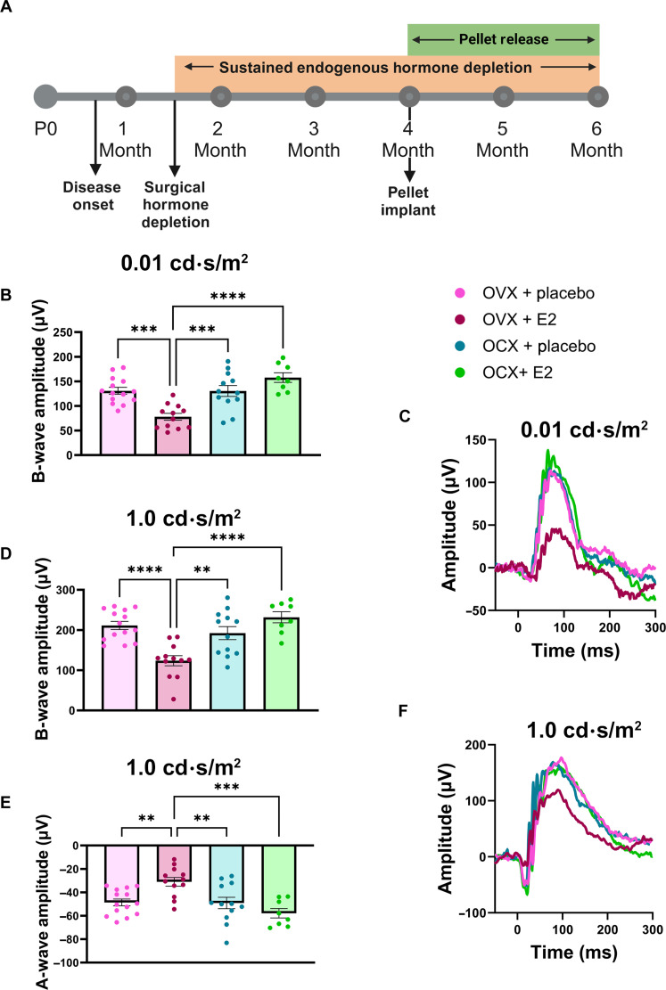
Neurodegenerative disorders such as Alzheimer's disease and macular degeneration represent major sources of human suffering, yet factors influencing disease severity remain poorly understood. Sex has been implicated as one modifying factor. Here, we show that female sex is a risk factor for worsened outcomes in a model of retinal degeneration and that this susceptibility is caused by the presence of female-specific sex hormones. The adverse effect of female sex hormones was specific to diseased retinal neurons, and depletion of these hormones ameliorated this phenotypic effect, while reintroduction worsened rates of disease in females. Transcriptional analysis of retinas showed significant differences between genes involved in pyroptosis, inflammatory responses, and endoplasmic reticulum stress-induced apoptosis between males and females with retinal degeneration. These findings provide crucial insights into the pathogenesis of neurodegenerative diseases and how sex hormones can affect disease severity. These findings have far-reaching implications for clinical trial design and the use of hormonal therapy in females with certain neurodegenerative disorders.


Figures

Fig. 1.
Fig. 1.. RHO P23H mice display sexually dimorphic loss of photoreceptor function and survival.
(A) Scotopic ERG (0.01 cd·s/m2) b-wave amplitude values for male (blue) and female (purple) RHO P23H mice from 1 to 7 months of age. (B to D) Representative 0.01 cd⋅s/m2 scotopic ERG traces at 1, 4, and 7 months of age. (E) Scotopic ERG (1.0 cd·s/m2) b- and (F) a-wave amplitudes from 1 to 7 months of age. (G to I) Representative 1.0 cd·s/m2 scotopic ERG traces at 1, 4, and 7 months of age. Error bars = SEM. *P < 0.05, **P < 0.01, ***P < 0.001, and ****P < 0.0001. Statistical analysis performed by multiple unpaired t test with a Holm-Šídák correction with α = 0.05. N ≥ 12 eyes per group. (J) Representative hematoxylin and eosin–stained histology from 7-month-old RHO P23H female and male mouse retinas. GCL, ganglion cell layer; INL, inner nuclear layer; RPE, retinal pigmented epithelium. Scale bars, 50 μm. (K) Quantification of ONL thickness measured every 100 μm on either side of the optic nerve head (ONH). Statistics for ONL quantification are available in data S1. N ≥ 3 mice per group with 38 measurements per retina.
Fig. 2.
Fig. 2.. Surgical gonadectomy depletes sex hormones in the serum and retina.
(A) Timeline depicting the surgical procedure at 6 weeks of age, after disease onset, followed by sustained hormone depletion through the duration of the study. P0, postnatal day 0. Figure panel created in BioRender (K.J.W., 2025; https://BioRender.com/f498903). Mass spectrometry analysis of (B) serum and (C) retina testosterone and progesterone in wild-type females (purple), males (blue), females with OVX (pink), and males with OCX (orange), 6 weeks postsurgery. Statistics performed via one-way analysis of variance (ANOVA) with Tukey’s multiple comparisons test. N ≥ 8 mice per group. Error bars = SEM. *P < 0.05, ***P < 0.001, and ****P < 0.0001.
Fig. 3.
Fig. 3.. OVX ameliorates the accelerated female photoreceptor neurodegeneration.
(A) Scotopic (0.01 cd·s/m2) b-wave amplitudes from 4 to 7 months of age in RHO P23H females (purple), males (blue), females with OVX (pink), and males with OCX (light blue). (B and C) Representative 0.01 cd·s/m2 scotopic traces. (D) Scotopic (1.0 cd·s/m2) b- and (E) a-wave amplitudes from 4 to 7 months of age and (F and G) representative traces. Statistics performed via two-way ANOVA with Tukey’s multiple comparisons test. Statistical results are shown for females compared to females + OVX and males compared to males + OCX. N ≥ 12 eyes. (H) Photopic amplitudes of 3.0 cd·s/m2 and (J) 10.0 cd·s/m2 at 7 months of age, (I and K) with respective representative traces. Statistics analyzed by one-way ANOVA with Tukey’s multiple comparisons test. N ≥ 10 eyes. Error bars = SEM. *P < 0.05, **P < 0.01, and ****P < 0.0001.
Fig. 4.
Fig. 4.. OVX ameliorates the loss of photoreceptor nuclei in females.
(A) Hematoxylin and eosin–stained histology from 7- and 10-month-old RHO P23H female and male mice with and without OVX or OCX, respectively. Scale bars, 50 μm. (B) Quantification of ONL thickness at 100-μm distances spanning from the ONH at both 7 and 10 months of age. Statistics performed via two-way ANOVA with Tukey’s multiple comparisons test. Statistics for ONL quantification are available in data S1. N ≥ 3 mice per group with 38 measurements per retina. Error bars = SEM.
Fig. 5.
Fig. 5.. Exogenous 17β-estradiol affects male and female retinas without altering progesterone or testosterone levels.
Mass spectrometry analysis of the serum and retina levels of estradiol, progesterone, and testosterone in 12-week-old wild-type mice that received either a vehicle injection (black) or an injection of estradiol (green). All mice received surgical hormone depletion (OVX and OCX) at 6 weeks of age. Statistics analyzed via t test (α = 0.05). Error bars = SD. **P < 0.01 and ***P < 0.001.
Fig. 6.
Fig. 6.. Reintroduction of estradiol at midstage of disease exacerbates visual function loss in females, but not males, with RP.
(A) Timeline depicting surgical procedure at 6 weeks of age, after disease onset, followed by sustained hormone depletion through the duration of the study and reintroduction of estradiol via implanted pellet at 4 months of age. Figure panel created in BioRender (K.J.W., 2025; https://BioRender.com/k14c552). (B) ERG results from female RHOP23H/+ (RP) mice implanted with either a placebo (pink) or estradiol (E2) (burgundy) slow-release pellet and male RP mice implanted with either placebo (blue) or E2 (green) pellets. Scotopic ERG (0.01 cd·s/m2) b-wave amplitudes, (D) scotopic ERG (1.0 cd·s/m2) b-wave, and (E) a-wave amplitudes. Representative ERG traces for the (C) 0.01 cd·s/m2 and (F) 1.0 cd·s/m2 light intensity settings. Statistics analyzed via one-way ANOVA (α = 0.05) with Tukey’s multiple comparisons test. N ≥ 8 eyes per group. Error bars = SEM. **P < 0.01, ***P < 0.001, and ****P < 0.0001.
Fig. 7.
Fig. 7.. RP females display a significant elevation of genes related to pyroptosis, inflammatory, and endoplasmic reticulum stress–induced apoptotic responses compared to RP males.
(A) TUNEL staining in 7-week-old RHOP23H/+ (RP) and wild-type (WT) retinas. DAPI (4′,6-diamidino-2-phenylindole) nuclear stain in blue and TUNEL in red. Scale bars, 25 μm. (B) Quantification of TUNEL-positive photoreceptor nuclei per region. N = 3 mice per group. Relative expression as measured by qPCR of the neural retina tissue for (C) Casp4 (caspase 4) and (D) Aif1 (allograft inflammatory factor 1). N = 3 retinas per group. WT females, burgundy; WT males, light blue; RP females, purple; RP males, blue. Statistics analyzed via one-way ANOVA (α = 0.05) with Tukey’s multiple comparisons test. Error bars = SEM. *P < 0.05, **P < 0.01, ***P < 0.001, and ****P < 0.0001.
Fig. 8.
Fig. 8.. Sex hormones affect cellular pathways underlying the RHO P23H mutation in photoreceptor neurons.
Graphical schematic depicting our findings that female systemic sex hormones interact within pathways found in diseased, but not healthy, retinal neurons to exacerbate neurodegeneration in a sexually dimorphic manner. Figure created in BioRender (K.J.W., 2025; https://BioRender.com/m93w003).

Update of

Similar articles

References

    1. Lopez-Lee C., Torres E. R. S., Carling G., Gan L., Mechanisms of sex differences in Alzheimer's disease. Neuron 112, 1208–1221 (2024). - PMC - PubMed
    1. Marin A. I., Poppelaars F., Wagner B. D., Palestine A. G., Patnaik J. L., Holers V. M., Frazer-Abel A. A., Mathias M. T., Manoharan N., Fonteh C. N., Mandava N., Lynch A. M., University of Colorado Retina Research Group , Sex and age-related differences in complement factors among patients with intermediate age-related macular degeneration. Transl. Vis. Sci. Technol. 11, 22 (2022). - PMC - PubMed
    1. Mielke M. M., Sex and gender differences in Alzheimer's disease dementia. Psychiatr. Times 35, 14–17 (2018). - PMC - PubMed
    1. Nuzzi R., Scalabrin S., Becco A., Panzica G., Gonadal hormones and retinal disorders: A review. Front. Endocrinol. 9, 66 (2018). - PMC - PubMed
    1. Sasaki M., Harada S., Kawasaki Y., Watanabe M., Ito H., Tanaka H., Takeuchi A., Tsubota K., Takebayashi T., Nishiwaki Y., Kawasaki R., Gender-specific association of early age-related macular degeneration with systemic and genetic factors in a Japanese population. Sci. Rep. 8, 785 (2018). - PMC - PubMed

Substances

LinkOut - more resources