. 2025 Apr 11;11(15):eadr2631.
doi: 10.1126/sciadv.adr2631. Epub 2025 Apr 11.

Generation of an inflammatory niche in a hydrogel depot through recruitment of key immune cells improves efficacy of mRNA vaccines

Affiliations

Generation of an inflammatory niche in a hydrogel depot through recruitment of key immune cells improves efficacy of mRNA vaccines

Emily L Meany et al. Sci Adv. .

Abstract

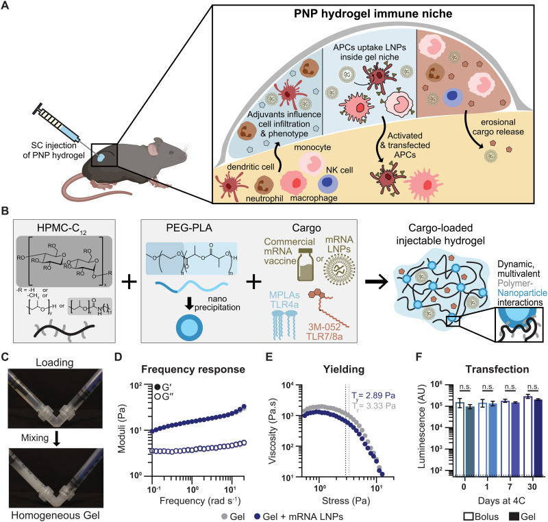
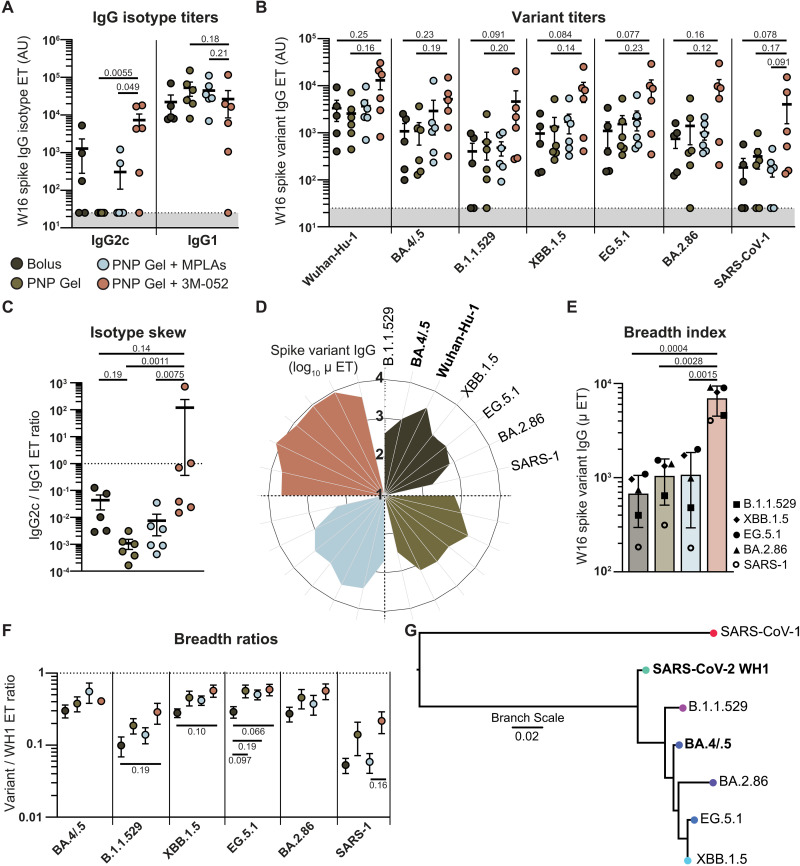
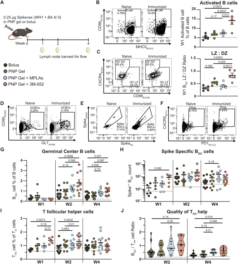
Messenger RNA (mRNA) delivered in lipid nanoparticles (LNPs) rose to the forefront of vaccine candidates during the COVID-19 pandemic due to scalability, adaptability, and potency. Yet, there remain critical areas for improvements of these vaccines in durability and breadth of humoral responses. In this work, we explore a modular strategy to target mRNA/LNPs to antigen-presenting cells with an injectable polymer-nanoparticle (PNP) hydrogel technology, which recruits key immune cells and forms an immunological niche in vivo. We characterize this niche on a single-cell level and find it is highly tunable through incorporation of adjuvants like MPLAs and 3M-052. Delivering commercially available severe acute respiratory syndrome coronavirus 2 mRNA vaccines in PNP hydrogels improves the durability and quality of germinal center reactions, and the magnitude, breadth, and durability of humoral responses. The tunable immune niche formed within PNP hydrogels effectively skews immune responses based on encapsulated adjuvants, creating opportunities to precisely modulate mRNA/LNP vaccines for various indications from infectious diseases to cancers.


Figures

Fig. 1.
Fig. 1.. Schematic of immune niche in PNP hydrogels comprising mRNA/LNPs.
(A) PNP hydrogels injected subcutaneously allow immune cell infiltration and interaction with loaded cargo. Incorporation of various adjuvants influences the immune niche. Cells within the niche take up mRNA/LNPs, express the delivered protein, and migrate to initiate immune responses in the draining LNs. (B) PNP hydrogels comprise HPMC-C12 and PEG-PLA NPs. Cargo, including mRNA/LNP vaccines and molecular adjuvants, can be easily mixed with the polymer components to form a dynamic hydrogel. (C) Hydrogel components are loaded into two syringes, attached with an elbow, and mixed to form a homogenous material, preloaded in a syringe and ready for administration. (D) Frequency shear rheology shows that PNP-0.5-5 material is solid-like across timescales, and mRNA/LNPs (50 μg ml−1) do not alter this. (E) Stress-ramp data show static yield stress, defined as the intersection of tangent lines for the plateau and yielding regimes, for PNP-0.5-5 hydrogel is not affected by LNPs. (F) Luminescent signal from RAW-Blue macrophages dosed with luciferase mRNA/LNPs in either PBS bolus or PNP hydrogel after storage in that condition for up to 30 days at 4°C. Hydrogels do not affect transfection or stability of mRNA/LNPs. Data shown as means ± SD, n = 3 to 4, and statistics are multiple unpaired two-tailed t tests run in GraphPad Prism with false discovery rate correction using two-stage step-up method of Benjamini, Krieger, and Yekutieli.
Fig. 2.
Fig. 2.. Characterization of in vivo immune niche in PNP hydrogels.
(A) PNP hydrogels loaded with mCherry mRNA/LNPs injected subcutaneously were excised at days 3 and 7 for dissociation and analysis of cells with flow cytometry. (B) Excised PNP hydrogel depot on day 7 showing size and no visible evidence of gross fibrosis. (C) Counts of CD45+ leukocytes per gel show robust infiltration on both days, with increased infiltration with adjuvant cargo. Data shown as means ± SEM, n = 6. Statistical values shown are P values <0.25 obtained from general linear model (GLM) fitting and Tukey post hoc multiple comparison test in JMP. (D) Changes in cell type populations visualized with UMAPs. (E) Quantification of cell types in each hydrogel niche as percentages of all gated CD45+ cells. Data shown as means + SEM, n = 6. (F) UMAP visualization showing relation of Ly6Chi macrophage subpopulation to macrophages and monocytes. (G) UMAP visualization of DC subpopulations highlighting differences between classical DCs (cDC1s and 2 s) and mDCs.
Fig. 3.
Fig. 3.. Characterization of mRNA expression in PNP hydrogel immune niche.
(A) Immune cells infiltrate injected PNP hydrogels loaded with mCherry mRNA/LNPs in vivo, take up LNPs, and express reporter mCherry protein. These cells can be quantified on days 3 and 7 following hydrogel excision with flow cytometry. (B) Representative flow plots showing mCherry signal in CD45+ cells with percentage of CD45+ parent and total cell counts. (C) The count of mCherry expressing CD45+ leukocytes per gel and (D) those cells as a percentage of all CD45+ cells. Adjuvants increase the absolute number and percentage of CD45+ cells expressing the delivered mRNA. Data shown as means ± SEM, n = 6. Statistical values shown are P values <0.25 obtained from GLM fitting and Tukey post hoc multiple comparison test in JMP. (E) Quantification of cell types in the mCherry+ subniche per hydrogel as percentages of all gated CD45+ mCherry+ cells. Data shown as means + SEM, n = 6. (F) Ratio of the proportion each cell type makes up of the mCherry+ subniche to its percentage of the full CD45+ niche. Values of one indicate that a cell is represented equally in the mCherry+ subniche as in the full gel. Values under one indicate that fewer cells are mCherry+ than expected based on that cell type’s percentage of the CD45+ niche, and vice versa for values over one (overrepresented in mCherry+ subniche). Data shown as means ± SEM, n = 6.
Fig. 4.
Fig. 4.. Humoral response to hydrogel-based mRNA/LNP vaccines.
(A) Mice were immunized with 0.25 μg of commercially available bivalent SARS-CoV-2 mRNA vaccine (0.125 μg of each variant) in PBS bolus or PNP-0.5-5 hydrogel with or without 1 μg of 3M-052 or 20 μg of MPLAs at week 0 and boosted with a homologous boost at week 8. Serum collected at designated time points was analyzed for anti-spike (Wuhan-Hu-1) antibodies via ELISA, as well as for other viral variant anti-spike antibodies, IgG subtypes, and neutralizing capacity. A separate cohort of animals primed and boosted in the same fashion were euthanized 2 weeks post-boost and spleens harvested for ELISpot evaluation of spike specific (WH1) T cells. Anti-WH1 spike IgG end point titers for all animals at (B) week 8, (C) week 26, and (D) over time. (E) AUC of antibody titer from week 1 to 8 (pre-boost) and 1 to 26 (full time course) per animal. (F) Decay half-life of antibody titer post-boost derived from parametric bootstrapping of titers following each treatment group’s post-boost peak. Data shown as means ± SEM, n = 1000 simulations. (G) Percent infectivity of BA.4/.5 pseudotyped lentivirus at week 13 post-prime and 1:50 serum dilution. (H) Spike-specific (WH1) IFN-γ–producing splenocytes, as a proxy for T cells, at week 2 post-boost. Data shown as means ± SEM, n = 4 to 6. Statistical values shown are P values <0.25 obtained from GLM fitting and Tukey post hoc multiple comparison test in JMP. In (D), comparisons shown are to bolus control for the groups of the text color.
Fig. 5.
Fig. 5.. Breadth and functionality of response to hydrogel-based mRNA/LNP vaccines.
(A) IgG isotype end point titers at week 16 post-prime. (B) Anti-spike end point ELISA titers against different variants at week 16 post-prime. (C) Ratio of IgG2c to IgG1 antibody isotypes indicating TH1 (higher values) or TH2 (lower values) skewing. (D) Radar plot of absolute IgG end point titer for each variant. Broader and larger petals indicate improved breadth and consistency of response. (E) Quantification of (D), where each data point is the average of n = 5 to 6 animals against a single spike variant. Data shown as means ± SD. (F) Ratio of end point IgG titer for each variant compared with WH1 as a metric of consistency and breadth of humoral response. (G) Phylogenetic tree showing lineage of variants assessed created using “Gene/Protein Tree” tool on the Bacterial and Viral Bioinformatics Resource Center (BV-BRC) website https://bv-brc.org/. Unless otherwise written, data shown as means ± SEM, n = 5 to 6. Statistical values shown are P values <0.25 obtained from GLM fitting and Tukey post hoc multiple comparison test in JMP.
Fig. 6.
Fig. 6.. Germinal center response to hydrogel-based mRNA/LNP vaccines.
(A) Mice were immunized with bivalent SARS-CoV-2 mRNA vaccine in PBS bolus or PNP-0.5-5 hydrogel with or without adjuvants and LNs harvested at weeks 1, 2, and 4. Week 1 representative flow plots and quantification of (B) activated B cells (B220+ MHCII+ CD86+) and (C) light and dark zone BGC cells (B220+ CD95 GL7+ CD38 CD86+ CXCR4 and CD86 CXCR4+). (D) Representative flow plots of BGC cells (B220+ CD95 GL7+), (E) spike-specific BGC cells (CD38 Spike++), and (F) TFH (CD19 CD3+ CD4+ CXCR5+ PD-1+). Percentages of parent population except Spike++ BGC cells, which are of grandparent (see supplemental gating strategy). (G) Quantification of BGC cell percentage of all B cells. (H) Counts of Spike++ BGC cells. (I) Quantification of TFH cell percentage of all CD4+ T cells. (J) Ratio of BGC cells to TFH cells as a metric of TFH cell help quality. All data shown as means ± SEM, n = 5 to 10. Statistical values shown are P values <0.25, obtained from GLM fitting and Tukey post hoc multiple comparison test in JMP.

Update of

Similar articles

Cited by

References

    1. Chaudhary N., Weissman D., Whitehead K. A., mRNA vaccines for infectious diseases: Principles, delivery and clinical translation. Nat. Rev. Drug Discov. 20, 817–838 (2021). - PMC - PubMed
    1. Hou X., Zaks T., Langer R., Dong Y., Lipid nanoparticles for mRNA delivery. Nat. Rev. Mater. 6, 1078–1094 (2021). - PMC - PubMed
    1. Paunovska K., Loughrey D., Dahlman J. E., Drug delivery systems for RNA therapeutics. Nat. Rev. Genet. 23, 265–280 (2022). - PMC - PubMed
    1. Echaide M., Chocarro de Erauso L., Bocanegra A., Blanco E., Kochan G., Escors D., mRNA vaccines against SARS-CoV-2: Advantages and caveats. Int. J. Mol. Sci. 24, 5944 (2023). - PMC - PubMed
    1. Pardi N., Hogan M. J., Weissman D., Recent advances in mRNA vaccine technology. Curr. Opin. Immunol. 65, 14–20 (2020). - PubMed

MeSH terms